Advertisement

Main menu

  • Home
  • Content
    • Current Issue
    • Accepted Manuscripts
    • Article Preview
    • Past Issue Archive
    • Video Articles
    • AJNR Case Collection
    • Case of the Week Archive
    • Case of the Month Archive
    • Classic Case Archive
  • Special Collections
    • AJNR Awards
    • ASNR Foundation Special Collection
    • Most Impactful AJNR Articles
    • Photon-Counting CT
    • Spinal CSF Leak Articles (Jan 2020-June 2024)
  • Multimedia
    • AJNR Podcasts
    • AJNR SCANtastic
    • Trainee Corner
    • MRI Safety Corner
    • Imaging Protocols
  • For Authors
    • Submit a Manuscript
    • Submit a Video Article
    • Submit an eLetter to the Editor/Response
    • Manuscript Submission Guidelines
    • Statistical Tips
    • Fast Publishing of Accepted Manuscripts
    • Graphical Abstract Preparation
    • Imaging Protocol Submission
    • Author Policies
  • About Us
    • About AJNR
    • Editorial Board
    • Editorial Board Alumni
  • More
    • Become a Reviewer/Academy of Reviewers
    • Subscribers
    • Permissions
    • Alerts
    • Feedback
    • Advertisers
    • ASNR Home

User menu

  • Alerts
  • Log in

Search

  • Advanced search
American Journal of Neuroradiology

American Journal of Neuroradiology

ASHNR American Society of Functional Neuroradiology ASHNR American Society of Pediatric Neuroradiology ASSR
  • Alerts
  • Log in

Advanced Search

  • Home
  • Content
    • Current Issue
    • Accepted Manuscripts
    • Article Preview
    • Past Issue Archive
    • Video Articles
    • AJNR Case Collection
    • Case of the Week Archive
    • Case of the Month Archive
    • Classic Case Archive
  • Special Collections
    • AJNR Awards
    • ASNR Foundation Special Collection
    • Most Impactful AJNR Articles
    • Photon-Counting CT
    • Spinal CSF Leak Articles (Jan 2020-June 2024)
  • Multimedia
    • AJNR Podcasts
    • AJNR SCANtastic
    • Trainee Corner
    • MRI Safety Corner
    • Imaging Protocols
  • For Authors
    • Submit a Manuscript
    • Submit a Video Article
    • Submit an eLetter to the Editor/Response
    • Manuscript Submission Guidelines
    • Statistical Tips
    • Fast Publishing of Accepted Manuscripts
    • Graphical Abstract Preparation
    • Imaging Protocol Submission
    • Author Policies
  • About Us
    • About AJNR
    • Editorial Board
    • Editorial Board Alumni
  • More
    • Become a Reviewer/Academy of Reviewers
    • Subscribers
    • Permissions
    • Alerts
    • Feedback
    • Advertisers
    • ASNR Home
  • Follow AJNR on Twitter
  • Visit AJNR on Facebook
  • Follow AJNR on Instagram
  • Join AJNR on LinkedIn
  • RSS Feeds

AJNR is seeking candidates for the AJNR Podcast Editor. Read the position description.

OtherPEDIATRICS

Hypophyseal Triplication: Case Report and Embryologic Considerations

R. Manara, V. Citton, M. Rossetto, A. Padoan and D. D’Avella
American Journal of Neuroradiology August 2009, 30 (7) 1328-1329; DOI: https://doi.org/10.3174/ajnr.A1520
R. Manara
  • Find this author on Google Scholar
  • Find this author on PubMed
  • Search for this author on this site
V. Citton
  • Find this author on Google Scholar
  • Find this author on PubMed
  • Search for this author on this site
M. Rossetto
  • Find this author on Google Scholar
  • Find this author on PubMed
  • Search for this author on this site
A. Padoan
  • Find this author on Google Scholar
  • Find this author on PubMed
  • Search for this author on this site
D. D’Avella
  • Find this author on Google Scholar
  • Find this author on PubMed
  • Search for this author on this site
  • Article
  • Figures & Data
  • Info & Metrics
  • Responses
  • References
  • PDF
Loading

Article Figures & Data

Figures

  • Fig 1.
    • Download figure
    • Open in new tab
    • Download powerpoint
    Fig 1.

    Unenhanced T1-weighted multiplanar reformatted MR images. A, Coronal image showing paired infundibula (white arrows) extending inferiorly to 2 pituitary glands symmetrically located in the lateral recesses of the pituitary fossa; the hypothalamus appears grossly thickened (black arrows). B, Coronal image along the pituitary stalk (white arrow) of the median aspect of the hypophysis, which appears as an oval, hyperintense-to-gray matter, homogeneous mass partially housed in the pituitary fossa (white arrowhead). Similar features might be found also in pedunculated hamartomas, which are usually characterized by the presence of clinical and laboratory signs of precocious puberty and by T1 isointensity compared with gray matter. C, In this oblique axial image, the 3 pituitary glands are contemporaneously visible; the paramedian glands present both the adenohypophyseal component (white stars) and the neurohypophyseal bright spot (black arrowheads), whereas the midline hypophysis (white arrowhead) is homogeneously isointense.
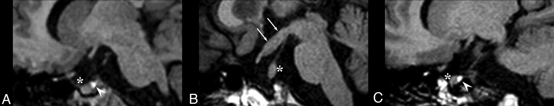
  • Fig 2.
    • Download figure
    • Open in new tab
    • Download powerpoint
    Fig 2.

    A,C, Paramedian sagittal T1-weighted images show 2 different pituitary glands, each with a posterior bright spot (white arrowhead) and an anterior pituitary lobe (white star). B, Midline sagittal T1-weighted image revealing a thickened third ventricle floor (white arrows) and an homogeneous, isointense median mass (white star) with its independent stalk.

PreviousNext
Back to top

In this issue

American Journal of Neuroradiology: 30 (7)
American Journal of Neuroradiology
Vol. 30, Issue 7
August 2009
  • Table of Contents
  • Index by author
Advertisement
Print
Download PDF
Email Article

Thank you for your interest in spreading the word on American Journal of Neuroradiology.

NOTE: We only request your email address so that the person you are recommending the page to knows that you wanted them to see it, and that it is not junk mail. We do not capture any email address.

Enter multiple addresses on separate lines or separate them with commas.
Hypophyseal Triplication: Case Report and Embryologic Considerations
(Your Name) has sent you a message from American Journal of Neuroradiology
(Your Name) thought you would like to see the American Journal of Neuroradiology web site.
CAPTCHA
This question is for testing whether or not you are a human visitor and to prevent automated spam submissions.
Cite this article
R. Manara, V. Citton, M. Rossetto, A. Padoan, D. D’Avella
Hypophyseal Triplication: Case Report and Embryologic Considerations
American Journal of Neuroradiology Aug 2009, 30 (7) 1328-1329; DOI: 10.3174/ajnr.A1520

Citation Manager Formats

  • BibTeX
  • Bookends
  • EasyBib
  • EndNote (tagged)
  • EndNote 8 (xml)
  • Medlars
  • Mendeley
  • Papers
  • RefWorks Tagged
  • Ref Manager
  • RIS
  • Zotero
0 Responses
Respond to this article
Share
Bookmark this article
Hypophyseal Triplication: Case Report and Embryologic Considerations
R. Manara, V. Citton, M. Rossetto, A. Padoan, D. D’Avella
American Journal of Neuroradiology Aug 2009, 30 (7) 1328-1329; DOI: 10.3174/ajnr.A1520
del.icio.us logo Twitter logo Facebook logo Mendeley logo
  • Tweet Widget
  • Facebook Like
  • Google Plus One
Purchase

Jump to section

  • Article
    • Abstract
    • Case Report
    • Discussion
    • References
  • Figures & Data
  • Info & Metrics
  • Responses
  • References
  • PDF

Related Articles

  • No related articles found.
  • PubMed
  • Google Scholar

Cited By...

  • No citing articles found.
  • Crossref (6)
  • Google Scholar

This article has been cited by the following articles in journals that are participating in Crossref Cited-by Linking.

  • Clinical Presentation of Epignathus Teratoma With Cleft Palate; and Duplication of Cranial Base, Tongue, Mandible, and Pituitary Gland
    Yujiro Maeda, Hideyuki Suenaga, Madoka Sugiyama, Hideto Saijo, Kazuto Hoshi, Yoshiyuki Mori, Tsuyoshi Takato
    Journal of Craniofacial Surgery 2013 24 4
  • Morphological Changes of Olfactory Bulbs and Grooves
    Lucien M. Levy, Andrew J. Degnan, Salil Sharma, Linda Kelahan, Robert I. Henkin
    Journal of Computer Assisted Tomography 2012 36 4
  • Fetal MRI
    Maria Theresa Schmook
    2010
  • Imaging Endocrine Diseases in Children
    Pierre Bessou, Sylviane Hanquinet, Jean-François Chateil
    2012
  • MRI of the Pituitary Gland
    Sonia Nagi
    2016
  • Neuro-Imagerie Diagnostique
    J.-L. Dietemann, M. Abu Eid, N. Rotaru, M. Koob, S. Draghici
    2018

More in this TOC Section

  • SyMRI & MR Fingerprinting in Brainstem Myelination
  • Comparison of Image Quality and Radiation Dose in Pediatric Temporal Bone CT Using Photon-Counting Detector CT and Energy-Integrating Detector CT
  • Neonatal Hypocalcemia: Cerebral MRI Abnormalities
Show more Pediatrics

Similar Articles

Advertisement

Indexed Content

  • Current Issue
  • Accepted Manuscripts
  • Article Preview
  • Past Issues
  • Editorials
  • Editor's Choice
  • Fellows' Journal Club
  • Letters to the Editor
  • Video Articles

Cases

  • Case Collection
  • Archive - Case of the Week
  • Archive - Case of the Month
  • Archive - Classic Case

Special Collections

  • AJNR Awards
  • ASNR Foundation Special Collection
  • Most Impactful AJNR Articles
  • Photon-Counting CT
  • Spinal CSF Leak Articles (Jan 2020-June 2024)

More from AJNR

  • Trainee Corner
  • Imaging Protocols
  • MRI Safety Corner

Multimedia

  • AJNR Podcasts
  • AJNR Scantastics

Resources

  • Turnaround Time
  • Submit a Manuscript
  • Submit a Video Article
  • Submit an eLetter to the Editor/Response
  • Manuscript Submission Guidelines
  • Statistical Tips
  • Fast Publishing of Accepted Manuscripts
  • Graphical Abstract Preparation
  • Imaging Protocol Submission
  • Evidence-Based Medicine Level Guide
  • Publishing Checklists
  • Author Policies
  • Become a Reviewer/Academy of Reviewers
  • News and Updates

About Us

  • About AJNR
  • Editorial Board
  • Editorial Board Alumni
  • Alerts
  • Permissions
  • Not an AJNR Subscriber? Join Now
  • Advertise with Us
  • Librarian Resources
  • Feedback
  • Terms and Conditions
  • AJNR Editorial Board Alumni

American Society of Neuroradiology

  • Not an ASNR Member? Join Now

© 2025 by the American Society of Neuroradiology All rights, including for text and data mining, AI training, and similar technologies, are reserved.
Print ISSN: 0195-6108 Online ISSN: 1936-959X

Powered by HighWire