Advertisement

Main menu

  • Home
  • Content
    • Current Issue
    • Accepted Manuscripts
    • Article Preview
    • Past Issue Archive
    • Video Articles
    • AJNR Case Collection
    • Case of the Week Archive
    • Case of the Month Archive
    • Classic Case Archive
  • Special Collections
    • AJNR Awards
    • ASNR Foundation Special Collection
    • Most Impactful AJNR Articles
    • Photon-Counting CT
    • Spinal CSF Leak Articles (Jan 2020-June 2024)
  • Multimedia
    • AJNR Podcasts
    • AJNR SCANtastic
    • Trainee Corner
    • MRI Safety Corner
    • Imaging Protocols
  • For Authors
    • Submit a Manuscript
    • Submit a Video Article
    • Submit an eLetter to the Editor/Response
    • Manuscript Submission Guidelines
    • Statistical Tips
    • Fast Publishing of Accepted Manuscripts
    • Graphical Abstract Preparation
    • Imaging Protocol Submission
    • Author Policies
  • About Us
    • About AJNR
    • Editorial Board
    • Editorial Board Alumni
  • More
    • Become a Reviewer/Academy of Reviewers
    • Subscribers
    • Permissions
    • Alerts
    • Feedback
    • Advertisers
    • ASNR Home

User menu

  • Alerts
  • Log in

Search

  • Advanced search
American Journal of Neuroradiology

American Journal of Neuroradiology

ASHNR American Society of Functional Neuroradiology ASHNR American Society of Pediatric Neuroradiology ASSR
  • Alerts
  • Log in

Advanced Search

  • Home
  • Content
    • Current Issue
    • Accepted Manuscripts
    • Article Preview
    • Past Issue Archive
    • Video Articles
    • AJNR Case Collection
    • Case of the Week Archive
    • Case of the Month Archive
    • Classic Case Archive
  • Special Collections
    • AJNR Awards
    • ASNR Foundation Special Collection
    • Most Impactful AJNR Articles
    • Photon-Counting CT
    • Spinal CSF Leak Articles (Jan 2020-June 2024)
  • Multimedia
    • AJNR Podcasts
    • AJNR SCANtastic
    • Trainee Corner
    • MRI Safety Corner
    • Imaging Protocols
  • For Authors
    • Submit a Manuscript
    • Submit a Video Article
    • Submit an eLetter to the Editor/Response
    • Manuscript Submission Guidelines
    • Statistical Tips
    • Fast Publishing of Accepted Manuscripts
    • Graphical Abstract Preparation
    • Imaging Protocol Submission
    • Author Policies
  • About Us
    • About AJNR
    • Editorial Board
    • Editorial Board Alumni
  • More
    • Become a Reviewer/Academy of Reviewers
    • Subscribers
    • Permissions
    • Alerts
    • Feedback
    • Advertisers
    • ASNR Home
  • Follow AJNR on Twitter
  • Visit AJNR on Facebook
  • Follow AJNR on Instagram
  • Join AJNR on LinkedIn
  • RSS Feeds

AJNR is seeking candidates for the AJNR Podcast Editor. Read the position description.

Review ArticleReview Articles
Open Access

MR Spectroscopy in Radiation Injury

P.C. Sundgren
American Journal of Neuroradiology September 2009, 30 (8) 1469-1476; DOI: https://doi.org/10.3174/ajnr.A1580
P.C. Sundgren
  • Find this author on Google Scholar
  • Find this author on PubMed
  • Search for this author on this site
  • Article
  • Figures & Data
  • Info & Metrics
  • Responses
  • References
  • PDF
Loading

Article Figures & Data

Figures

  • Fig 1.
    • Download figure
    • Open in new tab
    • Download powerpoint
    Fig 1.

    A, Axial noncontrast T1-weighted MR image obtained 18 months after resection and radiation of an anaplastic astrocytoma presenting with a hemorrhagic lesion in left parietal region. B, Postcontrast T1-weighted MR image demonstrates feathery Swiss cheese-like contrast enhancement surrounding the hemorrhagic lesion, suggestive of radiation injury. C, Axial T2-weighted MR image shows the extensive edema surrounding the lesion in the left hemisphere.

  • Fig 2.
    • Download figure
    • Open in new tab
    • Download powerpoint
    Fig 2.

    A, Postcontrast T1-weighted MR image obtained 12 months after resection and radiation of an ependymoma shows new contrast-enhancing lesions within the irradiated volume suspicious for tumor recurrence (arrow). B, 2D CSI MR spectroscopy (point-resolved spectroscopy sequence; TE, 144 ms, TR, 1500 ms) with manually placed voxels in the contrast-enhancing lesion and in the corresponding region in the contralateral hemisphere. C, 1H-MR spectrum shows moderately increased Cho and reduced NAA signal intensities (upper row), consistent with recurrent tumor, and normal signal intensities of NAA, Cho, and Cr in the right hemisphere (lower row).

  • Fig 3.
    • Download figure
    • Open in new tab
    • Download powerpoint
    Fig 3.

    A and B, Fluid-attenuated inversion recovery (FLAIR) (A) and postcontrast T1-weighted MR (B) images obtained 8 months after resection, radiation, and chemotherapy of an anaplastic oligodendroglioma in the left frontal lobe show a new area of hyperintensity on FLAIR (arrow, A) and a contrast-enhancing nodule (arrow, B) in the right frontal lobe within the irradiated volume, suspicious for radiation injury. C, Multivoxel 2D CSI MR spectroscopy (point-resolved spectroscopy sequence; TE, 144 ms; TR, 1500 ms) with manually placed voxels in the contrast-enhancing lesion and in the corresponding area in the left hemisphere. D, MR spectroscopy spectrum shows slightly decreased NAA and increased Cho signal intensities bilaterally, suggestive of radiation injury. A follow-up MR imaging (not shown) showed interval resolution of the enhancement and no new lesions.
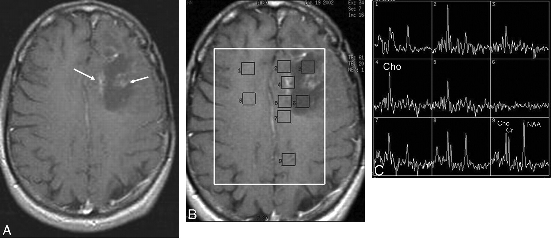
  • Fig 4.
    • Download figure
    • Open in new tab
    • Download powerpoint
    Fig 4.

    A, Postcontrast T1-weighted image obtained 12 months after resection, radiation, and chemotherapy of an astrocytoma in the left frontal lobe shows diffuse feathery contrast-enhancing areas in the vicinity of the resection cavity within the irradiated volume, suspicious for tumor recurrence. B, Multivoxel 2D CSI MR spectroscopy (point-resolved spectroscopy sequence; TE, 144 ms; TR, 1500 ms) with manually placed voxels in contrast-enhancing areas, in the cystic cavity, and in normal-appearing brain parenchyma in both left and right hemispheres. C, Significantly increased Cho and almost-absent NAA signal intensities in the contrast-enhancing areas, consistent with tumor recurrence verified at histopathology.

  • Fig 5.
    • Download figure
    • Open in new tab
    • Download powerpoint
    Fig 5.

    A, Postcontrast T1-weighted image obtained 10 months after resection and radiation of an astrocytoma in the left frontal lobe shows an irregular peripherally contrast-enhancing mass lesion with central necrosis surrounded by edema suspicious for tumor recurrence. The patient had the lesion resected, and histopathology revealed a high-grade astrocytoma. B, At follow-up MR imaging 6 months later and after additional radiation, a new diffuse contrast-enhancing lesion was present within the irradiated volume. 1H-MR spectroscopy by using SVS (point-resolved spectroscopy sequence; TE, 144 ms; TR, 2000 ms) was performed with the volume placed in over the contrast-enhancing lesion. C, Slightly increased Cho and normal NAA and Cr signal intensities are indicative of radiation injury, which was histopathologically confirmed after additional resection.

PreviousNext
Back to top

In this issue

American Journal of Neuroradiology: 30 (8)
American Journal of Neuroradiology
Vol. 30, Issue 8
1 Sep 2009
  • Table of Contents
  • Index by author
Advertisement
Print
Download PDF
Email Article

Thank you for your interest in spreading the word on American Journal of Neuroradiology.

NOTE: We only request your email address so that the person you are recommending the page to knows that you wanted them to see it, and that it is not junk mail. We do not capture any email address.

Enter multiple addresses on separate lines or separate them with commas.
MR Spectroscopy in Radiation Injury
(Your Name) has sent you a message from American Journal of Neuroradiology
(Your Name) thought you would like to see the American Journal of Neuroradiology web site.
CAPTCHA
This question is for testing whether or not you are a human visitor and to prevent automated spam submissions.
Cite this article
P.C. Sundgren
MR Spectroscopy in Radiation Injury
American Journal of Neuroradiology Sep 2009, 30 (8) 1469-1476; DOI: 10.3174/ajnr.A1580

Citation Manager Formats

  • BibTeX
  • Bookends
  • EasyBib
  • EndNote (tagged)
  • EndNote 8 (xml)
  • Medlars
  • Mendeley
  • Papers
  • RefWorks Tagged
  • Ref Manager
  • RIS
  • Zotero
0 Responses
Respond to this article
Share
Bookmark this article
MR Spectroscopy in Radiation Injury
P.C. Sundgren
American Journal of Neuroradiology Sep 2009, 30 (8) 1469-1476; DOI: 10.3174/ajnr.A1580
del.icio.us logo Twitter logo Facebook logo Mendeley logo
  • Tweet Widget
  • Facebook Like
  • Google Plus One
Purchase

Jump to section

  • Article
    • Abstract
    • Other Radiologic and Nuclear Medicine Methods to Discriminate Radiation Injury from Recurrent or Progressive Tumor
    • Effects of Radiation on Normal Brain
    • MR Spectroscopy in Radiation Injury
    • Measurements of Metabolites and Ratio Calculations
    • Need for Prediction Models for Clinical Decision Making
    • Conclusions
    • Acknowledgment
    • References
  • Figures & Data
  • Info & Metrics
  • Responses
  • References
  • PDF

Related Articles

  • No related articles found.
  • PubMed
  • Google Scholar

Cited By...

  • Differentiation between Radiation Necrosis and Tumor Progression Using Chemical Exchange Saturation Transfer
  • Pseudoprogression and Pseudoresponse: Imaging Challenges in the Assessment of Posttreatment Glioma
  • Crossref (107)
  • Google Scholar

This article has been cited by the following articles in journals that are participating in Crossref Cited-by Linking.

  • Pseudoprogression and Pseudoresponse: Imaging Challenges in the Assessment of Posttreatment Glioma
    L.C. Hygino da Cruz, I. Rodriguez, R.C. Domingues, E.L. Gasparetto, A.G. Sorensen
    American Journal of Neuroradiology 2011 32 11
  • Differentiating tumor recurrence from treatment necrosis: a review of neuro-oncologic imaging strategies
    Nishant Verma, Matthew C. Cowperthwaite, Mark G. Burnett, Mia K. Markey
    Neuro-Oncology 2013 15 5
  • Imaging of Brain Tumors: MR Spectroscopy and Metabolic Imaging
    Alena Horská, Peter B. Barker
    Neuroimaging Clinics of North America 2010 20 3
  • Radiation Necrosis in the Brain: Imaging Features and Differentiation from Tumor Recurrence
    Ritu Shah, Surjith Vattoth, Rojymon Jacob, Fathima Fijula Palot Manzil, Janis P. O’Malley, Peyman Borghei, Bhavik N. Patel, Joel K. Curé
    RadioGraphics 2012 32 5
  • Stereotactic radiosurgery for treatment of brain metastases
    Martin Kocher, Andrea Wittig, Marc Dieter Piroth, Harald Treuer, Heinrich Seegenschmiedt, Maximilian Ruge, Anca-Ligia Grosu, Matthias Guckenberger
    Strahlentherapie und Onkologie 2014 190 6
  • Postradiation Imaging Changes in the CNS: How can we Differentiate Between Treatment Effect and Disease Progression?
    Amanda J Walker, Jake Ruzevick, Ashkan A Malayeri, Daniele Rigamonti, Michael Lim, Kristin J Redmond, Lawrence Kleinberg
    Future Oncology 2014 10 7
  • Percent Change of Perfusion Skewness and Kurtosis: A Potential Imaging Biomarker for Early Treatment Response in Patients with Newly Diagnosed Glioblastomas
    Hye Jin Baek, Ho Sung Kim, Namkug Kim, Young Jun Choi, Young Joong Kim
    Radiology 2012 264 3
  • Differentiation between Radiation Necrosis and Tumor Progression Using Chemical Exchange Saturation Transfer
    Hatef Mehrabian, Kimberly L. Desmond, Hany Soliman, Arjun Sahgal, Greg J. Stanisz
    Clinical Cancer Research 2017 23 14
  • Differentiating Radiation-Induced Necrosis from Recurrent Brain Tumor Using MR Perfusion and Spectroscopy: A Meta-Analysis
    Ming-Tsung Chuang, Yi-Sheng Liu, Yi-Shan Tsai, Ying-Chen Chen, Chien-Kuo Wang, Jeroen Hendrikse
    PLOS ONE 2016 11 1
  • Distinguishing True Progression From Radionecrosis After Stereotactic Radiation Therapy for Brain Metastases With Machine Learning and Radiomics
    Luke Peng, Vishwa Parekh, Peng Huang, Doris D. Lin, Khadija Sheikh, Brock Baker, Talia Kirschbaum, Francesca Silvestri, Jessica Son, Adam Robinson, Ellen Huang, Heather Ames, Jimm Grimm, Linda Chen, Colette Shen, Michael Soike, Emory McTyre, Kristin Redmond, Michael Lim, Junghoon Lee, Michael A. Jacobs, Lawrence Kleinberg
    International Journal of Radiation Oncology*Biology*Physics 2018 102 4

More in this TOC Section

  • Mechanisms of Healing in Coiled Intracranial Aneurysms: A Review of the Literature
  • Ultra-High-Field MR Neuroimaging
  • 4D-CTA in Neurovascular Disease: A Review
Show more Review Articles

Similar Articles

Advertisement

Indexed Content

  • Current Issue
  • Accepted Manuscripts
  • Article Preview
  • Past Issues
  • Editorials
  • Editor's Choice
  • Fellows' Journal Club
  • Letters to the Editor
  • Video Articles

Cases

  • Case Collection
  • Archive - Case of the Week
  • Archive - Case of the Month
  • Archive - Classic Case

Special Collections

  • AJNR Awards
  • ASNR Foundation Special Collection
  • Most Impactful AJNR Articles
  • Photon-Counting CT
  • Spinal CSF Leak Articles (Jan 2020-June 2024)

More from AJNR

  • Trainee Corner
  • Imaging Protocols
  • MRI Safety Corner

Multimedia

  • AJNR Podcasts
  • AJNR Scantastics

Resources

  • Turnaround Time
  • Submit a Manuscript
  • Submit a Video Article
  • Submit an eLetter to the Editor/Response
  • Manuscript Submission Guidelines
  • Statistical Tips
  • Fast Publishing of Accepted Manuscripts
  • Graphical Abstract Preparation
  • Imaging Protocol Submission
  • Evidence-Based Medicine Level Guide
  • Publishing Checklists
  • Author Policies
  • Become a Reviewer/Academy of Reviewers
  • News and Updates

About Us

  • About AJNR
  • Editorial Board
  • Editorial Board Alumni
  • Alerts
  • Permissions
  • Not an AJNR Subscriber? Join Now
  • Advertise with Us
  • Librarian Resources
  • Feedback
  • Terms and Conditions
  • AJNR Editorial Board Alumni

American Society of Neuroradiology

  • Not an ASNR Member? Join Now

© 2025 by the American Society of Neuroradiology All rights, including for text and data mining, AI training, and similar technologies, are reserved.
Print ISSN: 0195-6108 Online ISSN: 1936-959X

Powered by HighWire