Advertisement

Main menu

  • Home
  • Content
    • Current Issue
    • Accepted Manuscripts
    • Article Preview
    • Past Issue Archive
    • Video Articles
    • AJNR Case Collection
    • Case of the Week Archive
    • Case of the Month Archive
    • Classic Case Archive
  • Special Collections
    • AJNR Awards
    • ASNR Foundation Special Collection
    • Most Impactful AJNR Articles
    • Photon-Counting CT
    • Spinal CSF Leak Articles (Jan 2020-June 2024)
  • Multimedia
    • AJNR Podcasts
    • AJNR SCANtastic
    • Trainee Corner
    • MRI Safety Corner
    • Imaging Protocols
  • For Authors
    • Submit a Manuscript
    • Submit a Video Article
    • Submit an eLetter to the Editor/Response
    • Manuscript Submission Guidelines
    • Statistical Tips
    • Fast Publishing of Accepted Manuscripts
    • Graphical Abstract Preparation
    • Imaging Protocol Submission
    • Author Policies
  • About Us
    • About AJNR
    • Editorial Board
    • Editorial Board Alumni
  • More
    • Become a Reviewer/Academy of Reviewers
    • Subscribers
    • Permissions
    • Alerts
    • Feedback
    • Advertisers
    • ASNR Home

User menu

  • Alerts
  • Log in

Search

  • Advanced search
American Journal of Neuroradiology

American Journal of Neuroradiology

ASHNR American Society of Functional Neuroradiology ASHNR American Society of Pediatric Neuroradiology ASSR
  • Alerts
  • Log in

Advanced Search

  • Home
  • Content
    • Current Issue
    • Accepted Manuscripts
    • Article Preview
    • Past Issue Archive
    • Video Articles
    • AJNR Case Collection
    • Case of the Week Archive
    • Case of the Month Archive
    • Classic Case Archive
  • Special Collections
    • AJNR Awards
    • ASNR Foundation Special Collection
    • Most Impactful AJNR Articles
    • Photon-Counting CT
    • Spinal CSF Leak Articles (Jan 2020-June 2024)
  • Multimedia
    • AJNR Podcasts
    • AJNR SCANtastic
    • Trainee Corner
    • MRI Safety Corner
    • Imaging Protocols
  • For Authors
    • Submit a Manuscript
    • Submit a Video Article
    • Submit an eLetter to the Editor/Response
    • Manuscript Submission Guidelines
    • Statistical Tips
    • Fast Publishing of Accepted Manuscripts
    • Graphical Abstract Preparation
    • Imaging Protocol Submission
    • Author Policies
  • About Us
    • About AJNR
    • Editorial Board
    • Editorial Board Alumni
  • More
    • Become a Reviewer/Academy of Reviewers
    • Subscribers
    • Permissions
    • Alerts
    • Feedback
    • Advertisers
    • ASNR Home
  • Follow AJNR on Twitter
  • Visit AJNR on Facebook
  • Follow AJNR on Instagram
  • Join AJNR on LinkedIn
  • RSS Feeds

AJNR is seeking candidates for the AJNR Podcast Editor. Read the position description.

Review ArticleReview Articles
Open Access

Imaging of Back Pain in Children

D.P. Rodriguez and T.Y. Poussaint
American Journal of Neuroradiology May 2010, 31 (5) 787-802; DOI: https://doi.org/10.3174/ajnr.A1832
D.P. Rodriguez
  • Find this author on Google Scholar
  • Find this author on PubMed
  • Search for this author on this site
T.Y. Poussaint
  • Find this author on Google Scholar
  • Find this author on PubMed
  • Search for this author on this site
  • Article
  • Figures & Data
  • Info & Metrics
  • Responses
  • References
  • PDF
Loading

Article Figures & Data

Figures

  • Tables
  • Fig 1.
    • Download figure
    • Open in new tab
    • Download powerpoint
    Fig 1.

    Spondylolisthesis in a 12-year-old girl. Sagittal 2D CT reconstruction image demonstrates grade 3 anterolisthesis of L5 on S1.

  • Fig 2.
    • Download figure
    • Open in new tab
    • Download powerpoint
    Fig 2.

    Spondylolysis in a 13-year-old girl. A, 99mTc-MDP SPECT scan demonstrates increased uptake in the region of the right pars interarticularis of L5. B, Axial helical CT image demonstrates bilateral spondylolysis at L5. C, Sagittal 2D reconstruction image shows extension of the right pars fracture into the right L5 superior facet.
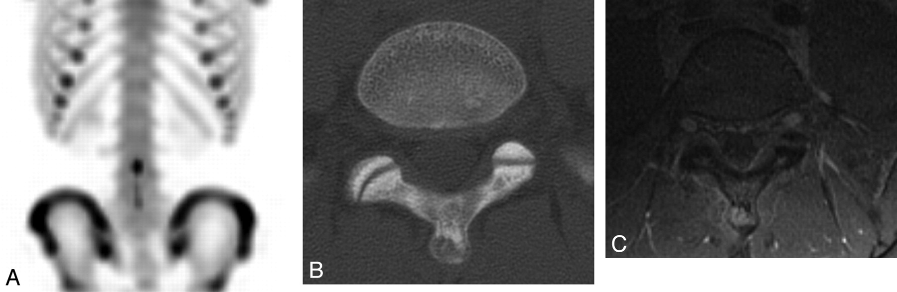
  • Fig 3.
    • Download figure
    • Open in new tab
    • Download powerpoint
    Fig 3.

    Spondylolysis in an 8-year-old boy. A, Axial T2-weighted image with fat saturation demonstrates hyperintense signal intensity consistent with bone marrow edema in the region of the pars defects bilaterally. B, Axial CT image demonstrates a complete pars defect of L5 on the right. C, Axial CT image demonstrates an incomplete pars defect of L5 on the left.
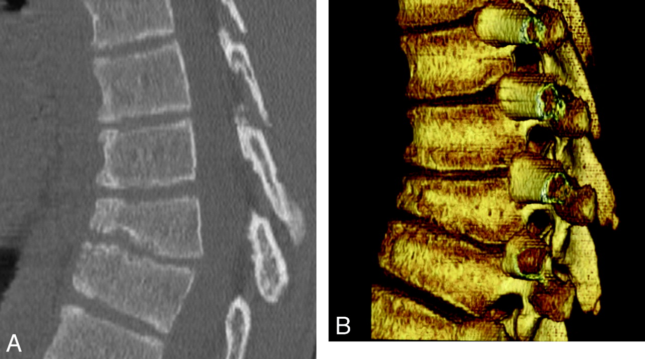
  • Fig 4.
    • Download figure
    • Open in new tab
    • Download powerpoint
    Fig 4.

    Scheuermann kyphosis in a 15-year-old boy. A, Sagittal 2D CT reconstruction image demonstrates midthoracic kyphosis with anterior wedging of at least 3 consecutive vertebrae with presence of Schmorl nodes. B, Sagittal 3D CT reconstruction image demonstrates midthoracic kyphosis with anterior wedging of at least 3 consecutive vertebrae with the presence of Schmorl nodes.
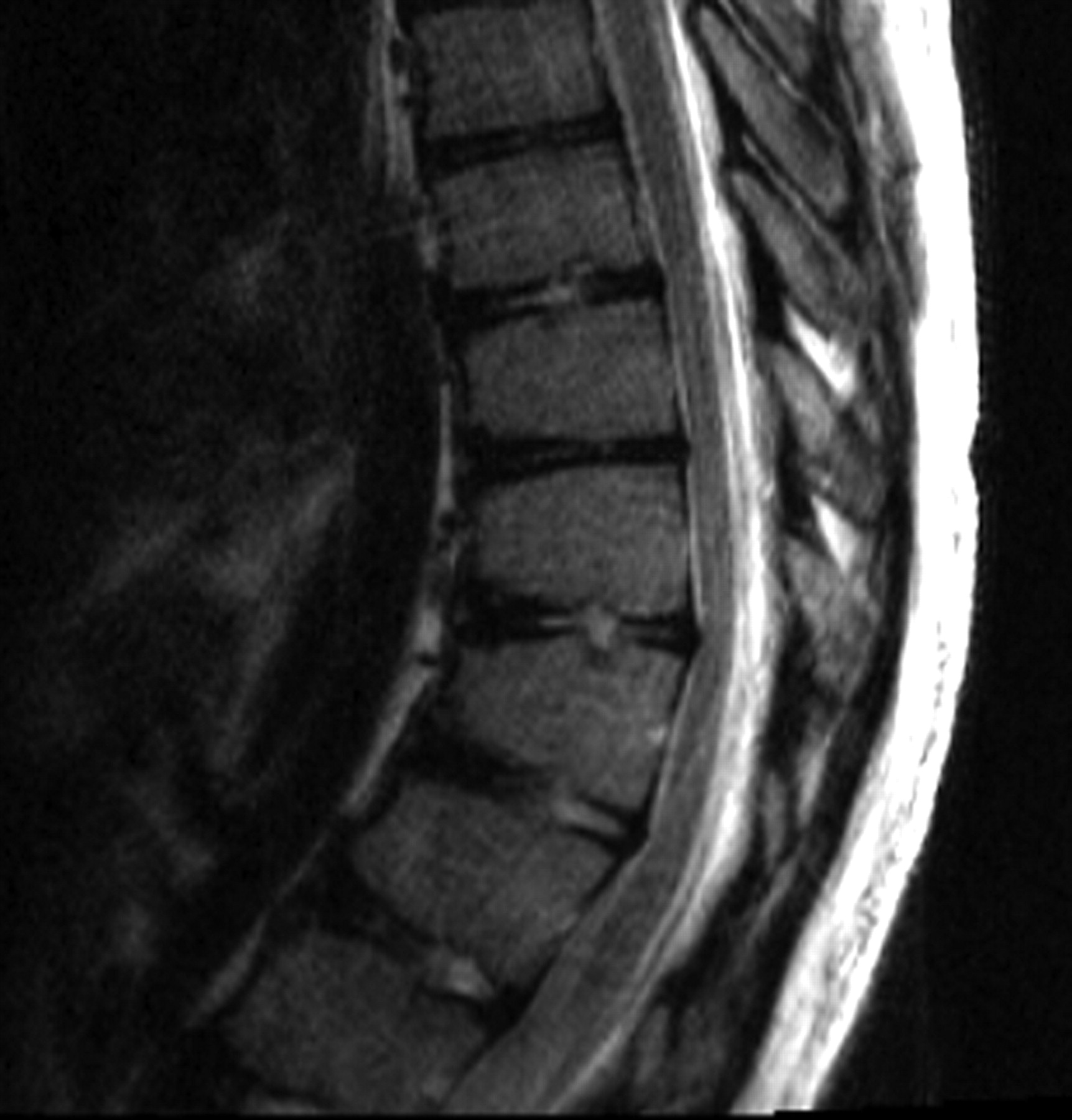
  • Fig 5.
    • Download figure
    • Open in new tab
    • Download powerpoint
    Fig 5.

    Scheuermann kyphosis in a 14-year-old boy. Sagittal T2-weighted image demonstrates thoracic kyphosis with mild anterior wedging of the T10–T12 vertebral bodies with slight disk space irregularity and Schmorl nodes. Minimal annular bulges slightly indent the ventral aspect of the thecal sac.

  • Fig 6.
    • Download figure
    • Open in new tab
    • Download powerpoint
    Fig 6.

    Disk calcification in a 6-year-old boy. A, Axial CT image demonstrates posterior extrusion of a calcified disk. B, Sagittal 2D CT reconstruction image demonstrates calcification in the central portion of the disk.

  • Fig 7.
    • Download figure
    • Open in new tab
    • Download powerpoint
    Fig 7.

    Vertebral osteomyelitis and diskitis in a 7-year-old boy. A, Sagittal T2-weighted image with fat saturation shows marked disk space narrowing at L2–L3 with hypointense T2 signal intensity within the disk. There is increased T2 prolongation in adjacent vertebral bodies. B, Sagittal T1-weighted MR image with fat saturation with gadolinium shows diffuse enhancement in the L2–L3 vertebral bodies and intervening disk space.
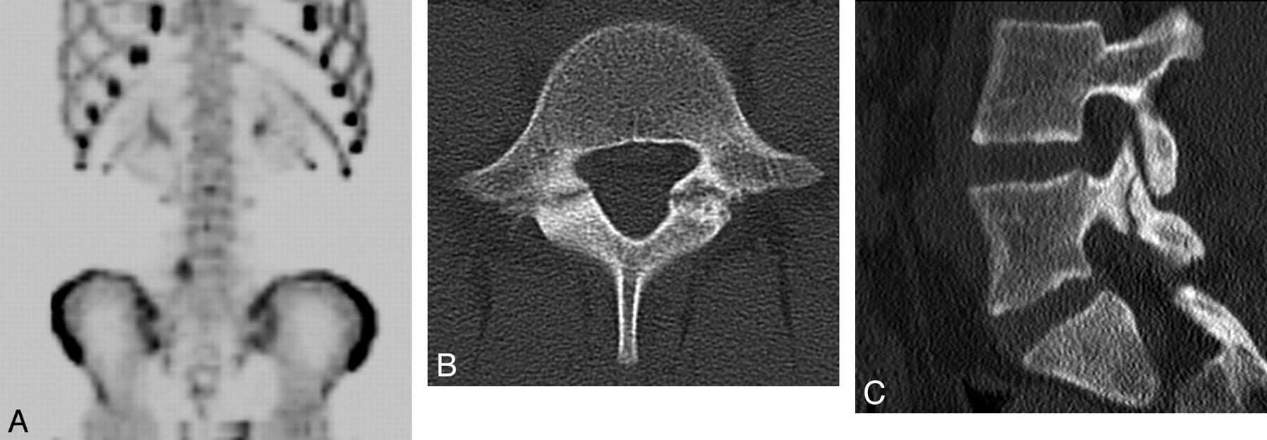
  • Fig 8.
    • Download figure
    • Open in new tab
    • Download powerpoint
    Fig 8.

    Osteoid osteoma in a 14-year-old girl. A, 99mTc-MDP SPECT scan demonstrates mild increased uptake in the spinous process of L5. B, Axial CT image shows a lucency in the tip of the spinous process of L5 with surrounding sclerosis and a tiny sclerotic nidus. C, Axial T1-weighted MR image with gadolinium demonstrates homogeneous enhancement of the lesion at the tip of the spinous process of L5.

  • Fig 9.
    • Download figure
    • Open in new tab
    • Download powerpoint
    Fig 9.

    Osteoblastoma in a 12-year-old boy. A, Axial CT image demonstrates an expansile lytic lesion in the pedicle of the C5 vertebra, which involves the C4–C5 facet joint and the left transverse foramen. B, Axial T1-weighted image with gadolinium and fat saturation demonstrates extensive enhancement in the adjacent bone and the left paraspinal soft tissues of the cervical spine, with extension into the epidural space.

  • Fig 10.
    • Download figure
    • Open in new tab
    • Download powerpoint
    Fig 10.

    LCH−vertebra plana in an 18-month-old boy. A, Sagittal 2D CT reconstruction image of the lumbar spine shows a collapsed L3 vertebral body. B, Sagittal T2-weighted MR image of the lumbar spine demonstrates a vertebra plana deformity with significant decreased height of the L3 vertebral body and preservation of the adjacent intervertebral disks.

  • Fig 11.
    • Download figure
    • Open in new tab
    • Download powerpoint
    Fig 11.

    Ewing sarcoma of the lumbar spine in a 17-year-old boy. A, Post-contrast-enhanced axial CT image demonstrates a large partially calcified mass in the paraspinal musculature at the L3–L5 levels. The mass involves the adjacent spinous process and extends into the spinal canal. B, Axial T1-weighted postgadolinium MR image shows extensive enhancement of the mass with epidural extension.

  • Fig 12.
    • Download figure
    • Open in new tab
    • Download powerpoint
    Fig 12.

    Lymphoblastic lymphoma in a 15-year-old boy. Sagittal T2-weighted MR image demonstrates an intraspinal extradural well-circumscribed hypointense lesion at the T2–T4 level with compression of the spinal cord with hypointensity diffusely in the vertebral bodies.

  • Fig 13.
    • Download figure
    • Open in new tab
    • Download powerpoint
    Fig 13.

    Acute lymphoblastic leukemia in a 3-year-old boy. A, Sagittal T1-weighted MR image demonstrates diffuse abnormal low signal intensity in the lumbar spine bone marrow consistent with a diffuse infiltrative process. B, Sagittal T1-weighted image of a healthy 3-year-old boy with normal higher T1 signal intensity of the vertebral body marrow relative to the intervertebral disks.

  • Fig 14.
    • Download figure
    • Open in new tab
    • Download powerpoint
    Fig 14.

    Astrocytoma in an 11-year-old boy. A, Sagittal T2 MR image of the cervical and upper thoracic spine demonstrates a partly cystic and solid intramedullary spinal cord tumor. B, Sagittal T2 MR image of the thoracic spine demonstrates a partly cystic and solid intramedullary spinal cord tumor. C, Sagittal T1-weighted postcontrast MR image shows enhancement of the solid portions of the tumor and peripheral enhancement of the cystic components.

  • Fig 15.
    • Download figure
    • Open in new tab
    • Download powerpoint
    Fig 15.

    Myxopapillary ependymoma in a 12-year-old boy. Sagittal T1-weighted postcontrast MR image demonstrates an intradural extramedullary mass lesion extending from the T12 to L3 levels and a second lesion at the L5–S2 level with diffuse enhancement. There is associated scalloping of the posterior aspects of the lumbar vertebral bodies.

Tables

  • Figures
    • View popup
    Table 1:

    Symptoms and signs that may indicate serious underlying pathology in children with back pain—red flags

    HistoryExamination
    Prepubertal children especially <5 yearsFever, tachycardia
    Functional disabilityWeight loss, bruising, lymphadenopathy, or abdominal mass
    Duration >4 weeksAltered spine shape or mobility
    Recurrent or worsening painVertebral or intervertebral tenderness
    Early morning stiffness and/or gellingLimp or altered gait
    Night painNeurologic symptoms
    Fever, weight loss, malaiseBladder or bowel dysfunction
    Postural changes: kyphosis or scoliosis
    Limp or altered gait
    • Reproduced with permission from BMJ Publishing Group Ltd & Royal College of Paediatrics and Child Health.15

    • View popup
    Table 2:

    Etiology of back pain in children and adolescents

    Back Pain
    1. Traumatic

      1. Spondylolysis/spondylolisthesis

      2. Vertebral column fractures

      3. Disk herniation

      4. Intraspinal hematoma

      5. Spinal cord injury

    2. Musculoskeletal

      1. Scheuermann disease

      2. Scoliosis

      3. Intervertebral disk degeneration

      4. Intervertebral disk herniation

      5. Intervertebral disk calcification

      6. Nonspecific musculoskeletal back pain

    3. Infectious

      1. Diskitis

      2. Vertebral osteomyelitis

      3. Epidural abscess

      4. Sacroiliac joint infection

      5. Chronic recurrent multifocal osteomyelitis

      6. Nonspinal infection

        1. Pyelonephritis

        2. Pneumonia

        3. Pelvic inflammatory disease

        4. Paraspinal muscle abscess

    4. Inflammatory

      1. Ankylosing spondylitis

      2. Juvenile idiopathic arthritis

      3. Arthritis

        1. Psoriatic arthritis

        2. Reactive arthritis

        3. Inflammatory bowel disease−associated arthritis

    5. Neoplastic disorders

      1. Spinal column

        1. Primary neoplasms

          1. Osteoid osteoma

          2. Osteoblastoma

          3. Aneurysmal bone cyst

          4. Giant cell tumor

          5. Chordoma

          6. Osteogenic sarcoma

          7. Ewing sarcoma

          8. Osteochondroma

          9. Histiocytosis

        2. Secondary neoplasms

          1. Leukemia

          2. Lymphoma

          3. Neuroblastoma

          4. Metastatic disease

      2. Spinal cord

        1. Intramedullary

          1. Astrocytoma

          2. Ependymoma

          3. Ganglioglioma

          4. Gangliocytoma

        2. Extradural tumors

          1. Neuroblastoma

          2. Ganglioneuroblastoma

          3. Ganglioneuroma

          4. Lymphoma

          5. Peripheral primitive neuroectodermal tumor

        3. Intradural-extramedullary

          1. Schwannoma

          2. Neurofibroma

          3. Meningioma

          4. CSF dissemination of intracranial neoplasms

    6. Congenital

      1. Syringomyelia

      2. Tethered cord syndrome

    7. Other

      1. Sickle cell pain crisis

      2. Cholecystitis

      3. Chronic pain syndromes

      4. Osteopo

PreviousNext
Back to top

In this issue

American Journal of Neuroradiology: 31 (5)
American Journal of Neuroradiology
Vol. 31, Issue 5
1 May 2010
  • Table of Contents
  • Index by author
Advertisement
Print
Download PDF
Email Article

Thank you for your interest in spreading the word on American Journal of Neuroradiology.

NOTE: We only request your email address so that the person you are recommending the page to knows that you wanted them to see it, and that it is not junk mail. We do not capture any email address.

Enter multiple addresses on separate lines or separate them with commas.
Imaging of Back Pain in Children
(Your Name) has sent you a message from American Journal of Neuroradiology
(Your Name) thought you would like to see the American Journal of Neuroradiology web site.
CAPTCHA
This question is for testing whether or not you are a human visitor and to prevent automated spam submissions.
Cite this article
D.P. Rodriguez, T.Y. Poussaint
Imaging of Back Pain in Children
American Journal of Neuroradiology May 2010, 31 (5) 787-802; DOI: 10.3174/ajnr.A1832

Citation Manager Formats

  • BibTeX
  • Bookends
  • EasyBib
  • EndNote (tagged)
  • EndNote 8 (xml)
  • Medlars
  • Mendeley
  • Papers
  • RefWorks Tagged
  • Ref Manager
  • RIS
  • Zotero
0 Responses
Respond to this article
Share
Bookmark this article
Imaging of Back Pain in Children
D.P. Rodriguez, T.Y. Poussaint
American Journal of Neuroradiology May 2010, 31 (5) 787-802; DOI: 10.3174/ajnr.A1832
del.icio.us logo Twitter logo Facebook logo Mendeley logo
  • Tweet Widget
  • Facebook Like
  • Google Plus One
Purchase

Jump to section

  • Article
    • Abstract
    • Abbreviations
    • Epidemiology
    • Clinical Evaluation
    • Imaging Evaluation
    • Traumatic/Musculoskeletal Disorders
    • Imaging Evaluation
    • Infectious Disorders
    • Neoplastic Disorders
    • Spinal Column Tumors
    • Secondary Neoplasms
    • Acknowledgments
    • References
  • Figures & Data
  • Info & Metrics
  • Responses
  • References
  • PDF

Related Articles

  • No related articles found.
  • PubMed
  • Google Scholar

Cited By...

  • Spondylodiscitis in infancy: A potentially fatal condition that can lead to major spinal complications
  • Crossref (48)
  • Google Scholar

This article has been cited by the following articles in journals that are participating in Crossref Cited-by Linking.

  • Evaluation of a Systematic Approach to Pediatric Back Pain
    Norman Ramirez, John M. Flynn, Brian W. Hill, Jose A. Serrano, Carlos E. Calvo, Rafael Bredy, Raul E. Macchiavelli
    Journal of Pediatric Orthopaedics 2015 35 1
  • Review for the generalist: evaluation of low back pain in children and adolescents
    Kristin M Houghton
    Pediatric Rheumatology 2010 8 1
  • Backpack-back pain complexity and the need for multifactorial safe weight recommendation
    Ademola James Adeyemi, Jafri Mohd Rohani, Mat Rebi Abdul Rani
    Applied Ergonomics 2017 58
  • Back pain and scoliosis in children: When to image, what to consider
    Sonia F Calloni, Thierry AGM Huisman, Andrea Poretti, Bruno P Soares
    The Neuroradiology Journal 2017 30 5
  • Spondylodiscitis in infancy
    A. I. Tsirikos, F. Tome-Bermejo
    The Journal of Bone and Joint Surgery. British volume 2012 94-B 10
  • Cervical intervertebral disc calcification combined with ossification of posterior longitudinal ligament in an-11-year old girl: case report and review of literature
    Guoqiang Wang, Yijun Kang, Fei Chen, Bing Wang
    Child's Nervous System 2016 32 2
  • ACR Appropriateness Criteria ® Back Pain—Child
    Timothy N. Booth, Ramesh S. Iyer, Richard A. Falcone, Laura L. Hayes, Jeremy Y. Jones, Nadja Kadom, Abhaya V. Kulkarni, John S. Myseros, Sonia Partap, Charles Reitman, Richard L. Robertson, Maura E. Ryan, Gaurav Saigal, Bruno P. Soares, Aylin Tekes, Andrew T. Trout, Nicholas A. Zumberge, Brian D. Coley, Susan Palasis
    Journal of the American College of Radiology 2017 14 5
  • Etiology, Risk Factors, and Diagnosis of Back Pain in Children and Adolescents: Evidence- and Consensus-Based Interdisciplinary Recommendations
    Michael Frosch, Maximilian D. Mauritz, Stefan Bielack, Susanne Blödt, Uta Dirksen, Michael Dobe, Florian Geiger, Renate Häfner, Lea Höfel, Bettina Hübner-Möhler, Thekla von Kalle, Burkhard Lawrenz, Andreas Leutner, Frauke Mecher, Kiril Mladenov, Heike Norda, Lorin Stahlschmidt, Marc Steinborn, Ralf Stücker, Ralf Trauzeddel, Regina Trollmann, Julia Wager, Boris Zernikow
    Children 2022 9 2
  • Evaluation of Low Back Pain in Athletes
    James M. Daniels, Gina Pontius, Saadiq El-Amin, Keith Gabriel
    Sports Health: A Multidisciplinary Approach 2011 3 4
  • Review of spinal involvement in Chronic recurrent multifocal osteomyelitis (CRMO): What radiologists need to know about CRMO and its imitators
    Amisha Shah, Margalit Rosenkranz, Mahesh Thapa
    Clinical Imaging 2022 81

More in this TOC Section

  • Mechanisms of Healing in Coiled Intracranial Aneurysms: A Review of the Literature
  • Ultra-High-Field MR Neuroimaging
  • 4D-CTA in Neurovascular Disease: A Review
Show more Review Articles

Similar Articles

Advertisement

Indexed Content

  • Current Issue
  • Accepted Manuscripts
  • Article Preview
  • Past Issues
  • Editorials
  • Editor's Choice
  • Fellows' Journal Club
  • Letters to the Editor
  • Video Articles

Cases

  • Case Collection
  • Archive - Case of the Week
  • Archive - Case of the Month
  • Archive - Classic Case

Special Collections

  • AJNR Awards
  • ASNR Foundation Special Collection
  • Most Impactful AJNR Articles
  • Photon-Counting CT
  • Spinal CSF Leak Articles (Jan 2020-June 2024)

More from AJNR

  • Trainee Corner
  • Imaging Protocols
  • MRI Safety Corner

Multimedia

  • AJNR Podcasts
  • AJNR Scantastics

Resources

  • Turnaround Time
  • Submit a Manuscript
  • Submit a Video Article
  • Submit an eLetter to the Editor/Response
  • Manuscript Submission Guidelines
  • Statistical Tips
  • Fast Publishing of Accepted Manuscripts
  • Graphical Abstract Preparation
  • Imaging Protocol Submission
  • Evidence-Based Medicine Level Guide
  • Publishing Checklists
  • Author Policies
  • Become a Reviewer/Academy of Reviewers
  • News and Updates

About Us

  • About AJNR
  • Editorial Board
  • Editorial Board Alumni
  • Alerts
  • Permissions
  • Not an AJNR Subscriber? Join Now
  • Advertise with Us
  • Librarian Resources
  • Feedback
  • Terms and Conditions
  • AJNR Editorial Board Alumni

American Society of Neuroradiology

  • Not an ASNR Member? Join Now

© 2025 by the American Society of Neuroradiology All rights, including for text and data mining, AI training, and similar technologies, are reserved.
Print ISSN: 0195-6108 Online ISSN: 1936-959X

Powered by HighWire