Advertisement

Main menu

  • Home
  • Content
    • Current Issue
    • Publication Preview--Ahead of Print
    • Past Issue Archive
    • Case of the Week Archive
    • Classic Case Archive
    • Case of the Month Archive
    • COVID-19 Content and Resources
  • For Authors
    • Author Policies
    • Manuscript Submission Guidelines
  • About Us
    • About AJNR
    • American Society of Neuroradiology
  • Submit a Manuscript
  • Podcasts
    • Podcasts
    • Subscribe on iTunes
  • More
    • Subscribers
    • Permissions
    • Advertisers
    • Alerts
    • Feedback

User menu

  • Subscribe
  • Alerts
  • Log in

Search

  • Advanced search
American Journal of Neuroradiology

American Journal of Neuroradiology

  • Subscribe
  • Alerts
  • Log in

Advanced Search

  • Home
  • Content
    • Current Issue
    • Publication Preview--Ahead of Print
    • Past Issue Archive
    • Case of the Week Archive
    • Classic Case Archive
    • Case of the Month Archive
    • COVID-19 Content and Resources
  • For Authors
    • Author Policies
    • Manuscript Submission Guidelines
  • About Us
    • About AJNR
    • American Society of Neuroradiology
  • Submit a Manuscript
  • Podcasts
    • Podcasts
    • Subscribe on iTunes
  • More
    • Subscribers
    • Permissions
    • Advertisers
    • Alerts
    • Feedback
  • Follow AJNR on Twitter
  • Visit AJNR on Facebook
  • Follow AJNR on Instagram
  • Join AJNR on LinkedIn
  • RSS Feeds
LetterLetter

Extracranial Internal Carotid Artery Stenosis as a Cause of Cortical Subarachnoid Hemorrhage

R.V. Chandra, T.M. Leslie-Mazwi, D. Oh, B. Mehta and A.J. Yoo
American Journal of Neuroradiology March 2011, 32 (3) E51-E52; DOI: https://doi.org/10.3174/ajnr.A2456
R.V. Chandra
  • Find this author on Google Scholar
  • Find this author on PubMed
  • Search for this author on this site
T.M. Leslie-Mazwi
  • Find this author on Google Scholar
  • Find this author on PubMed
  • Search for this author on this site
D. Oh
  • Find this author on Google Scholar
  • Find this author on PubMed
  • Search for this author on this site
B. Mehta
  • Find this author on Google Scholar
  • Find this author on PubMed
  • Search for this author on this site
A.J. Yoo
  • Find this author on Google Scholar
  • Find this author on PubMed
  • Search for this author on this site
  • Article
  • Figures & Data
  • Info & Metrics
  • References
  • PDF
Loading

We commend Cuvinciuc et al1 on their recent review of nontraumatic cortical subarachnoid hemorrhage (cSAH). While not an uncommon clinical presentation, there is an overall paucity of literature on this subject, with the largest reported series by Kumar et al2 comprising only 29 patients. Cuvinciuc et al1 suggested that cSAH may occur in the setting of high-grade atherosclerotic stenosis of the extracranial cerebral arteries, potentially by a mechanism similar to cSAH in Moyamoya disease (eg, rupture of fragile dilated pial collateral vessels). Recently 3 cases of cSAH have been described in association with bilateral cervical internal carotid artery (ICA) stenosis,3 and 2 further cases, in the setting of acute ICA occlusion during ischemic stroke.4 We describe a rare case of unilateral extracranial ICA stenosis associated with cSAH and discuss the angiographic findings that support the postulated mechanism of fragile pial collateral vessels.

A 70-year-old man experienced a sudden onset of transient aphasia and dysarthria, along with right face and arm numbness at a systolic blood pressure of 200 mm Hg. Noncontrast CT demonstrated isolated cSAH in the left central sulcus (Fig 1), and CT angiography (CTA) revealed an 80% left ICA-origin stenosis without evidence for arterial or venous cause of the cSAH. He continued to have recurrent events in the hospital and was managed for possible transient ischemia or seizure with intravenous anticoagulation and seizure prophylaxis, respectively. MR imaging excluded acute infarction, posterior reversible leukoencephalopathy, amyloid angiopathy, or an underlying mass. There was no clinical evidence of endocarditis, with negative findings on blood cultures and a normal finding on transthoracic echocardiogram. Continuous electroencephalographic monitoring during these episodes did not reveal epileptiform correlation during briefly symptomatic episodes that remained similar to those on the initial presentation. Given the frequency of transient symptoms, carotid endarterectomy (CEA) was considered for the ipsilateral stenosis. However, it was imperative to definitively rule out any vascular lesions responsible for the cSAH, which could worsen from potential hyperperfusion syndrome following CEA. Cerebral angiography was performed and demonstrated no evidence of vasculitis, reversible cerebral vasoconstriction syndrome, mycotic aneurysm, or arteriovenous malformation.

Fig 1.
  • Download figure
  • Open in new tab
  • Download powerpoint
Fig 1.

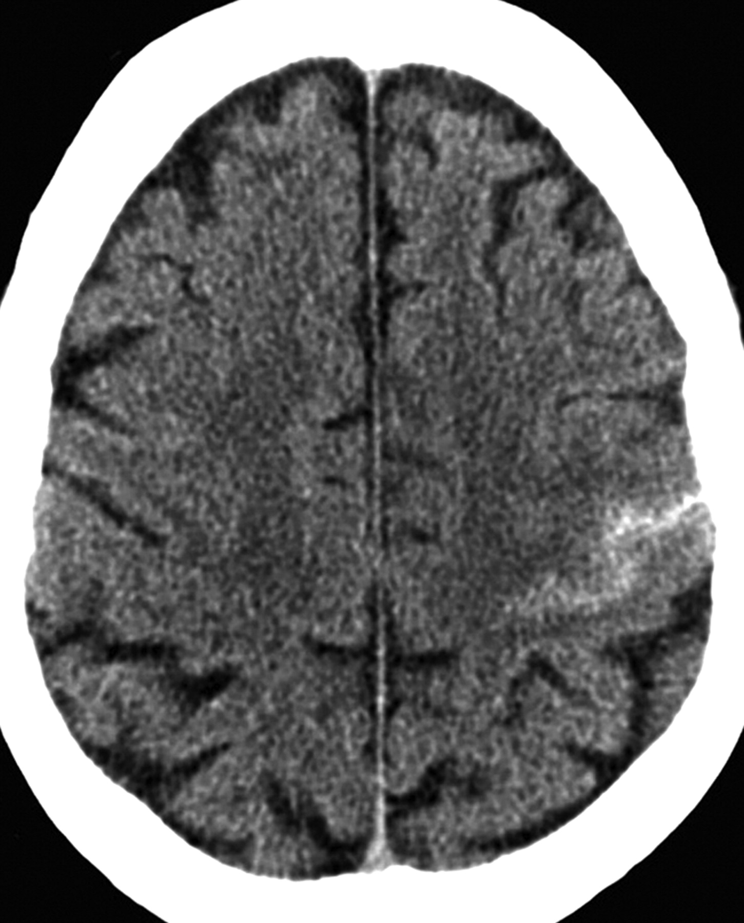
Noncontrast axial CT scan demonstrates subarachnoid hemorrhage in the left central sulcus.

Of note, the 80% left ICA-origin stenosis (Fig 2) resulted in a shift of the angiographic watershed between the left middle cerebral artery (MCA) and posterior cerebral artery (PCA) territories toward the central sulcus (Fig 3). Most importantly, there was minimal collateral support via the circle of Willis. A hypoplastic precommunicating right anterior cerebral artery (ACA) segment provided minimal flow across the anterior communicating artery. No left posterior communicating artery was discernible. Subsequently, the patient underwent emergent ipsilateral CEA with resolution of his symptoms.

Fig 2.
  • Download figure
  • Open in new tab
  • Download powerpoint
Fig 2.

Left common carotid artery angiogram demonstrated 80% ICA stenosis.

Fig 3.
  • Download figure
  • Open in new tab
  • Download powerpoint
Fig 3.

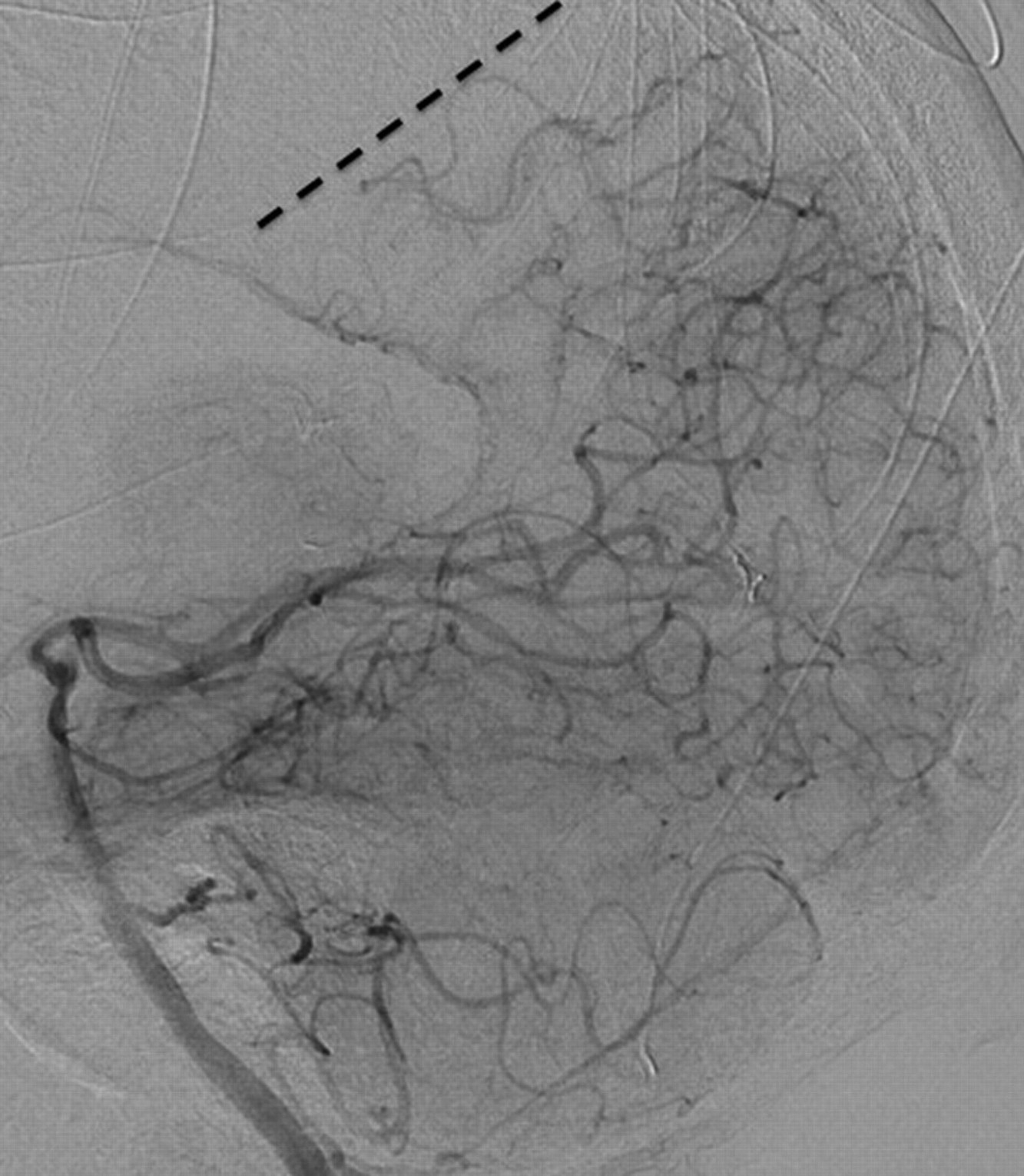
Left vertebral artery angiogram reveals a shift of the left PCA/MCA watershed (dotted line) toward the central sulcus. Perisplenial PCA collaterals also retrogradely fill the pericallosal ACA territory, indicating the severe left anterior circulation hemodynamic compromise.

Isolated cSAH in the present case, the 3 cases with bilateral ICA stenosis described by Kleinig et al,3 and the 2 cases of acute ICA occlusion described by Geraldes et al,4 all occurred on the symptomatic side or the side with the more severe anatomic narrowing (stenosis or occlusion). All 6 patients were acutely symptomatic suggesting that cSAH may derive from an acute alteration in hemodynamics (eg, plaque rupture and further atherothrombotic narrowing or acute hypertension). We propose that this hemodynamic stress may damage already maximally dilated pial collateral vasculature and produce cSAH.

This idea is supported by our angiographic findings, in which the subarachnoid hemorrhage colocalized to the watershed region between the MCA and PCA territories. In the setting of severe ICA stenosis or occlusion, it is the MCA-PCA watershed rather than the MCA-ACA watershed that may be particularly prone to this phenomenon, because the ipsilateral ACA and MCA have reduced perfusion pressure and are less able to respond to acute hemodynamic failure, particularly in the absence of sufficient blood flow via the anterior or posterior communicating arteries as in this case. On the other hand, the normally perfused PCA territory is able to mount a compensatory increase in perfusion pressure, which may rupture fragile pial collateral vessels in the watershed. Consistent with this idea, 4 of the 6 described cases occurred in a perirolandic location, at the watershed between the MCA and PCA territories.

The rarity of cSAH in the setting of unilateral extracranial ICA stenosis may reflect the collateral pathways across the circle of Willis that are usually present. As in our case, the absence of adequate collaterals may provide a clue to this mechanism of cSAH. Contrary to Cuvinciuc et al,1 who advocated only brain vascular imaging, CTA evaluation of cSAH must include the cervical vasculature, lest a high-risk yet treatable lesion is missed.

References

  1. 1.↵
    1. Cuvinciuc V,
    2. Viguier A,
    3. Calviere L,
    4. et al
    . Isolated acute nontraumatic cortical subarachnoid hemorrhage. AJNR Am J Neuroradiol 2010;31:1355–62
    Abstract/FREE Full Text
  2. 2.↵
    1. Kumar S,
    2. Goddeau RP Jr.,
    3. Selim MH,
    4. et al
    . Atraumatic convexal subarachnoid hemorrhage: clinical presentation, imaging patterns, and etiologies. Neurology 2010;74:893–99
    Abstract/FREE Full Text
  3. 3.↵
    1. Kleinig TJ,
    2. Kimber TE,
    3. Thompson PD
    . Convexity subarachnoid haemorrhage associated with bilateral internal carotid artery stenoses. J Neurol 2009;256:669–71
    CrossRefPubMed
  4. 4.↵
    1. Geraldes R,
    2. Santos C,
    3. Canhao P
    . Atraumatic localized convexity subarachnoid hemorrhage associated with acute carotid artery occlusion. Eur J Neurol 2011;2:e28–e29
  • Copyright © American Society of Neuroradiology
PreviousNext
Back to top

In this issue

American Journal of Neuroradiology: 32 (3)
American Journal of Neuroradiology
Vol. 32, Issue 3
1 Mar 2011
  • Table of Contents
  • Index by author
Advertisement
Print
Download PDF
Email Article

Thank you for your interest in spreading the word on American Journal of Neuroradiology.

NOTE: We only request your email address so that the person you are recommending the page to knows that you wanted them to see it, and that it is not junk mail. We do not capture any email address.

Enter multiple addresses on separate lines or separate them with commas.
Extracranial Internal Carotid Artery Stenosis as a Cause of Cortical Subarachnoid Hemorrhage
(Your Name) has sent you a message from American Journal of Neuroradiology
(Your Name) thought you would like to see the American Journal of Neuroradiology web site.
CAPTCHA
This question is for testing whether or not you are a human visitor and to prevent automated spam submissions.
Citation Tools
Extracranial Internal Carotid Artery Stenosis as a Cause of Cortical Subarachnoid Hemorrhage
R.V. Chandra, T.M. Leslie-Mazwi, D. Oh, B. Mehta, A.J. Yoo
American Journal of Neuroradiology Mar 2011, 32 (3) E51-E52; DOI: 10.3174/ajnr.A2456

Citation Manager Formats

  • BibTeX
  • Bookends
  • EasyBib
  • EndNote (tagged)
  • EndNote 8 (xml)
  • Medlars
  • Mendeley
  • Papers
  • RefWorks Tagged
  • Ref Manager
  • RIS
  • Zotero
Share
Extracranial Internal Carotid Artery Stenosis as a Cause of Cortical Subarachnoid Hemorrhage
R.V. Chandra, T.M. Leslie-Mazwi, D. Oh, B. Mehta, A.J. Yoo
American Journal of Neuroradiology Mar 2011, 32 (3) E51-E52; DOI: 10.3174/ajnr.A2456
del.icio.us logo Digg logo Reddit logo Twitter logo CiteULike logo Facebook logo Google logo Mendeley logo
  • Tweet Widget
  • Facebook Like
  • Google Plus One
Purchase

Jump to section

  • Article
    • References
  • Figures & Data
  • Info & Metrics
  • References
  • PDF

Related Articles

  • Reply:
  • PubMed
  • Google Scholar

Cited By...

  • Convexity subarachnoid haemorrhage secondary to internal carotid stenosis: an indication for revascularisation
  • Reply:
  • Central Sulcus Focal Subarachnoid Hemorrhage in the Elderly: Cerebral Amyloid Angiopathy Is the Most Frequent Cause
  • Crossref (18)
  • Google Scholar

This article has been cited by the following articles in journals that are participating in Crossref Cited-by Linking.

  • Nontraumatic Convexity Subarachnoid Hemorrhage: Different Etiologies and Outcomes
    Ruth Geraldes, Paulo R. Sousa, Ana C. Fonseca, Filipa Falcão, Patrícia Canhão, Teresa Pinho e Melo
    Journal of Stroke and Cerebrovascular Diseases 2014 23 1
  • Nontraumatic Convexal Subarachnoid Hemorrhage Concomitant with Acute Ischemic Stroke
    Makoto Nakajima, Yuichiro Inatomi, Toshiro Yonehara, Teruyuki Hirano, Yukio Ando
    Journal of Stroke and Cerebrovascular Diseases 2014 23 6
  • Cerebral convexity subarachnoid hemorrhage: various causes and role of diagnostic imaging
    Rajiv Mangla, Douglas Drumsta, Jeevak Alamst, Manisha Mangla, Michael Potchen
    Emergency Radiology 2015 22 2
  • Hémorragies sous-arachnoïdiennes corticales focales : présentation clinique, radiologique et diagnostic étiologique dans une série de 23 patients
    J. Mas, S. Bouly, I. Mourand, D. Renard, N. de Champfleur, P. Labauge
    Revue Neurologique 2013 169 1
  • Acute ischemic stroke with contralateral convexal subarachnoid hemorrhage: two cases report
    Yayun Cao, Jie Cao, Suqiong Ji, Shabei Xu, Chenchen Liu
    BMC Neurology 2019 19 1
  • Incidence and possible causes of nontraumatic convexal subarachnoid haemorrhage in Chinese patients: A retrospective review
    Haiyan Zhao, Jintao Han, Ming Lu, Yingshuang Zhang, Dongsheng Fan
    Journal of International Medical Research 2017 45 6
  • Central Sulcus Focal Subarachnoid Hemorrhage in the Elderly: Cerebral Amyloid Angiopathy Is the Most Frequent Cause
    American Journal of Neuroradiology 2011 32 8
  • Isolated Acute Nontraumatic Cortical Subarachnoid Hemorrhage: Etiologies Based on MRI Findings
    Eun Cho, Myongjin Kang, Sanghyeon Kim
    Journal of the Korean Society of Radiology 2016 74 6
  • Subarachnoid Hemorrhage of Unknown Etiology along the Cortical Convexity
    Yasuo Murai, Shiro Kobayashi, Akira Teramoto
    Journal of Nippon Medical School 2012 79 4
  • Subarachnoid Hemorrhage with Progressive Cerebral Steno-Occlusive Disease: Report of 2 Cases
    Soichiro Takamiya, Tetsuyuki Yoshimoto, Katsuhiko Maruichi
    Journal of Stroke and Cerebrovascular Diseases 2019 28 4

More in this TOC Section

  • Regarding “Altered Blood Flow in the Ophthalmic and Internal Carotid Arteries in Patients with Age-Related Macular Degeneration Measured Using Noncontrast MR Angiography at 7T”
  • Fair Performance of CT in Diagnosing Unilateral Vocal Fold Paralysis
  • Reply:
Show more LETTERS

Similar Articles

Advertisement

News and Updates

  • Lucien Levy Best Research Article Award
  • Thanks to our 2022 Distinguished Reviewers

Resources

  • Evidence-Based Medicine Level Guide
  • AJNR Podcast Archive
  • Librarian Resources
  • Terms and Conditions

Opportunities

  • Get Peer Review Credit from Publons

American Society of Neuroradiology

  • Neurographics
  • ASNR Annual Meeting
  • Fellowship Portal

© 2023 by the American Society of Neuroradiology | Print ISSN: 0195-6108 Online ISSN: 1936-959X

Powered by HighWire