Advertisement

Main menu

  • Home
  • Content
    • Current Issue
    • Publication Preview--Ahead of Print
    • Past Issue Archive
    • Case of the Week Archive
    • Classic Case Archive
    • Case of the Month Archive
    • COVID-19 Content and Resources
  • For Authors
    • Author Policies
    • Manuscript Submission Guidelines
  • About Us
    • About AJNR
    • American Society of Neuroradiology
  • Submit a Manuscript
  • Podcasts
    • Podcasts
    • Subscribe on iTunes
  • More
    • Subscribers
    • Permissions
    • Advertisers
    • Alerts
    • Feedback

User menu

  • Subscribe
  • Alerts
  • Log in

Search

  • Advanced search
American Journal of Neuroradiology

American Journal of Neuroradiology

  • Subscribe
  • Alerts
  • Log in

Advanced Search

  • Home
  • Content
    • Current Issue
    • Publication Preview--Ahead of Print
    • Past Issue Archive
    • Case of the Week Archive
    • Classic Case Archive
    • Case of the Month Archive
    • COVID-19 Content and Resources
  • For Authors
    • Author Policies
    • Manuscript Submission Guidelines
  • About Us
    • About AJNR
    • American Society of Neuroradiology
  • Submit a Manuscript
  • Podcasts
    • Podcasts
    • Subscribe on iTunes
  • More
    • Subscribers
    • Permissions
    • Advertisers
    • Alerts
    • Feedback
  • Follow AJNR on Twitter
  • Visit AJNR on Facebook
  • Follow AJNR on Instagram
  • Join AJNR on LinkedIn
  • RSS Feeds
Research ArticleTechnical Note
Open Access

Fast Whole-Brain 4D Contrast-Enhanced MR Angiography with Velocity Encoding Using Undersampled Radial Acquisition and Highly Constrained Projection Reconstruction: Image-Quality Assessment in Volunteer Subjects

Y. Wu, W. Chang, K.M. Johnson, J. Velikina, H. Rowley, C. Mistretta and P. Turski
American Journal of Neuroradiology March 2011, 32 (3) E47-E50; DOI: https://doi.org/10.3174/ajnr.A2048
Y. Wu
  • Find this author on Google Scholar
  • Find this author on PubMed
  • Search for this author on this site
W. Chang
  • Find this author on Google Scholar
  • Find this author on PubMed
  • Search for this author on this site
K.M. Johnson
  • Find this author on Google Scholar
  • Find this author on PubMed
  • Search for this author on this site
J. Velikina
  • Find this author on Google Scholar
  • Find this author on PubMed
  • Search for this author on this site
H. Rowley
  • Find this author on Google Scholar
  • Find this author on PubMed
  • Search for this author on this site
C. Mistretta
  • Find this author on Google Scholar
  • Find this author on PubMed
  • Search for this author on this site
P. Turski
  • Find this author on Google Scholar
  • Find this author on PubMed
  • Search for this author on this site
  • Article
  • Figures & Data
  • Info & Metrics
  • References
  • PDF
Loading

Abstract

SUMMARY: We report on the image quality obtained by using fast contrast-enhanced whole-brain 4D radial MRA with 0.75-second temporal resolution, isotropic submillimeter spatial resolution, and velocity encoding (HYPRFlow). Images generated by HYPR-LR by using the velocity-encoded data as the constraining image were of diagnostic quality. In addition, we demonstrate that measurements of shear stress within the middle cerebral artery can be derived from the high-resolution 3D velocity data.

Abbreviations

AV
arteriovenous
CE-VIPR
contrast-enhanced VIPR
HYPR
highly constrained back-projection
HYPRFlow
HYPR with phase contrast as constraint
HYPR-LR
HYPR local reconstruction
MCA
middle cerebral artery
MIP
maximum intensity projection
MRA
MR angiography
PC-VIPR
phase-contrast VIPR
SNR
signal intensity–to-noise ratio
VIPR
vastly undersampled isotropic projection reconstruction
WSS
wall shear stress

Whole-brain 4D contrast-enhanced MRA has been previously described by using Cartesian acquisitions, partial-Fourier encoding, and parallel imaging with multiple coils.1–3 However, performing high-resolution whole-brain 4D contrast-enhanced MRA examinations is challenging using conventional imaging methods because acquisition time increases proportionally with matrix size. Radial acquisitions like 2D projection reconstruction4–6 and PC-VIPR7,8 can be effective in accelerating MR angiography; however, image quality is limited by undersampling artifacts and decreased SNR at high acceleration factors. Innovative reconstruction algorithms, such as HYPR and HYPR-LR9–11), which exploit prior information to constrain individual images, are able to reduce the severity of undersampling artifacts and SNR loss even at high levels of acceleration. Combining undersampled radial acquisition with HYPR reconstruction can dramatically improve frame rates and image coverage without degradation of image quality.12

HYPRFlow is one of the hybrid HYPR techniques that uses the phase-contrast image from a separate scan as the vascular constraint to provide whole-brain 4D contrast-enhanced MRA with subsecond temporal resolution and submillimeter isotropic spatial resolution as well as flow dynamics.12–14 In this article, we investigate the clinical utility of this technique by assessing the image quality of HYPRFlow and estimate WSS in the middle cerebral arteries by using the time-average velocity through a single cardiac cycle.

Technique

Volunteer studies were performed in compliance with the Health Insurance Portability and Accountability Act regulations and by using a protocol approved by the local institutional review board. Eight adult subjects ranging from 24 to 40 years of age were imaged (4 women, 4 men) with a clinical 3T MR imaging system (Excite HD, TwinSpeed, Signa; GE Healthcare, Waukesha, Wisconsin) with an 8-channel head coil (HD Brain Coil, GE Healthcare). Each HYPRFlow study involved 2 encoding methods. First, during contrast injection, spatial encoding was performed by using a time-resolved multiecho 3D radial acquisition (CE-VIPR).15 Subsequently, velocity encoding was performed by using a high-resolution dual-echo 3D radial phase-contrast acquisition (PC-VIPR). The PC-VIPR data were used to form a composite image (angiographic constraint) for HYPR-LR reconstruction and for hemodynamic evaluation.

MR Imaging Protocol

Imaging parameters for CE-VIPR were the following: FOV = 26 × 26 × 26 cm3, TR/TE = 3.0/0.4 ms, bandwidth = 125 kHz, 64 points from the center to the edge of the k-space for each projection, frame update time = 0.5 seconds. Scanning parameters for postcontrast PC-VIPR were the following: FOV = 22 × 22 × 22 cm3, TR/TE = 12.5/4.8 ms, velocity encoding = 60 cm/s, bandwidth = 62.5 kHz. The readout matrix was 320 points per projection, and the spatial resolution for the composite image was 0.7 × 0.7 × 0.7 mm3. Seven thousand projections were acquired within 5 minutes. Gadobenate dimeglumine (MultiHance, Bracco Diagnostics, Princeton, New Jersey) was injected at 3 mL/s, and the contrast dose was 0.1 mm/kg followed by a 20-mL saline flush.

Image Reconstruction

In the HYPRFlow method, complex difference images were reconstructed from the postcontrast PC-VIPR (spatial resolution = 0.7 × 0.7 × 0.7 mm3) and were used as a composite for the HYPR-LR algorithm. The temporal weighting images were used to define the contrast kinetics obtained by convolving timeframe images (reconstructed from the CE-VIPR) with a locally averaging kernel.8 Adaptive k-space filtering with a 0.75-second temporal window at the cutoff frequency was used to suppress the high-spatial-frequency undersampling artifacts and increase the SNR while maintaining a high temporal resolution.16 Following HYPR-LR reconstruction, the result was a time series of contrast-enhanced MRAs with both high temporal resolution (0.75 seconds) and high spatial resolution (0.7 × 0.7 × 0.7 mm3).

SNR Comparison

To quantitatively compare the SNR improvement of HYPRFlow, we drew, for each volunteer, 3 target regions of interest from the internal carotid artery, basilar artery, and sinus veins at peak arterial and peak venous phases. Corresponding background regions of interest were drawn near the target regions of interest at the same timeframes. SNRs were measured from HYPRFlow images and CE-VIPR images.

Image-Quality Analysis

HYPRFlow arterial and venous phase images were scored by 2 experienced observers for image quality in the carotid siphons, A1, M1, P1, basilar artery, deep veins, and dural sinuses. Images were scored on a scale of 1–4 (1, not visible; 2, nondiagnostic; 3, diagnostic; 4, well-delineated). The examinations were graded for temporal image quality by scoring AV separation (1, no AV separation; 2, AV separation but vessels obscured by overlap; 3, AV separation adequate for diagnostic purposes; 4, excellent AV separation with minimal arterial signal intensity).

Results

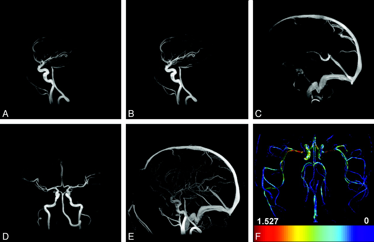
The top row of Figure 1 shows 3 representative HYPRFlow timeframes (out of 120 timeframes in total) demonstrating early arterial phase (Fig 1A), late arterial phase (Figure 1B), and late late venous phase (Fig 1C) in sagittal view. Figure 1D shows the late arterial phase in coronal plane. Figure 1E is the sagittal MIP of the complex different images acquired from the phase contrast scan and served as the spatial constraint for the HYPRFlow reconstruction. Figure 1F shows a map of estimated WSS derived from the velocity data. Average HYPRFlow spatial image quality scores were 3.50 for the arterial phase and 3.81 for the venous phase, and AV separation was scored as 3.56. PC-VIPR image quality was 3.39 for arteries and 3.78 for venous structures. The difference in image quality between HYPRFlow and PC-VIPR was not statistically significant (P = .1075, Wilcoxon rank sum test). Interobserver κ analysis showed 78.6% concordance with κ = 0.714, which is considered good agreement. The interobserver error for observers 1 and 2 was 3.8% (SD differences, 13.3%–18.4%). The average SNR measurements were 50.5 ± 19.5 for HYPRFlow images and 22.6 ± 13.1 for CE-VIPR images. The difference was statistically significant (P < .0001). WSS values measured in the proximal M1 segment ranged from 0.8 to 1.6 N/m2, and there was no significant difference in WSS between the right and left M1 segments of the MCA (P = .44).

Fig 1.
  • Download figure
  • Open in new tab
  • Download powerpoint
Fig 1.

A, Sagittal HYPRFlow timeframe in the early arterial phase. B, Sagittal HYPRFlow timeframe in the late arterial phase. C, Sagittal HYPRFlow timeframe in the venous phase. D, Coronal HYPRFlow timeframe in the arterial phase. E, Sagittal PC-VIPR MIP. F, WSS map of the axial whole brain generated from PC-VIPR velocity data.

Discussion

Previous methods of whole-brain time-resolved contrast-enhanced MRA using Cartesian acquisitions were limited by long scanning times, relatively low spatial resolution from few phase encodes, or low temporal resolution due to view sharing. Parallel imaging techniques such as sensitivity encoding allow acceleration via unfolding of k-space (at the expense of SNR), but the acceleration factor is limited to the number of coils (8 in standard head coils) and noise at that acceleration factor would be prohibitive.17,18 HYPRFlow avoids these limitations by using 2 separate scans to achieve high temporal and spatial resolution and by exploiting the sparsity inherent in brain MRA to achieve high acceleration factors while maintaining an acceptable SNR.

Phase-contrast radial acquisition is particularly well-suited to cerebrovascular imaging because most tissue in the FOV is stationary and is subtracted during reconstruction, leading to enhanced sparsity, which allows a high level of undersampling because streak artifacts are diffuse, reduced, and projected outside the region of interest at the center of image space. The contrast-enhanced time-resolved image is also sparse because a mask is acquired before the contrast injection and then subtracted, with only the areas with contrast left. Thus, the contrast-enhanced time-resolved acquisition and the phase-contrast data used as the composite are sparse; this feature allows HYPR-LR to provide near-exact reconstruction even at high levels of acceleration.8

Shear stress is an important physiologic parameter, which is associated with the development of atherosclerotic plaque.19 Prior studies of estimated WSS by using phase-contrast techniques were limited to 2D sections or very time-consuming 3D phase-contrast acquisitions. Prior reports have shown a high degree of correlation between PC-VIPR and catheter-based endovascular measurements of physiologic parameters.20,21 In our investigations, relatively high-resolution HYPRFlow velocity data were used to estimate WSS in the proximal middle cerebral artery. We recognize that MR imaging underestimates WSS compared with techniques such as computational fluid dynamics22 because of insufficient resolution at the boundary and could yield slightly different results compared with other MR imaging WSS techniques, such as spline interpolation; we plan to use estimated WSS as a surrogate parameter to distinguish areas of high and low WSS.

Because HYPRFlow requires 2 acquisition modes (dynamic and velocity encoding), patient motion is a concern. However, when the subject moves between modes, image registration can be used before HYPR-LR reconstruction to reduce this problem. Another challenge is that the phase-contrast images are sensitive to flow. In the regions of complex and/or slow flow, phase-contrast MRA images may not sufficiently delineate the vasculature, causing diagnostically obscuring noise amplification in the final HYPRFlow images. Magnitude images from phase-contrast MRA, which are effectively postcontrast time-of-flight images, provide better depiction of the vasculature but are substantially less sparse; these features make HYPR processing more difficult. Further work is required to investigate alternative reconstruction schemes, such as iterative HYPR and compressed sensing techniques,23–26 which may provide better decoupling of the constraining image and data being reconstructed. This should allow the use of a composite that is a weighted combination of angiographic and magnitude images from postcontrast PC-VIPR.

Future research will include automated reconstruction software, investigating the use of HYPRFlow velocity data to generate pressure fields, velocity vector plots, streamline flow, and tracing particle paths through the flow field. Development of dual-velocity encoded methods is currently underway.

Appendix

WSS Estimation

To measure WSS, we accept that the achievable temporal and spatial resolution with MR imaging is not sufficient to depict the vessel boundary layer. However, we aim to use MR imaging–derived WSS as a surrogate parameter that can be used to compare the heterogeneous WSS distribution. For blood, the WSS can be approximated as Embedded Image where μ is the viscosity (assumed to be 4.0 cP), VT is the velocity tangent to the wall, and n^ is the normal vector of the wall. The algorithm to compute 3D WSS has 3 basic steps: 1) segmentation of the vessel boundaries, 2) calculation of the normal vector, and 3) velocity fitting.27,28 Segmentation used an isosurface of the angiographic data with a manually chosen value as the minimum value that did not introduce significant noise. Image normal vectors were determined by using the gradient of the angiogram at the segmented vessel wall locations. Second-order polynomials were used to fit the velocity data and determine the velocity derivative. An average WSS map was generated by taking the average velocity through a cardiac cycle. Small segments (∼100 voxels inside each segment, corresponding to 35-mm3 volume) in the middle of the MCAs were selected to represent the WSS estimation of the MCA.

Footnotes

  • P. Turski was supported by the National Institutes of Health grant R21 EB009441.

Indicates open access to non-subscribers at www.ajnr.org

References

  1. 1.↵
    1. Willinek W,
    2. Hadizadeh D,
    3. Falkenhausen,
    4. et al
    . 4D time-resolved MR angiography with keyhole (4D-TRAK): more than 60 times accelerated MRA using a combination of CENTRA, keyhole, and SENSE at 3.0T. J Magn Reson Imaging 2008;27:1455–60
    CrossRefPubMed
  2. 2.↵
    1. Fink C,
    2. Ley S,
    3. Kroeker R,
    4. et al
    . Time-resolved contrast-enhanced three-dimensional magnetic resonance angiography of the chest: combination of parallel imaging with view sharing (TREAT). Invest Radiol 2005;40:40–48
    PubMed
  3. 3.↵
    1. Parmar H,
    2. Ivancevic M,
    3. Dudek,
    4. et al
    . Neuroradiologic applications of dynamic MR angiography at 3T. Magn Reson Imaging Clin N Am 2009;17:63–75
    CrossRefPubMed
  4. 4.↵
    1. Peters D,
    2. Korosec F,
    3. Grist T,
    4. et al
    . Undersampled projection reconstruction applied to MR angiography. Magn Reson Med 2000;43:91–101
    CrossRefPubMed
  5. 5.↵
    1. Barger A,
    2. Bloch W,
    3. Toropov Y,
    4. et al
    . Time-resolved contrast-enhanced imaging with isotropic resolution and broad coverage using an undersampled 3D projection trajectory. Magn Reson Med 2002:48:297–305
    CrossRefPubMed
  6. 6.↵
    1. Lu A,
    2. Wieben O,
    3. Grist T,
    4. et al
    . Vastly undersampled isotropic projection reconstruction imaging with multi-half-echo (VIPR ME). In: Proceedings of the 11th Annual Meeting of International Society of Magnetic Resonance in Medicine, Toronto, Ontario, Canada. July 10–16, 2003;50
  7. 7.↵
    1. Gu T,
    2. Korosec F,
    3. Block W,
    4. et al
    . PC VIPR: a high-speed 3D phase-contrast method for flow quantification and high-resolution angiography. AJNR Am J Neuroradiol 2005;26:743–49
    Abstract/FREE Full Text
  8. 8.↵
    1. Johnson K,
    2. Lum D,
    3. Turski P,
    4. et al
    . Improved 3D phase contrast MRI with off-resonance corrected dual echo VIPR. Magn Reson Med 2008;60:1329–36
    CrossRefPubMed
  9. 9.↵
    1. Mistretta C,
    2. Wieben O,
    3. Velikina J,
    4. et al
    . Highly constrained backprojection for time-resolved MRI. Magn Reson Med 2006;55:30–40
    CrossRefPubMed
  10. 10.↵
    1. Johnson K,
    2. Velikina J,
    3. Wu Y,
    4. et al
    . Improved waveform fidelity using local HYPR reconstruction (HYPR LR). Magn Reson Med 2008;59:456–62
    CrossRefPubMed
  11. 11.↵
    1. Supanich M,
    2. Tao Y,
    3. Nett B,
    4. et al
    . Radiation dose reduction in time-resolved CT angiography using highly constrained back projection reconstruction. Phys Med Biol 2009;54:4575–93
    CrossRefPubMed
  12. 12.↵
    1. Mistretta C
    . Undersampled radial MR acquisition and highly constrained back projection (HYPR) reconstruction: potential medical imaging applications in the post-Nyquist era. J Magn Reson Imaging 2009;29:501–16
    PubMed
  13. 13.↵
    1. Velikina J,
    2. Johnson K,
    3. Wieben O,
    4. et al
    . PC HYPRFlow: a technique for rapid imaging of contrast dynamics. J Magn Reson Imaging 2010;37:447–56
  14. 14.↵
    1. Wu Y,
    2. Johnson K,
    3. Velikina J,
    4. et al
    . Clinical experience of HYPR Flow. In: Proceedings of the 16th Annual Meeting of International Society of Magnetic Resonance in Medicine, Toronto, Ontario, Canada. May 3–9, 2008;20
  15. 15.↵
    1. Brodsky EK,
    2. Lu A,
    3. Thornton FJ,
    4. et al
    . Using multiple half-echoes to improve sampling efficiency and fat suppression in time-resolved MRA. In: Proceedings of the 11th Annual Meeting of International Society of Magnetic Resonance in Medicine, Toronto, Ontario, Canada. July 10–16, 2003;24
  16. 16.↵
    1. Du J,
    2. Carroll T,
    3. Brodsky E,
    4. et al
    . Contrast-enhanced peripheral magnetic resonance angiography using time-resolved vastly undersampled isotropic projection reconstruction. J Magn Reson Imaging 2004;20:894–900
    CrossRefPubMed
  17. 17.↵
    1. Golay X,
    2. Brown S,
    3. Itoh R,
    4. et al
    . Time-resolved contrast-enhanced carotid MR angiography using sensitivity encoding (SENSE). AJNR Am J Neuroradiol 2001;22:1615–19
    Abstract/FREE Full Text
  18. 18.↵
    1. Glockner JF,
    2. Hu HH,
    3. Stanley DW,
    4. et al
    . Parallel MR imaging: a user's guide. Radiographics 2005;25:1279–97
    PubMed
  19. 19.↵
    1. Melek AM,
    2. Alpers SI,
    3. Izumo S
    . Hemodynamic shear stress and its role in atherosclerosis. JAMA 1999;282:2035–42
    CrossRefPubMed
  20. 20.↵
    1. Turk AS,
    2. Johnson K,
    3. Lum D,
    4. et al
    . Physiologic and anatomic assessment of a canine carotid artery stenosis model utilizing phase contrast with vastly undersampled isotropic projection imaging. AJNR Am J Neuroradiol 2007;28:111–15
    Abstract/FREE Full Text
  21. 21.↵
    1. Moftakhar R,
    2. Aagaard-Kienitz B,
    3. Johnson K,
    4. et al
    . Noninvasive measurement of intra-aneurysmal pressure and flow pattern using phase contrast with vastly undersampled isotropic projection imaging. AJNR Am J Neuroradiol 2008;28:1710–14
    CrossRef
  22. 22.↵
    1. Wood N,
    2. Weston S,
    3. Kilner P,
    4. et al
    . Combined MR imaging and CFD simulation of flow in the human descending aorta. J Magn Reson Imaging 2001;13:699–713
    CrossRefPubMed
  23. 23.↵
    1. Velikina J,
    2. Samsonov A
    . HYPR- l0: a hybrid technique for CE-MRA with extreme undersampling factors. Presented at: International Society of Magnetic Resonance in Medicine Workshop on Data Sampling and Image Reconstruction, Sedona, Arizona, January 25–28, 2009
  24. 24.↵
    1. O'Halloran RL,
    2. Wen Z,
    3. Holmes JH,
    4. et al
    . Iterative projection reconstruction of time-resolved images using highly-constrained back-projection (HYPR). Magn Reson Med 2008;59:132–39
    CrossRefPubMed
  25. 25.↵
    1. Griswold M,
    2. Barkauskas K,
    3. Blaimer M,
    4. et al
    . More optimal HYPR reconstructions using a combination of hyper- and conjugate-gradient minimization. In: Proceedings of the International Society of Magnetic Resonance in Medicine, Berlin, Germany. May 19–25, 2007;188
  26. 26.↵
    1. Lustig M,
    2. Donoho D,
    3. Pauly JM
    . Sparse MRI: the application of compressed sensing for rapid MR imaging. Magn Reson Med 2007;58:1182–95
    CrossRefPubMed
  27. 27.↵
    1. Papathanasopoulou P,
    2. Zhao S,
    3. Kohler U,
    4. et al
    . MRI measurement of time-resolved wall shear stress vectors in a carotid bifurcation model, and comparison with CFD predictions. J Magn Reson Imaging 2002;17:153–62
  28. 28.↵
    1. Oshinski JN,
    2. Ku DN,
    3. Mukundan S Jr.,
    4. et al
    . Determination of wall shear stress in the aorta with the use of MR phase velocity mapping. J Magn Reson Imaging 1995;5:640–47
    PubMed
  • Received August 20, 2009.
  • Accepted after revision December 14, 2009.
  • Copyright © American Society of Neuroradiology
View Abstract
PreviousNext
Back to top

In this issue

American Journal of Neuroradiology: 32 (3)
American Journal of Neuroradiology
Vol. 32, Issue 3
1 Mar 2011
  • Table of Contents
  • Index by author
Advertisement
Print
Download PDF
Email Article

Thank you for your interest in spreading the word on American Journal of Neuroradiology.

NOTE: We only request your email address so that the person you are recommending the page to knows that you wanted them to see it, and that it is not junk mail. We do not capture any email address.

Enter multiple addresses on separate lines or separate them with commas.
Fast Whole-Brain 4D Contrast-Enhanced MR Angiography with Velocity Encoding Using Undersampled Radial Acquisition and Highly Constrained Projection Reconstruction: Image-Quality Assessment in Volunteer Subjects
(Your Name) has sent you a message from American Journal of Neuroradiology
(Your Name) thought you would like to see the American Journal of Neuroradiology web site.
CAPTCHA
This question is for testing whether or not you are a human visitor and to prevent automated spam submissions.
Citation Tools
Fast Whole-Brain 4D Contrast-Enhanced MR Angiography with Velocity Encoding Using Undersampled Radial Acquisition and Highly Constrained Projection Reconstruction: Image-Quality Assessment in Volunteer Subjects
Y. Wu, W. Chang, K.M. Johnson, J. Velikina, H. Rowley, C. Mistretta, P. Turski
American Journal of Neuroradiology Mar 2011, 32 (3) E47-E50; DOI: 10.3174/ajnr.A2048

Citation Manager Formats

  • BibTeX
  • Bookends
  • EasyBib
  • EndNote (tagged)
  • EndNote 8 (xml)
  • Medlars
  • Mendeley
  • Papers
  • RefWorks Tagged
  • Ref Manager
  • RIS
  • Zotero
Share
Fast Whole-Brain 4D Contrast-Enhanced MR Angiography with Velocity Encoding Using Undersampled Radial Acquisition and Highly Constrained Projection Reconstruction: Image-Quality Assessment in Volunteer Subjects
Y. Wu, W. Chang, K.M. Johnson, J. Velikina, H. Rowley, C. Mistretta, P. Turski
American Journal of Neuroradiology Mar 2011, 32 (3) E47-E50; DOI: 10.3174/ajnr.A2048
del.icio.us logo Digg logo Reddit logo Twitter logo CiteULike logo Facebook logo Google logo Mendeley logo
  • Tweet Widget
  • Facebook Like
  • Google Plus One
Purchase

Jump to section

  • Article
    • Abstract
    • Abbreviations
    • Technique
    • Results
    • Discussion
    • Appendix
    • Footnotes
    • References
  • Figures & Data
  • Info & Metrics
  • References
  • PDF

Related Articles

  • No related articles found.
  • PubMed
  • Google Scholar

Cited By...

  • Fast Contrast-Enhanced 4D MRA and 4D Flow MRI Using Constrained Reconstruction (HYPRFlow): Potential Applications for Brain Arteriovenous Malformations
  • Measuring Pulsatile Flow in Cerebral Arteries Using 4D Phase-Contrast MR Imaging
  • Hemodynamic Changes in Patients with Arteriovenous Malformations Assessed Using High-Resolution 3D Radial Phase-Contrast MR Angiography
  • Crossref (16)
  • Google Scholar

This article has been cited by the following articles in journals that are participating in Crossref Cited-by Linking.

  • MRI temporal acceleration techniques
    Jeffrey Tsao, Sebastian Kozerke
    Journal of Magnetic Resonance Imaging 2012 36 3
  • Measuring Pulsatile Flow in Cerebral Arteries Using 4D Phase-Contrast MR Imaging
    A. Wåhlin, K. Ambarki, R. Birgander, O. Wieben, K.M. Johnson, J. Malm, A. Eklund
    American Journal of Neuroradiology 2013 34 9
  • Time‐resolved angiography: Past, present, and future
    Thomas M. Grist, Charles A. Mistretta, Charles M. Strother, Patrick A. Turski
    Journal of Magnetic Resonance Imaging 2012 36 6
  • Hemodynamic Changes in Patients with Arteriovenous Malformations Assessed Using High-Resolution 3D Radial Phase-Contrast MR Angiography
    W. Chang, M.W. Loecher, Y. Wu, D.B. Niemann, B. Ciske, B. Aagaard-Kienitz, S. Kecskemeti, K.M. Johnson, O. Wieben, C. Mistretta, P. Turski
    American Journal of Neuroradiology 2012 33 8
  • Velocity Measurements in the Middle Cerebral Arteries of Healthy Volunteers Using 3D Radial Phase-Contrast HYPRFlow: Comparison with Transcranial Doppler Sonography and 2D Phase-Contrast MR Imaging
    W. Chang, B. Landgraf, K.M. Johnson, S. Kecskemeti, Y. Wu, J. Velikina, H. Rowley, O. Wieben, C. Mistretta, P. Turski
    American Journal of Neuroradiology 2011 32 1
  • Fast Contrast-Enhanced 4D MRA and 4D Flow MRI Using Constrained Reconstruction (HYPRFlow): Potential Applications for Brain Arteriovenous Malformations
    W. Chang, Y. Wu, K. Johnson, M. Loecher, O. Wieben, M. Edjlali, C. Oppenheim, P. Roca, J. Hald, B. Aagaard-Kienitz, D. Niemann, C. Mistretta, P. Turski
    American Journal of Neuroradiology 2015 36 6
  • 4D Contrast-enhanced MR Angiography with the Keyhole Technique in Children: Technique and Clinical Applications
    Rajesh Krishnamurthy, Sara M. Bahouth, Raja Muthupillai
    RadioGraphics 2016 36 2
  • Neurovascular 4DFlow MRI (Phase Contrast MRA): emerging clinical applications
    Patrick Turski, Andrew Scarano, Eric Hartman, Zachary Clark, Tilman Schubert, Leonardo Rivera, Yijing Wu, Oliver Wieben, Kevin Johnson
    Neurovascular Imaging 2016 2 1
  • Emerging Techniques for Evaluation of the Hemodynamics of Intracranial Vascular Pathology
    Warren Chang, Melissa Huang, Aichi Chien
    The Neuroradiology Journal 2015 28 1
  • Current Status and Future Perspectives of Magnetic Resonance High-Field Imaging: A Summary
    Vivek Prabhakaran, Veena A. Nair, Benjamin P. Austin, Christian La, Thomas A. Gallagher, Yijing Wu, Donald G. McLaren, Guofan Xu, Patrick Turski, Howard Rowley
    Neuroimaging Clinics of North America 2012 22 2

More in this TOC Section

  • 3D Fast Spin-Echo T1 Black-Blood Imaging for the Diagnosis of Cervical Artery Dissection
  • The Kety-Schmidt Technique for Quantitative Perfusion and Oxygen Metabolism Measurements in the MR Imaging Environment
  • A Swine Model to Analyze Arterial Structural Changes Induced by Mechanical Thrombectomy
Show more Technical Note

Similar Articles

Advertisement

News and Updates

  • Lucien Levy Best Research Article Award
  • Thanks to our 2022 Distinguished Reviewers

Resources

  • Evidence-Based Medicine Level Guide
  • AJNR Podcast Archive
  • Librarian Resources
  • Terms and Conditions

Opportunities

  • Get Peer Review Credit from Publons

American Society of Neuroradiology

  • Neurographics
  • ASNR Annual Meeting
  • Fellowship Portal

© 2023 by the American Society of Neuroradiology | Print ISSN: 0195-6108 Online ISSN: 1936-959X

Powered by HighWire