Advertisement

Main menu

  • Home
  • Content
    • Current Issue
    • Publication Preview--Ahead of Print
    • Past Issue Archive
    • Case of the Week Archive
    • Classic Case Archive
    • Case of the Month Archive
    • COVID-19 Content and Resources
  • For Authors
  • About Us
    • About AJNR
    • Editors
    • American Society of Neuroradiology
  • Submit a Manuscript
  • Podcasts
    • Subscribe on iTunes
    • Subscribe on Stitcher
  • More
    • Subscribers
    • Permissions
    • Advertisers
    • Alerts
    • Feedback
  • Other Publications
    • ajnr

User menu

  • Subscribe
  • Alerts
  • Log in

Search

  • Advanced search
American Journal of Neuroradiology

American Journal of Neuroradiology

  • Subscribe
  • Alerts
  • Log in

Advanced Search

  • Home
  • Content
    • Current Issue
    • Publication Preview--Ahead of Print
    • Past Issue Archive
    • Case of the Week Archive
    • Classic Case Archive
    • Case of the Month Archive
    • COVID-19 Content and Resources
  • For Authors
  • About Us
    • About AJNR
    • Editors
    • American Society of Neuroradiology
  • Submit a Manuscript
  • Podcasts
    • Subscribe on iTunes
    • Subscribe on Stitcher
  • More
    • Subscribers
    • Permissions
    • Advertisers
    • Alerts
    • Feedback
  • Follow AJNR on Twitter
  • Visit AJNR on Facebook
  • Follow AJNR on Instagram
  • Join AJNR on LinkedIn
  • RSS Feeds
Research ArticleTechnical Note

Transcranial Access Using Fluoroscopic Flat Panel Detector CT Navigation

D.L. Cooke, M. Levitt, L.J. Kim, D.K. Hallam and B. Ghodke
American Journal of Neuroradiology April 2011, 32 (4) E69-E70; DOI: https://doi.org/10.3174/ajnr.A2066
D.L. Cooke
  • Find this author on Google Scholar
  • Find this author on PubMed
  • Search for this author on this site
M. Levitt
  • Find this author on Google Scholar
  • Find this author on PubMed
  • Search for this author on this site
L.J. Kim
  • Find this author on Google Scholar
  • Find this author on PubMed
  • Search for this author on this site
D.K. Hallam
  • Find this author on Google Scholar
  • Find this author on PubMed
  • Search for this author on this site
B. Ghodke
  • Find this author on Google Scholar
  • Find this author on PubMed
  • Search for this author on this site
  • Article
  • Figures & Data
  • Supplemental
  • Info & Metrics
  • References
  • PDF
Loading

Abstract

SUMMARY: FPCT and navigation software on contemporary fluoroscopic units perform imaging of a quality comparable with conventional CT. They can accurately guide percutaneous procedures, providing live instrument visualization and the capability to re-image without patient transfer. FPCT navigation was used in the placement of a ventricular drain in a 62-year-old woman for subarachnoid-related hydrocephalus by using an otherwise standard bedside technique. Ventriculostomy catheter placement was technically successful without complication with a catheter at the foramen of Monro.

Abbreviations

DSA
digital subtraction angiography
FPCT
flat panel detector CT

FPCT and integrated guidance software available on contemporary fluoroscopic systems emit a lower radiation dose while producing imaging of a quality comparable with that of conventional CT.1–5 They also have the ability to perform percutaneous procedures within the body, spine, and head and neck safely and accurately.4–11 FPCT demonstrates excellent spatial resolution and, though limited in subtle contrast differentiation, is of diagnostic accuracy in the assessment of general cerebral anatomy and higher attenuation features such as hemorrhage.5–8,10,11 Beyond the ability of FPCT-based navigation to quickly and accurately target a particular focus is the provision of real-time instrument visualization using live fluoroscopic overlay and the capability of re-imaging without moving the patient, limitations of current neuro-navigational programs.10

Ventricular drain catheter placement in the setting of subarachnoid hemorrhage−related hydrocephalus is a common neurosurgical procedure. Performed at the bedside by using anatomic landmarks, ventriculostomy placement is safe and accurate with a low rate of complications.12–15 Given the frequent need for ventricular drain placement and the relative technical ease of the procedure, we chose it as means to demonstrate proof of principle regarding transcranial FPCT guidance. We herein describe the use of the navigation system in the placement of a ventriculostomy catheter.

Case Report

A 62-year-old woman presented with altered mental status. Physical examination revealed an intubated obtunded patient with left-sided hemiplegia and equal and reactive pupils. CT of the head showed diffuse subarachnoid hemorrhage, hydrocephalus, and significant hemorrhage in the basal cisterns. CT angiography revealed a 6 × 7 mm right-sided posterior communicating artery aneurysm and a 3 × 2 mm left-sided middle cerebral artery aneurysm.

The patient underwent emergent FPCT-guided ventriculostomy placement in the angiography suite. To minimize movement, we carefully taped her head to the table. The left scalp was prepped, and a 1-cm incision was made at the Kocher point, approximately 11 cm posterior to the glabella and 3 cm lateral to midline in the midpupillary line. A twist-drill craniostomy was performed. The dura and pia mater were cauterized and incised.

Before catheter placement, an FPCT (XperCT and XperGuide; Philips Healthcare, Andover, Massachusetts) was performed and the data were analyzed on the 3D workstation. XperGuide analysis software was used to define the target as the ipsilateral foramen of Monro and the entry point at the craniostomy site. Using a progress view in the lateral plane, we passed a ventriculostomy catheter along the defined trajectory to a depth of 6.75 cm, with the tip reaching the target at the foramen of Monro (Fig 1 , on-line video). The surgeon was able to advance the catheter without prolonged fluoroscopic exposure and could stand comfortably adjacent to the C-arm. We noted that during catheter-guidance planning, the bull's eye or “entry” view generated by the navigational software often necessitated a working angle outside the range of the C-arm when using a standard Kocher point entry site. To remedy this, we found that 10°-15° of head flexion or the reverse Trendelenburg position was adequate to address this problem.

Fig 1.
  • Download figure
  • Open in new tab
  • Download powerpoint
Fig 1.

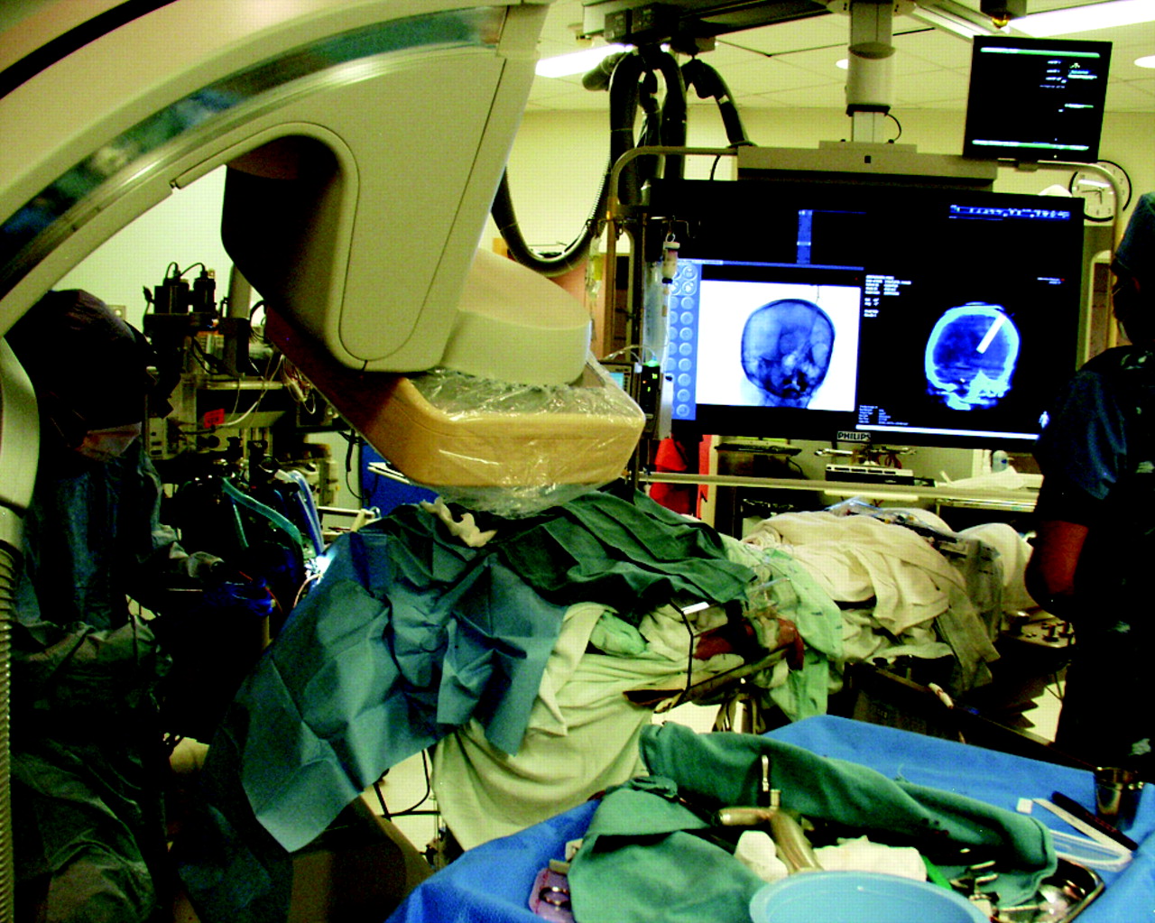
Ventricular drain placement. The surgeon is working adjacent to the C-arm during placement of the ventriculostomy catheter.

CSF was noted to be under high pressure. The catheter was secured and the incision was closed. A second FPCT was then performed, confirming accurate catheter placement, which was also shown on a subsequent formal head CT (Fig 2 ). The ventriculostomy catheter tip terminated in the expected location at the foramen of Monro, and no placement-related complications were noted on post−head CT. The procedure required 12.3 minutes of fluoroscopy and produced 273 Gy/cm2 of dose area product. Overall, guidance added approximately 30–40 minutes to the traditional bedside technique. The patient then underwent diagnostic cerebral angiography and eventual balloon-assisted coiling.

Fig 2.
  • Download figure
  • Open in new tab
  • Download powerpoint
Fig 2.

Axial images from a FPCT (left) and a standard noncontrast CT (right) demonstrate the tip of the ventricular catheter at the foramen of Monro.

Discussion

To the authors' knowledge, this is the first report of the use of FPCT navigation for transcranial access. There are reports documenting the use of standard CT and virtual navigation systems for the placement of external ventricular catheters, though neither provides the ability to immediately re-image the patient while also providing real-time visualization during catheter placement.16,17

The main limitation of FPCT navigation is the need for the patient to remain stationary during the procedure. Most neurosurgical guidance systems necessitate a Mayfield clamp to immobilize the head, a device not currently compatible with a conventional angiography table. In the setting of an FPCT-guided procedure, securely taping a patient's head and the use of general anesthesia help minimize potential patient motion, though not to the degree provided by a true surgical clamp. In light of such a limitation, the FPCT guidance system has the ability to reorient the CT image onto live fluoroscopy by using the patient's osseous anatomy as reference landmarks, much in the manner that fiducials are used in other neuro-navigation systems.10 Ultimately, if there is concern that the patient or the intracranial anatomy has moved significantly, a new FPCT and trajectory can be acquired without transferring the patient.

The use of FPCT navigation will not likely supplant the traditional placement of ventricular drains, given the increase in time and cost, though it could prove helpful in those patients with slit-like ventricles or significantly distorted intracranial anatomy. With optimization, such a navigational system could be used for tissue resection (therapeutic and diagnostic in nature), general fluid aspiration (CSF, abscess, hemorrhage), and drug or device delivery for any number of neurologic pathologies. For example, there is evidence demonstrating the safety and technical success of frameless stereotactic intracerebral hemorrhagic aspiration.18–21 The evacuation of deep intracerebral hemorrhage requiring conventional neuro-navigational software could be further optimized by using FPCT navigation techniques. Additionally, the ability to simultaneously perform endovascular intervention opens the door to true tandem procedures, (eg, stereotactic aspiration of an epidural hematoma and meningeal artery embolization).

Footnotes

  • Indicates article with supplemental online video.

References

  1. 1.↵
    1. Braak S,
    2. van Strijen M,
    3. Van Leersum M,
    4. et al
    . Effective dose during needle interventions: real time 3D fluoroscopy guidance versus CT fluoroscopy. In: Proceedings of the 94th Scientific Assembly and Annual Meeting of the Radiological Society of North America, Chicago, Illinois. November 30-December 5, 2008
  2. 2.↵
    1. Racadio J,
    2. Yoshizumi T,
    3. Toncheva G,
    4. et al
    . Radiation dosimetry evaluation of C-arm cone beam CT for pediatric interventional radiology procedures: a comparison with MDCT. In: Proceedings of the 94th Scientific Assembly and Annual Meeting of the Radiological Society of North America, Chicago, Illinois. November 30-December 5, 2008
  3. 3.↵
    1. Noordhoek NJ,
    2. van de Haar P,
    3. Timmer J
    . Direct comparison of commercially available C-arm CT to multi-slice CT image quality. In: Proceedings of the 94th Scientific Assembly and Annual Meeting of the Radiological Society of North America, Chicago, Illinois. November 30-December 5, 2008
  4. 4.↵
    1. Miracle A,
    2. Mukherji S
    . Conebeam CT of the head and neck. Part 1. Physical principles. AJNR Am J Neuroradiol 2009;30:1088–95.Epub 2009 May 13
    Abstract/FREE Full Text
  5. 5.↵
    1. Miracle A,
    2. Mukherji S
    . Conebeam CT of the head and neck. Part 2. Clinical applications. AJNR Am J Neuroradiol 2009;30:1285–92.Epub 2009 May 20
    Abstract/FREE Full Text
  6. 6.↵
    1. Heran N,
    2. Song J,
    3. Namba K,
    4. et al
    . The utility of DynaCT in neuroendovascular procedures. AJNR Am J Neuroradiol 2006;27:330–32
    Abstract/FREE Full Text
  7. 7.↵
    1. Daly M,
    2. Siewerdsen J,
    3. Moseley D,
    4. et al
    . Intraoperative cone-beam CT for guidance of head and neck surgery: assessment of dose and image quality using a C-arm prototype. Med Phys 2006;33:3767–80
    CrossRefPubMed
  8. 8.↵
    1. Doelken M,
    2. Struffert T,
    3. Richter G,
    4. et al
    . Flat-panel detector volumetric CT for visualization of subarachnoid hemorrhage and ventricles: preliminary results compared to conventional CT. Neuroradiology 2008;50:517–23
    CrossRefPubMed
  9. 9.↵
    1. Söderman M,
    2. Babic D,
    3. Holmin S,
    4. et al
    . Brain imaging with a flat detector C-arm: technique and clinical interest of XperCT. Neuroradiology 2008;50:863–68.Epub 2008 Jun 17
    CrossRefPubMed
  10. 10.↵
    1. Racadio J,
    2. Babic D,
    3. Homan R,
    4. et al
    . Live 3D guidance in the interventional radiology suite. AJR Am J Roentgenol 2007;189:W357–64
    CrossRefPubMed
  11. 11.↵
    1. Dörfler A,
    2. Struffert T,
    3. Engelhorn T,
    4. et al
    . Rotational flat-panel computed tomography in diagnostic and interventional neuroradiology. Rofo 2008;180:891–98
    PubMed
  12. 12.↵
    1. Huyette D,
    2. Turnbow B,
    3. Kaufman C,
    4. et al
    . Accuracy of the freehand pass technique for ventriculostomy catheter placement: retrospective assessment using computed tomography scans. J Neurosurg 2008;108:88–91
    CrossRefPubMed
  13. 13.↵
    1. Binz DD,
    2. Toussaint LG 3rd.,
    3. Friedman JA
    . Hemorrhagic complications of ventriculostomy placement: a meta-analysis. Neurocrit Care 2009;10:253–56.Epub 2009 Feb 18
    CrossRefPubMed
  14. 14.↵
    1. Kakarla UL,
    2. Kim LJ,
    3. Chang SW,
    4. et al
    . Safety and accuracy of bedside external ventricular drain placement. Neurosurgery 2008;63 (1 suppl 1):ONS162–66,discussion ONS166–67
    CrossRefPubMed
  15. 15.↵
    1. Saladino A,
    2. White J,
    3. Wijdicks E,
    4. et al
    . Malplacement of ventricular catheters by neurosurgeons: a single institution experience. Neurocrit Care 2009;10:248–52
    CrossRefPubMed
  16. 16.↵
    1. Banerjee P,
    2. Luciano C,
    3. Lemole GJ,
    4. et al
    . Accuracy of ventriculostomy catheter placement using a head- and hand-tracked high-resolution virtual reality simulator with haptic feedback. J Neurosurg 2007;107:515–21
    CrossRefPubMed
  17. 17.↵
    1. Krötz M,
    2. Linsenmaier U,
    3. Kanz K,
    4. et al
    . Evaluation of minimally invasive percutaneous CT-controlled ventriculostomy in patients with severe head trauma. Eur Radiol 2004;14:227–33
    CrossRefPubMed
  18. 18.↵
    1. Barrett R,
    2. Hussain R,
    3. Coplin W,
    4. et al
    . Frameless stereotactic aspiration and thrombolysis of spontaneous intracerebral hemorrhage. Neurocrit Care 2005;3:237–45
    CrossRefPubMed
  19. 19.↵
    1. Morgan T,
    2. Zuccarello M,
    3. Narayan R,
    4. et al
    . Preliminary findings of the minimally-invasive surgery plus rtPA for intracerebral hemorrhage evacuation (MISTIE) clinical trial. Acta Neurochir Suppl 2008;105:147–51
    CrossRefPubMed
  20. 20.↵
    1. Vespa P,
    2. McArthur D,
    3. Miller C,
    4. et al
    . Frameless stereotactic aspiration and thrombolysis of deep intracerebral hemorrhage is associated with reduction of hemorrhage volume and neurological improvement. Neurocrit Care 2005;2:274–81
    CrossRefPubMed
  21. 21.↵
    1. Thiex R,
    2. Rohde V,
    3. Rohde I,
    4. et al
    . Frame-based and frameless stereotactic hematoma puncture and subsequent fibrinolytic therapy for the treatment of spontaneous intracerebral hemorrhage. J Neurol 2004;251:1443–50
    CrossRefPubMed
  • Received December 4, 2009.
  • Accepted after revision January 10, 2010.
  • Copyright © American Society of Neuroradiology
View Abstract
PreviousNext
Back to top

In this issue

American Journal of Neuroradiology: 32 (4)
American Journal of Neuroradiology
Vol. 32, Issue 4
1 Apr 2011
  • Table of Contents
  • Index by author
Advertisement
Print
Download PDF
Email Article

Thank you for your interest in spreading the word on American Journal of Neuroradiology.

NOTE: We only request your email address so that the person you are recommending the page to knows that you wanted them to see it, and that it is not junk mail. We do not capture any email address.

Enter multiple addresses on separate lines or separate them with commas.
Transcranial Access Using Fluoroscopic Flat Panel Detector CT Navigation
(Your Name) has sent you a message from American Journal of Neuroradiology
(Your Name) thought you would like to see the American Journal of Neuroradiology web site.
CAPTCHA
This question is for testing whether or not you are a human visitor and to prevent automated spam submissions.
Citation Tools
Transcranial Access Using Fluoroscopic Flat Panel Detector CT Navigation
D.L. Cooke, M. Levitt, L.J. Kim, D.K. Hallam, B. Ghodke
American Journal of Neuroradiology Apr 2011, 32 (4) E69-E70; DOI: 10.3174/ajnr.A2066

Citation Manager Formats

  • BibTeX
  • Bookends
  • EasyBib
  • EndNote (tagged)
  • EndNote 8 (xml)
  • Medlars
  • Mendeley
  • Papers
  • RefWorks Tagged
  • Ref Manager
  • RIS
  • Zotero
Share
Transcranial Access Using Fluoroscopic Flat Panel Detector CT Navigation
D.L. Cooke, M. Levitt, L.J. Kim, D.K. Hallam, B. Ghodke
American Journal of Neuroradiology Apr 2011, 32 (4) E69-E70; DOI: 10.3174/ajnr.A2066
Reddit logo Twitter logo Facebook logo Mendeley logo
  • Tweet Widget
  • Facebook Like
  • Google Plus One
Purchase

Jump to section

  • Article
    • Abstract
    • Abbreviations
    • Case Report
    • Discussion
    • Footnotes
    • References
  • Figures & Data
  • Supplemental
  • Info & Metrics
  • References
  • PDF

Related Articles

  • PubMed
  • Google Scholar

Cited By...

  • Accuracy of image-guided percutaneous injection into a phantom spinal cord utilizing flat panel detector CT with MR fusion and integrated navigational software
  • Accuracy of flat panel detector CT with integrated navigational software with and without MR fusion for single-pass needle placement
  • Minimally invasive cone beam CT-guided evacuation of parenchymal and ventricular hemorrhage using the Apollo system: proof of concept in a cadaver model
  • Crossref
  • Google Scholar

This article has not yet been cited by articles in journals that are participating in Crossref Cited-by Linking.

More in this TOC Section

  • 3D Fast Spin-Echo T1 Black-Blood Imaging for the Diagnosis of Cervical Artery Dissection
  • The Kety-Schmidt Technique for Quantitative Perfusion and Oxygen Metabolism Measurements in the MR Imaging Environment
  • A Swine Model to Analyze Arterial Structural Changes Induced by Mechanical Thrombectomy
Show more Technical Note

Similar Articles

Advertisement

News and Updates

  • Lucien Levy Best Research Article Award
  • Thanks to our 2022 Distinguished Reviewers
  • Press Releases

Resources

  • Evidence-Based Medicine Level Guide
  • How to Participate in a Tweet Chat
  • AJNR Podcast Archive
  • Ideas for Publicizing Your Research
  • Librarian Resources
  • Terms and Conditions

Opportunities

  • Share Your Art in Perspectives
  • Get Peer Review Credit from Publons
  • Moderate a Tweet Chat

American Society of Neuroradiology

  • Neurographics
  • ASNR Annual Meeting
  • Fellowship Portal
  • Position Statements

© 2023 by the American Society of Neuroradiology | Print ISSN: 0195-6108 Online ISSN: 1936-959X

Powered by HighWire