
Article Figures & Data
Figures
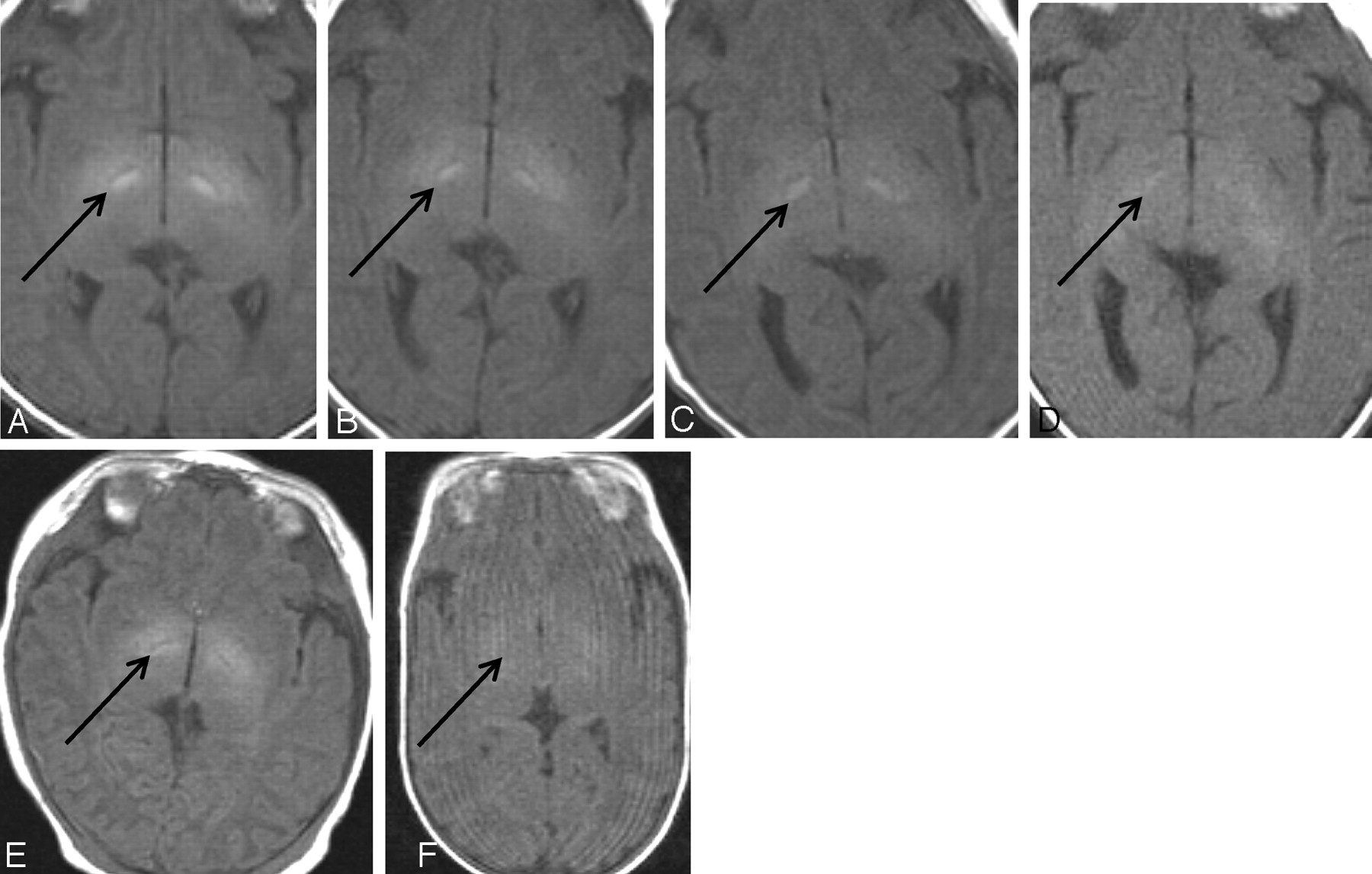
- Fig 1.
Original cases showing transient hyperintensities in STN on T1-weighted image. A-D show the brain of a female infant born at 42 weeks 4 days gestational age at examination. At postnatal age at examination 5 days, the STN (arrow) shows extremely high signal intensity (A). This high signal intensity has diminished at follow-up examinations at postnatal 12 days (B), 23 days (C), and 2 months (D). The patient showed no clinical abnormalities at any stage. E and F show the brains of 2 patients of similar gestational age at MR imaging examination (40 weeks 6 days versus 40 weeks 5 days) but with differing signal intensities. E, High signal intensity can be seen in the STN (arrow) (gestational age at birth, 39 weeks 0 day; postnatal age at time of MR imaging examination, 13 days). F, No significant difference in signal intensity between the STN (arrow) and surrounding structures can be seen (gestational age at birth, 25 weeks 5 days; postnatal age at time of MR imaging examination, 105 days).
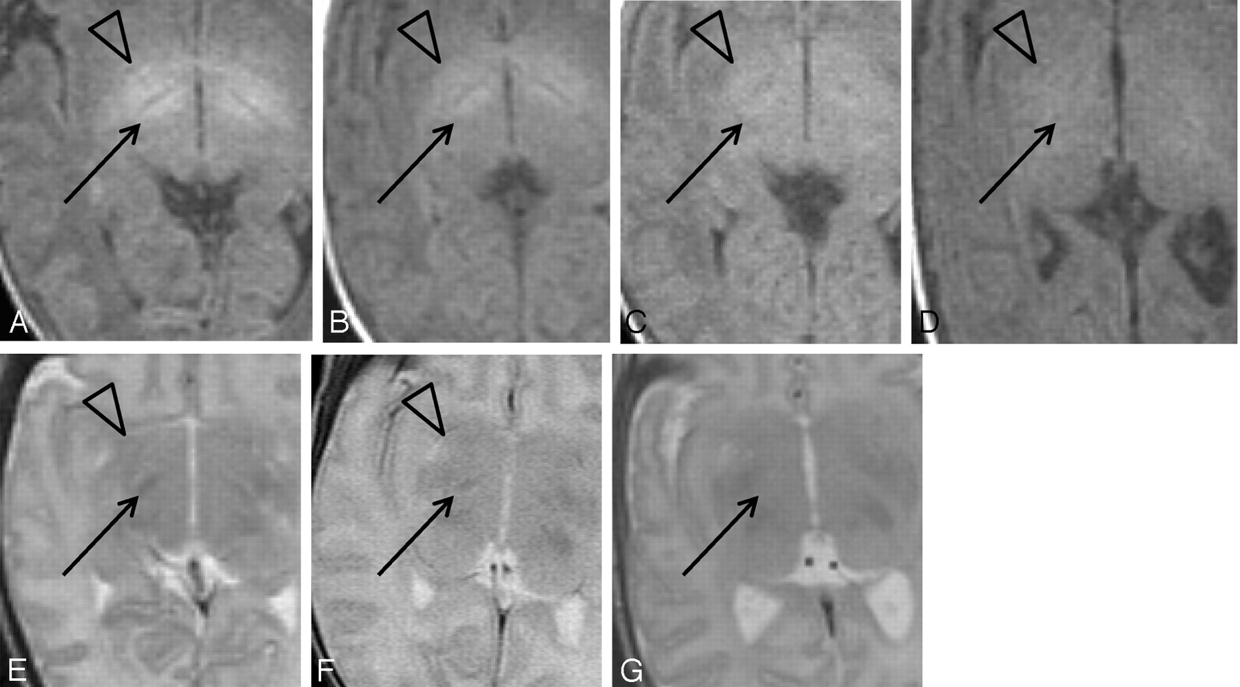
- Fig 2.
Qualitative evaluation of STN and globus pallidus. T1WI qualitatively evaluated by grading the signal intensities of the STN (arrows) and globus pallidus (arrowheads) as follows: grade A+, signal intensity higher than that of the cortex and close to that of the fat (A); grade A, signal intensity higher than but close to that of the cortex (B); grade B, signal intensity lower than that of the cortex and higher than that of the adjacent white matter (C); and grade C, signal intensity indistinguishable from the surrounding structures (D). T2WI qualitatively evaluated by grading the signal intensities of the STN (arrows) and globus pallidus (arrowheads) as follows: grade A, signal intensity lower than that of the cortex (E); grade B, signal intensity higher than that of the cortex and lower than that of the adjacent white matter (F); and grade C, signal intensity indistinguishable from surrounding structure. There are no cases with globus pallidus grade C; therefore, G shows a case with STN grade C and globus pallidus grade B.
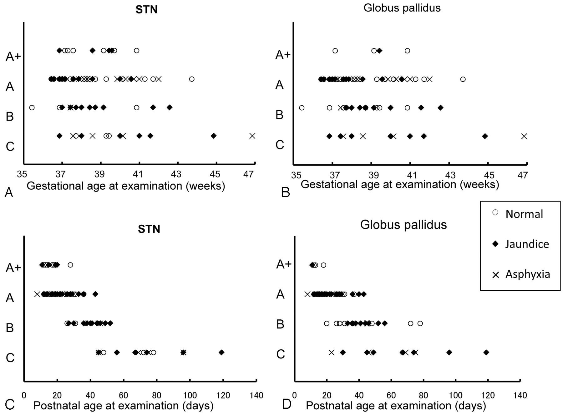
- Fig 3.
Qualitative evaluation of STN and globus pallidus signal intensities on T1-weighted images. Relationships between age and quantitative grade of signal intensity are shown. A, Gestational age at examination and STN. B, Gestational age at examination and globus pallidus. C, Postnatal age at examination and STN. D, Postnatal age at examination and globus pallidus. A and B show considerable overlap in gestational ages at examination among the grades. The grades for the STN and globus pallidus tend to be lower in subjects whose gestational age at examination was ≥44 weeks; however, no other marked trends are observed among the 3 groups. C and D show a distinct tendency for the signal intensities of both STN and globus pallidus to decrease among the 3 groups as postnatal age increases. Normal, jaundice, and asphyxia subjects are plotted separately.
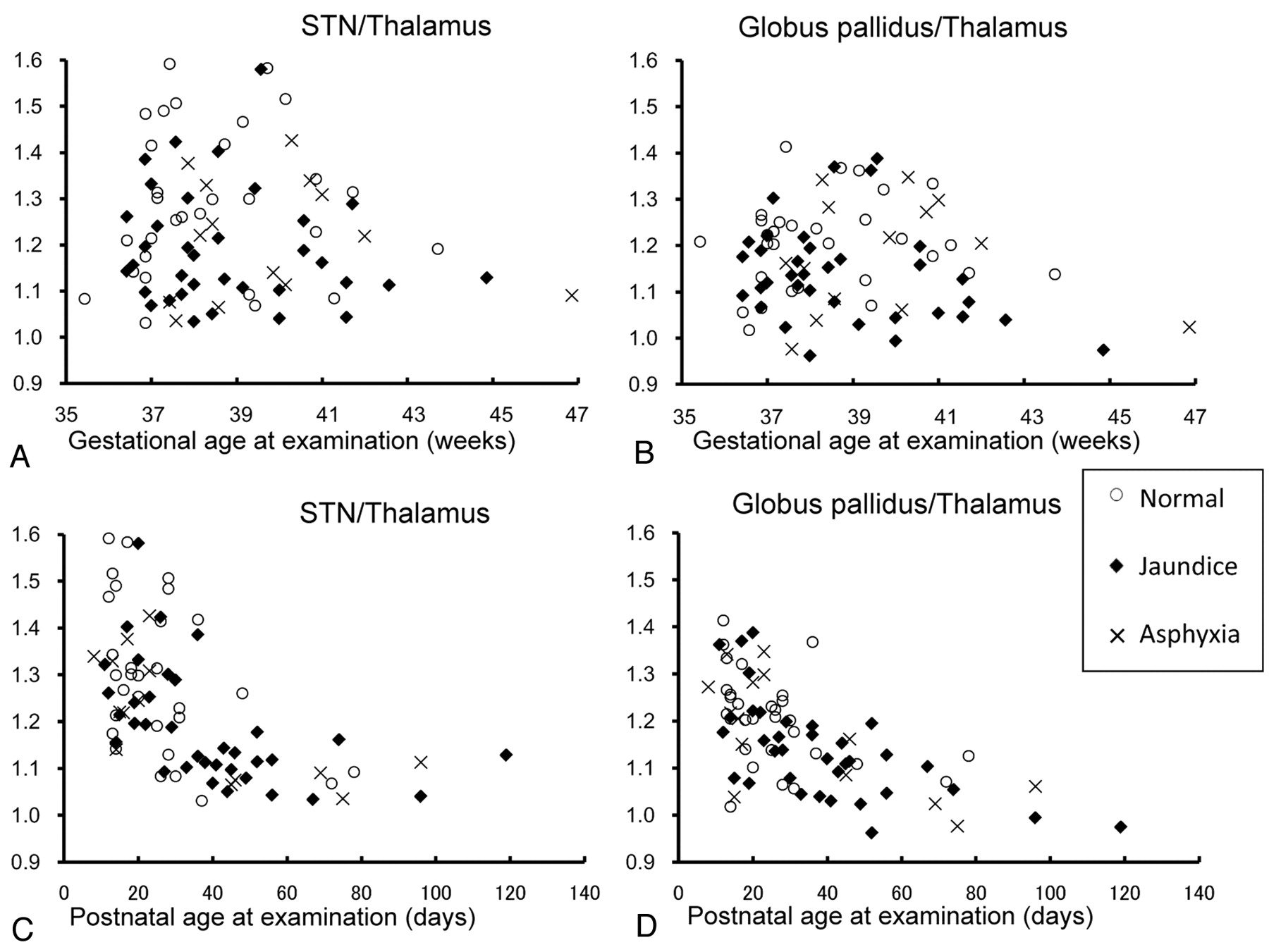
- Fig 4.
Quantitative evaluation of STN and globus pallidus signal intensities compared with the thalamus on T1-weighted images. Relationships between age and signal intensity ratio to the thalamus are shown. A, Gestational age at examination and STN/thalamus. B, Gestational age at examination and globus pallidus/thalamus. C, Postnatal age at examination and STN/thalamus. D, Postnatal age at examination and globus pallidus/thalamus. The ratios of STN/thalamus and globus pallidus/thalamus tend to be lower in subjects whose gestational age at examination was ≥44 weeks; however, no other marked trends are observed among the 3 groups (A and B). C and D show a distinct tendency among the 3 groups for the signal intensity ratio of both STN/thalamus and globus pallidus/thalamus to decrease as postnatal age at examination increases. Normal, jaundice, and asphyxia groups are plotted separately.
- Fig 5.
Statistical analysis of STN/thalamus and globus pallidus/thalamus ratios on T1-weighted images. A and B, Mean STN/thalamus and globus pallidus/thalamus ratios according to gestational age at examination (≤37, 38–39, and ≥40 weeks). There is no statistically significant difference among the 3 age groups. C and D, Mean STN/thalamus and globus pallidus/thalamus ratios according to postnatal age at examination (≤19, 20–39, and ≥40 days). ANOVA shows a statistically significant difference (P < .01) between <19 and >40 days and 20–39 and >40 days.
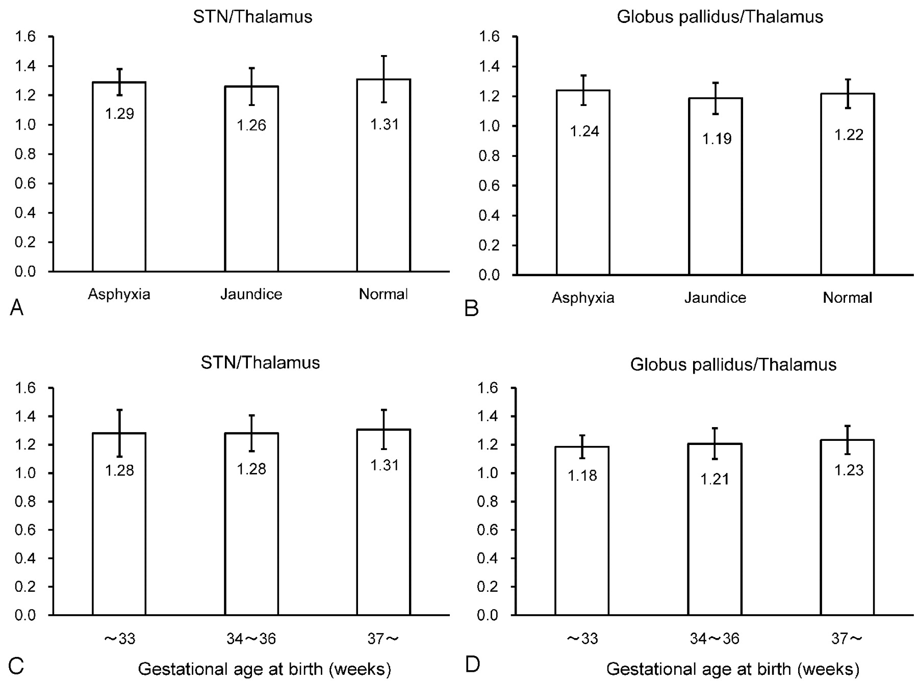
- Fig 6.
Statistical analysis on T1-weighted images based on clinical status groups and gestational ages at birth. Statistical analysis of STN/thalamus (A) and globus pallidus/thalamus (B) ratios on T1-weighted images among the 3 clinical status groups (normal, jaundice, and asphyxia). There are no statistically significant differences among the 3 groups. Statistical analysis of the differences in STN/thalamus (C) and globus pallidus/thalamus (D) ratios on T1WI among the groups whose gestational age at birth are ≤33, 34–36, and ≥37 weeks. There are no statistically significant differences among the 3 groups.
- Fig 7.
Qualitative evaluation of STN and globus pallidus signal intensities on T2-weighted images. Relationships between age and quantitative grade of signal intensity are shown. A, Gestational age at examination and STN. B, Gestational age at examination and globus pallidus. C, Postnatal age at examination and STN. D, Postnatal age at examination and globus pallidus. Trends in signal intensity changes are not observed among the 3 groups for either gestational age at examination or postnatal age at examination. Most subjects show grade A for the STN (62.0%; 49/79) and grade B for the globus pallidus (87.3%; 69/79) regardless of the gestational age at examination or postnatal age at examination. Normal, jaundice, and asphyxia subjects are plotted separately.
- Fig 8.
Quantitative evaluation of STN and globus pallidus signal intensities compared with the thalamus on T2-weighted images. Relationships between age and signal intensity ratio to the thalamus are shown. A, Gestational age at examination and STN/thalamus. B, Gestational age at examination and globus pallidus/thalamus. C, Postnatal age at examination and STN/thalamus. D, Postnatal age at examination and globus pallidus/thalamus. No chronologic changes in ratios are observed among the 3 groups for either gestational age at examination or postnatal age at examination. Statistical evaluations also demonstrate no significant differences. Normal, jaundice, and asphyxia subjects are plotted separately.


In this issue


Jump to section
Related Articles
Cited By...
- No citing articles found.