
Article Figures & Data
Figures
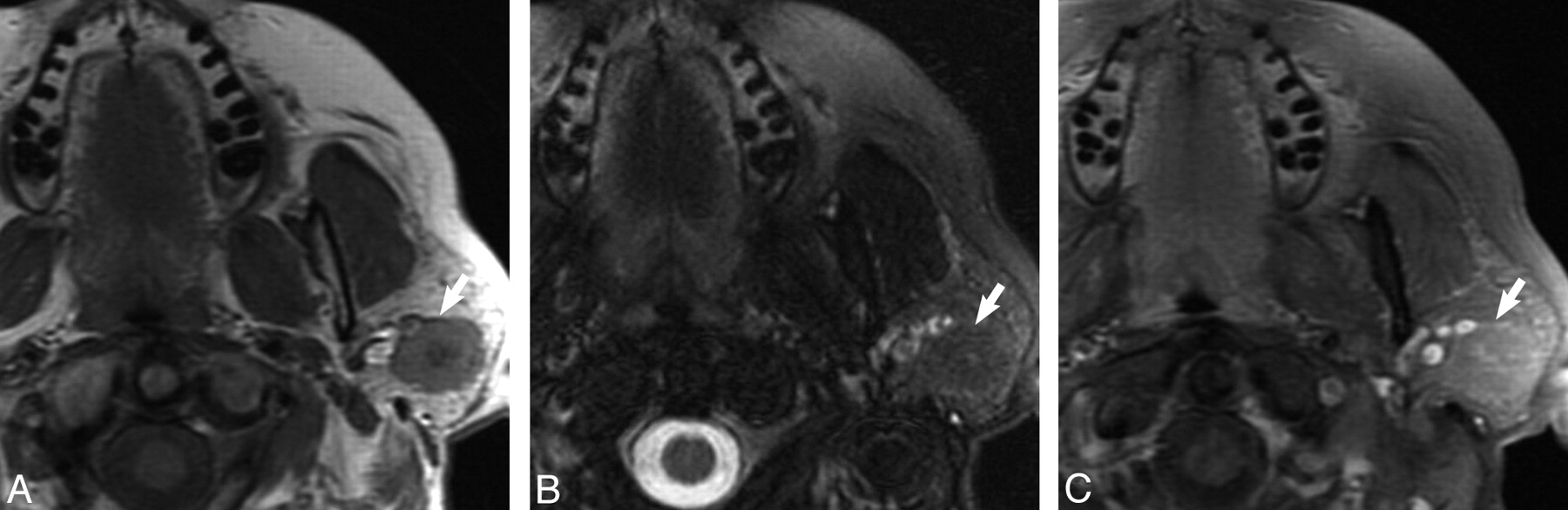
- Fig 1.
Case 1, 62-year-old female with 2-year history of firm, painless left parotid mass. A, Axial T1-weighted MR image shows a well-demarcated mass within the superficial lobe of the left parotid gland (white arrow) that is T1 hypointense. B, Axial T2-weighted MR image with fat saturation shows the mass (white arrow) to be isointense to the native parotid gland. C, Axial T1-weighted postgadolinium sequences illustrates the mass (white arrow) as isointense to the native parotid gland.
- Fig 2.
Case 5, 55-year-old woman with painless “lump” behind her left ear. A, Axial T1-weighted MR image shows a well-demarcated mass within the superficial and deep lobe of the left parotid gland (white arrow) that is T1 hypointense. B, Axial T2-weighted MR image with fat saturation illustrates the mass (white arrow) to be isointense to the native parotid gland. C, Axial T1-weighted postgadolinium sequence shows the mass (white arrow) as isointense to the native parotid gland.
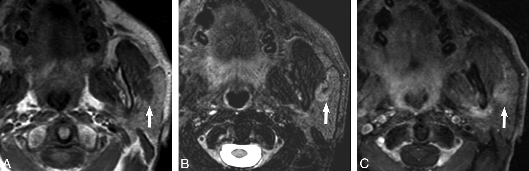
- Fig 3.
Case 7, 75-year-old man with history of squamous cell carcinoma of the larynx status postradiation found to have an enlarging mass in the left parotid gland on surveillance imaging. A, Axial T1-weighted MR image shows a well-demarcated mass within the superficial lobe of the left parotid gland (white arrow) that is T1 hypointense. B, Axial T2-weighted MR image with fat saturation shows the mass to be hyperintense but heterogeneous to the native parotid gland. C, Axial T1-weighted postgadolinium sequence shows the mass (white arrow) to demonstrate heterogeneous enhancement to the native parotid gland.
- Fig 4.
Typical histologic features of a parotid gland oncocytoma including eosinophilic cells with finely granular cytoplasm and uniform, round, centrally placed nuclei (hematoxylin-eosin stain, original magnification, ×400).


Tables
Presentation and MR imaging features of parotid gland oncocytomas
Patient No. Age/Sex Presentation Intraparotid Location Size (cm) Tumor Margin T1WI T2 FS WI T1 FS +C WI 1 62/F Painless mass Superficial 2.0 Smooth Hypointense Isointense Isointense 2 66/F Painless mass Superficial and deep 3.4 Lobular Hypointense Isointense Isointense 3 46/F Right ear pain Superficial and deep 1.3 Lobular Hypointense Isointense Isointense 4 36/M Painless mass Superficial 3.0 Smooth Hypointense Isointense Isointense 5 55/F Painless mass Superficial and deep 2.6 Smooth Hypointense Isointense Isointense 6 54/F Painless mass Superficial 2.5 Smooth Hypointense Isointense Isointense 7 75/M Painless mass Superficial 2.5 Lobular Hypointense Hyperintense Heterogeneous enhancement 8 83/F Painless mass Superficial 3.3 Lobular Hypointense Isointense Isointense 9 74/M Painless mass Superficial 3.0 Smooth Hypointense Isointense Isointense
In this issue


Jump to section
Related Articles
Cited By...
- No citing articles found.