Advertisement

Main menu

  • Home
  • Content
    • Current Issue
    • Accepted Manuscripts
    • Article Preview
    • Past Issue Archive
    • Video Articles
    • AJNR Case Collection
    • Case of the Week Archive
    • Case of the Month Archive
    • Classic Case Archive
  • Special Collections
    • AJNR Awards
    • ASNR Foundation Special Collection
    • Most Impactful AJNR Articles
    • Photon-Counting CT
    • Spinal CSF Leak Articles (Jan 2020-June 2024)
  • Multimedia
    • AJNR Podcasts
    • AJNR SCANtastic
    • Trainee Corner
    • MRI Safety Corner
    • Imaging Protocols
  • For Authors
    • Submit a Manuscript
    • Submit a Video Article
    • Submit an eLetter to the Editor/Response
    • Manuscript Submission Guidelines
    • Statistical Tips
    • Fast Publishing of Accepted Manuscripts
    • Graphical Abstract Preparation
    • Imaging Protocol Submission
    • Author Policies
  • About Us
    • About AJNR
    • Editorial Board
    • Editorial Board Alumni
  • More
    • Become a Reviewer/Academy of Reviewers
    • Subscribers
    • Permissions
    • Alerts
    • Feedback
    • Advertisers
    • ASNR Home

User menu

  • Alerts
  • Log in

Search

  • Advanced search
American Journal of Neuroradiology

American Journal of Neuroradiology

ASHNR American Society of Functional Neuroradiology ASHNR American Society of Pediatric Neuroradiology ASSR
  • Alerts
  • Log in

Advanced Search

  • Home
  • Content
    • Current Issue
    • Accepted Manuscripts
    • Article Preview
    • Past Issue Archive
    • Video Articles
    • AJNR Case Collection
    • Case of the Week Archive
    • Case of the Month Archive
    • Classic Case Archive
  • Special Collections
    • AJNR Awards
    • ASNR Foundation Special Collection
    • Most Impactful AJNR Articles
    • Photon-Counting CT
    • Spinal CSF Leak Articles (Jan 2020-June 2024)
  • Multimedia
    • AJNR Podcasts
    • AJNR SCANtastic
    • Trainee Corner
    • MRI Safety Corner
    • Imaging Protocols
  • For Authors
    • Submit a Manuscript
    • Submit a Video Article
    • Submit an eLetter to the Editor/Response
    • Manuscript Submission Guidelines
    • Statistical Tips
    • Fast Publishing of Accepted Manuscripts
    • Graphical Abstract Preparation
    • Imaging Protocol Submission
    • Author Policies
  • About Us
    • About AJNR
    • Editorial Board
    • Editorial Board Alumni
  • More
    • Become a Reviewer/Academy of Reviewers
    • Subscribers
    • Permissions
    • Alerts
    • Feedback
    • Advertisers
    • ASNR Home
  • Follow AJNR on Twitter
  • Visit AJNR on Facebook
  • Follow AJNR on Instagram
  • Join AJNR on LinkedIn
  • RSS Feeds

AJNR is seeking candidates for the AJNR Podcast Editor. Read the position description.

Research ArticleBrainF
Open Access

Differentiation of Tumefactive Demyelinating Lesions from High-Grade Gliomas with the Use of Diffusion Tensor Imaging

C.H Toh, K.-C. Wei, S.-H. Ng, Y.-L. Wan, M. Castillo and C.-P. Lin
American Journal of Neuroradiology May 2012, 33 (5) 846-851; DOI: https://doi.org/10.3174/ajnr.A2871
C.H Toh
  • Find this author on Google Scholar
  • Find this author on PubMed
  • Search for this author on this site
K.-C. Wei
  • Find this author on Google Scholar
  • Find this author on PubMed
  • Search for this author on this site
S.-H. Ng
  • Find this author on Google Scholar
  • Find this author on PubMed
  • Search for this author on this site
Y.-L. Wan
  • Find this author on Google Scholar
  • Find this author on PubMed
  • Search for this author on this site
M. Castillo
  • Find this author on Google Scholar
  • Find this author on PubMed
  • Search for this author on this site
C.-P. Lin
  • Find this author on Google Scholar
  • Find this author on PubMed
  • Search for this author on this site
  • Article
  • Figures & Data
  • Info & Metrics
  • Responses
  • References
  • PDF
Loading

Article Figures & Data

Figures

  • Tables
  • Fig 1.
    • Download figure
    • Open in new tab
    • Download powerpoint
    Fig 1.

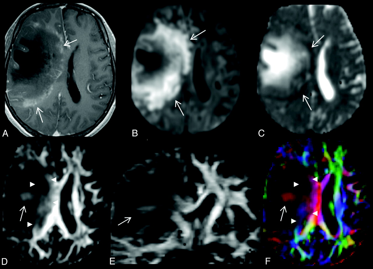
    A 28-year-old woman with a large TDL in the right frontal lobe. A, There is heterogeneous enhancement at the medial margin (arrows) of the lesion on the axial contrast-enhanced T1-weighted image. B and C, On the axial DWI (B) and MD map (C), the peripheral portion of the lesion shows restricted diffusion (arrows). D and E, Intralesional hyperintensities (arrow) are seen in both the lesion center (arrow) and periphery (arrowheads) on axial (D) and coronal (E) FA maps. F, Directional-coded FA map displays the directionality of these intralesional hyperintensities. The FA values are 0.06 for central nonenhancing portion and 0.21 for peripheral enhancing portion. The MD values (× 10−3 mm2/s) are 1.966 for central nonenhancing portion and 0.704 for peripheral enhancing portion.

  • Fig 2.
    • Download figure
    • Open in new tab
    • Download powerpoint
    Fig 2.

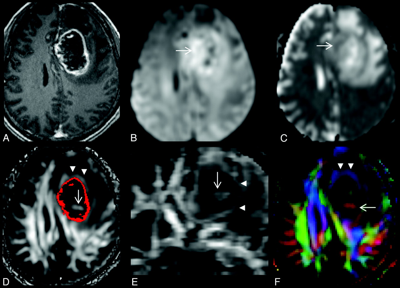
    A, A 68-year-old man with a left frontal glioblastoma, which appears as a rim-enhancing mass on the axial contrast-enhanced T1-weighted image. B and C, On the axial DWI (B) and MD map (C), restricted diffusion is present at the lesion periphery (arrow). D and E, Intralesional hyperintensities (arrow) are seen on axial (D) and coronal (E) FA maps. A red scattered ROI representing a contrast-enhancing rim is overlaid on the FA map (D). F, Note that there is a hyperintense FA rim (arrowheads in D, E, and F) in the perifocal edema, external to the enhancing rim. Directional-coded (F) FA map displays the directionality of hyperintense FA rim and intralesional hyperintensity. The FA values are 0.09 for the central nonenhancing portion and 0.13 for the peripheral enhancing portion. The MD values (× 10−3 mm2/s) are 1.530 for the central nonenhancing portion and 1.252 for the peripheral enhancing portion.

Tables

  • Figures
    • View popup
    Table 1:

    Results of visual inspection of TDLs and high-grade gliomas

    Imaging SignsTDLsHigh-Grade GliomasP ValueORSENSPEAccuracy
    Intralesional hyperintensities on FA maps6 (75%)4 (30.8%).0496.875%69.2%71.4%
    Restricted diffusion in lesion periphery7 (87.5%)7 (53.8%).112
    Perilesional hyperintense FA rim1 (12.5%)12 (92.3%)<.0018492.3%87.5%90.4%
    • Note:—OR indicates odds ratio; SEN, sensitivity; SPE, specificity.

    • View popup
    Table 2:

    MD and FA of TDLs and high-grade gliomasa

    Region/MetricsTDLsHigh-Grade GliomasP Value95% CI
    Nonenhancing portion
        FA0.06 ± 0.020.07 ± 0.03.526−0.035–0.018
        MD1.702 ± 0.4141.806 ± 0.578.663−0.597–0.388
    Enhancing portion
        FA0.18 ± 0.050.12 ± 0.01.0040.020-.088
        MD0.887 ± 0.2651.237 ± 0.124.001−0.528–0.172
    Perilesional edema
        FA0.11 ± 0.010.17 ± 0.03.001−0.091–0.028
        MD1.411 ± 0.2621.270 ± 0.121.158−0.062–0.346
    • Note:—CI indicates confidence interval.

    • ↵a Data are the mean. Unit is × 10−3 mm2/s for MD values.

PreviousNext
Back to top

In this issue

American Journal of Neuroradiology: 33 (5)
American Journal of Neuroradiology
Vol. 33, Issue 5
1 May 2012
  • Table of Contents
  • Index by author
Advertisement
Print
Download PDF
Email Article

Thank you for your interest in spreading the word on American Journal of Neuroradiology.

NOTE: We only request your email address so that the person you are recommending the page to knows that you wanted them to see it, and that it is not junk mail. We do not capture any email address.

Enter multiple addresses on separate lines or separate them with commas.
Differentiation of Tumefactive Demyelinating Lesions from High-Grade Gliomas with the Use of Diffusion Tensor Imaging
(Your Name) has sent you a message from American Journal of Neuroradiology
(Your Name) thought you would like to see the American Journal of Neuroradiology web site.
CAPTCHA
This question is for testing whether or not you are a human visitor and to prevent automated spam submissions.
Cite this article
C.H Toh, K.-C. Wei, S.-H. Ng, Y.-L. Wan, M. Castillo, C.-P. Lin
Differentiation of Tumefactive Demyelinating Lesions from High-Grade Gliomas with the Use of Diffusion Tensor Imaging
American Journal of Neuroradiology May 2012, 33 (5) 846-851; DOI: 10.3174/ajnr.A2871

Citation Manager Formats

  • BibTeX
  • Bookends
  • EasyBib
  • EndNote (tagged)
  • EndNote 8 (xml)
  • Medlars
  • Mendeley
  • Papers
  • RefWorks Tagged
  • Ref Manager
  • RIS
  • Zotero
0 Responses
Respond to this article
Share
Bookmark this article
Differentiation of Tumefactive Demyelinating Lesions from High-Grade Gliomas with the Use of Diffusion Tensor Imaging
C.H Toh, K.-C. Wei, S.-H. Ng, Y.-L. Wan, M. Castillo, C.-P. Lin
American Journal of Neuroradiology May 2012, 33 (5) 846-851; DOI: 10.3174/ajnr.A2871
del.icio.us logo Twitter logo Facebook logo Mendeley logo
  • Tweet Widget
  • Facebook Like
  • Google Plus One
Purchase

Jump to section

  • Article
    • Abstract
    • ABBREVIATIONS:
    • Materials and Methods
    • Results
    • Discussion
    • Conclusions
    • Footnotes
    • References
  • Figures & Data
  • Info & Metrics
  • Responses
  • References
  • PDF

Related Articles

  • No related articles found.
  • PubMed
  • Google Scholar

Cited By...

  • MRI Findings in Tumefactive Demyelinating Lesions: A Systematic Review and Meta-Analysis
  • Combining Diffusion Tensor Metrics and DSC Perfusion Imaging: Can It Improve the Diagnostic Accuracy in Differentiating Tumefactive Demyelination from High-Grade Glioma?
  • Diagnostic Utility of Diffusion Tensor Imaging in Differentiating Glioblastomas from Brain Metastases
  • Assessment of Angiographic Vascularity of Meningiomas with Dynamic Susceptibility Contrast-Enhanced Perfusion-Weighted Imaging and Diffusion Tensor Imaging
  • Crossref (23)
  • Google Scholar

This article has been cited by the following articles in journals that are participating in Crossref Cited-by Linking.

  • MRI Findings in Tumefactive Demyelinating Lesions: A Systematic Review and Meta-Analysis
    C.H. Suh, H.S. Kim, S.C. Jung, C.G. Choi, S.J. Kim
    American Journal of Neuroradiology 2018 39 9
  • Diagnostic Utility of Diffusion Tensor Imaging in Differentiating Glioblastomas from Brain Metastases
    S. Wang, S. J. Kim, H. Poptani, J. H. Woo, S. Mohan, R. Jin, M. R. Voluck, D. M. O'Rourke, R. L. Wolf, E. R. Melhem, S. Kim
    American Journal of Neuroradiology 2014 35 5
  • A Review of Clinical and Imaging Findings in Tumefactive Demyelination
    Mariko Nakayama, Shotaro Naganawa, Michelle Ouyang, Karra A. Jones, John Kim, Aristides A. Capizzano, Toshio Moritani
    American Journal of Roentgenology 2021 217 1
  • Histogram Analysis of Apparent Diffusion Coefficient Maps for Differentiating Primary CNS Lymphomas From Tumefactive Demyelinating Lesions
    Shan Shan Lu, Sang Joon Kim, Namkug Kim, Ho Sung Kim, Choong Gon Choi, Young Min Lim
    American Journal of Roentgenology 2015 204 4
  • Analysis of fractional anisotropy facilitates differentiation of glioblastoma and brain metastases in a clinical setting
    Stefanie Bette, Thomas Huber, Benedikt Wiestler, Tobias Boeckh-Behrens, Jens Gempt, Florian Ringel, Bernhard Meyer, Claus Zimmer, Jan S. Kirschke
    European Journal of Radiology 2016 85 12
  • Combining Diffusion Tensor Metrics and DSC Perfusion Imaging: Can It Improve the Diagnostic Accuracy in Differentiating Tumefactive Demyelination from High-Grade Glioma?
    S.B. Hiremath, A. Muraleedharan, S. Kumar, C. Nagesh, C. Kesavadas, M. Abraham, T.R. Kapilamoorthy, B. Thomas
    American Journal of Neuroradiology 2017 38 4
  • Distinguishing brain inflammation from grade II glioma in population without contrast enhancement: a radiomics analysis based on conventional MRI
    Yu Han, Yang Yang, Zhe-sheng Shi, An-ding Zhang, Lin-feng Yan, Yu-chuan Hu, Lan-lan Feng, Jiao Ma, Wen Wang, Guang-bin Cui
    European Journal of Radiology 2021 134
  • Assessment of Angiographic Vascularity of Meningiomas with Dynamic Susceptibility Contrast-Enhanced Perfusion-Weighted Imaging and Diffusion Tensor Imaging
    C. H. Toh, K.- C. Wei, C. N. Chang, Y.- W. Peng, S.- H. Ng, H.- F. Wong, C.- P. Lin
    American Journal of Neuroradiology 2014 35 2
  • Differential imaging of atypical demyelinating lesions of the central nervous system
    Matteo Paoletti, Shaun Ivan Muzic, Francesca Marchetti, Lisa Maria Farina, Stefano Bastianello, Anna Pichiecchio
    La radiologia medica 2021 126 6
  • Restriction spectrum imaging of white matter and its relation to neurological disability in multiple sclerosis
    Piotr Sowa, Hanne F Harbo, Nathan S White, Elisabeth G Celius, Hauke Bartsch, Pål Berg-Hansen, Stine M Moen, Atle Bjørnerud, Lars T Westlye, Ole A Andreassen, Anders M Dale, Mona K Beyer
    Multiple Sclerosis Journal 2019 25 5

More in this TOC Section

  • Usefulness of Quantitative Susceptibility Mapping for the Diagnosis of Parkinson Disease
  • White Matter Alterations in the Brains of Patients with Active, Remitted, and Cured Cushing Syndrome: A DTI Study
  • Qualitative and Quantitative Analysis of MR Imaging Findings in Patients with Middle Cerebral Artery Stroke Implanted with Mesenchymal Stem Cells
Show more BRAIN

Similar Articles

Advertisement

Indexed Content

  • Current Issue
  • Accepted Manuscripts
  • Article Preview
  • Past Issues
  • Editorials
  • Editor's Choice
  • Fellows' Journal Club
  • Letters to the Editor
  • Video Articles

Cases

  • Case Collection
  • Archive - Case of the Week
  • Archive - Case of the Month
  • Archive - Classic Case

Special Collections

  • AJNR Awards
  • ASNR Foundation Special Collection
  • Most Impactful AJNR Articles
  • Photon-Counting CT
  • Spinal CSF Leak Articles (Jan 2020-June 2024)

More from AJNR

  • Trainee Corner
  • Imaging Protocols
  • MRI Safety Corner

Multimedia

  • AJNR Podcasts
  • AJNR Scantastics

Resources

  • Turnaround Time
  • Submit a Manuscript
  • Submit a Video Article
  • Submit an eLetter to the Editor/Response
  • Manuscript Submission Guidelines
  • Statistical Tips
  • Fast Publishing of Accepted Manuscripts
  • Graphical Abstract Preparation
  • Imaging Protocol Submission
  • Evidence-Based Medicine Level Guide
  • Publishing Checklists
  • Author Policies
  • Become a Reviewer/Academy of Reviewers
  • News and Updates

About Us

  • About AJNR
  • Editorial Board
  • Editorial Board Alumni
  • Alerts
  • Permissions
  • Not an AJNR Subscriber? Join Now
  • Advertise with Us
  • Librarian Resources
  • Feedback
  • Terms and Conditions
  • AJNR Editorial Board Alumni

American Society of Neuroradiology

  • Not an ASNR Member? Join Now

© 2025 by the American Society of Neuroradiology All rights, including for text and data mining, AI training, and similar technologies, are reserved.
Print ISSN: 0195-6108 Online ISSN: 1936-959X

Powered by HighWire