Advertisement

Main menu

  • Home
  • Content
    • Current Issue
    • Accepted Manuscripts
    • Article Preview
    • Past Issue Archive
    • Video Articles
    • AJNR Case Collection
    • Case of the Week Archive
    • Case of the Month Archive
    • Classic Case Archive
  • Special Collections
    • AJNR Awards
    • ASNR Foundation Special Collection
    • Most Impactful AJNR Articles
    • Photon-Counting CT
    • Spinal CSF Leak Articles (Jan 2020-June 2024)
  • Multimedia
    • AJNR Podcasts
    • AJNR SCANtastic
    • Trainee Corner
    • MRI Safety Corner
    • Imaging Protocols
  • For Authors
    • Submit a Manuscript
    • Submit a Video Article
    • Submit an eLetter to the Editor/Response
    • Manuscript Submission Guidelines
    • Statistical Tips
    • Fast Publishing of Accepted Manuscripts
    • Graphical Abstract Preparation
    • Imaging Protocol Submission
    • Author Policies
  • About Us
    • About AJNR
    • Editorial Board
    • Editorial Board Alumni
  • More
    • Become a Reviewer/Academy of Reviewers
    • Subscribers
    • Permissions
    • Alerts
    • Feedback
    • Advertisers
    • ASNR Home

User menu

  • Alerts
  • Log in

Search

  • Advanced search
American Journal of Neuroradiology

American Journal of Neuroradiology

ASHNR American Society of Functional Neuroradiology ASHNR American Society of Pediatric Neuroradiology ASSR
  • Alerts
  • Log in

Advanced Search

  • Home
  • Content
    • Current Issue
    • Accepted Manuscripts
    • Article Preview
    • Past Issue Archive
    • Video Articles
    • AJNR Case Collection
    • Case of the Week Archive
    • Case of the Month Archive
    • Classic Case Archive
  • Special Collections
    • AJNR Awards
    • ASNR Foundation Special Collection
    • Most Impactful AJNR Articles
    • Photon-Counting CT
    • Spinal CSF Leak Articles (Jan 2020-June 2024)
  • Multimedia
    • AJNR Podcasts
    • AJNR SCANtastic
    • Trainee Corner
    • MRI Safety Corner
    • Imaging Protocols
  • For Authors
    • Submit a Manuscript
    • Submit a Video Article
    • Submit an eLetter to the Editor/Response
    • Manuscript Submission Guidelines
    • Statistical Tips
    • Fast Publishing of Accepted Manuscripts
    • Graphical Abstract Preparation
    • Imaging Protocol Submission
    • Author Policies
  • About Us
    • About AJNR
    • Editorial Board
    • Editorial Board Alumni
  • More
    • Become a Reviewer/Academy of Reviewers
    • Subscribers
    • Permissions
    • Alerts
    • Feedback
    • Advertisers
    • ASNR Home
  • Follow AJNR on Twitter
  • Visit AJNR on Facebook
  • Follow AJNR on Instagram
  • Join AJNR on LinkedIn
  • RSS Feeds

AJNR is seeking candidates for the AJNR Podcast Editor. Read the position description.

Research ArticleBrainE

Detection of Microhemorrhage in Posterior Reversible Encephalopathy Syndrome Using Susceptibility-Weighted Imaging

A.M. McKinney, B. Sarikaya, C. Gustafson and C.L. Truwit
American Journal of Neuroradiology May 2012, 33 (5) 896-903; DOI: https://doi.org/10.3174/ajnr.A2886
A.M. McKinney
  • Find this author on Google Scholar
  • Find this author on PubMed
  • Search for this author on this site
B. Sarikaya
  • Find this author on Google Scholar
  • Find this author on PubMed
  • Search for this author on this site
C. Gustafson
  • Find this author on Google Scholar
  • Find this author on PubMed
  • Search for this author on this site
C.L. Truwit
  • Find this author on Google Scholar
  • Find this author on PubMed
  • Search for this author on this site
  • Article
  • Figures & Data
  • Info & Metrics
  • Responses
  • References
  • PDF
Loading

Article Figures & Data

Figures

  • Fig 1.
    • Download figure
    • Open in new tab
    • Download powerpoint
    Fig 1.

    Patients with PRES on SWI, with the frequency of MH and other subtypes of hemorrhage.

  • Fig 2.
    • Download figure
    • Open in new tab
    • Download powerpoint
    Fig 2.

    A 50-year-old woman with seizure and a history of hypertension who presented with “mild” PRES-related cortical and subcortical edema (dashed arrows) on a 3T FLAIR MR image (A), with a small underlying MH (arrow) on SWI (B). On a follow-up 3T FLAIR MR image (C), the PRES-related edema had mostly resolved, while the tiny MH persisted on SWI (D).

  • Fig 3.
    • Download figure
    • Open in new tab
    • Download powerpoint
    Fig 3.

    A 51-year-old hypertensive woman with unilateral moderate edema from PRES on 3T FLAIR images. This likely occurred unilaterally because the patient had a severe (>90%) left carotid bulb stenosis, which presumably prevented hyperperfusion of the left cerebral hemisphere. B, There is a small amount of SAH (arrows) on SWI. On a follow-up 3T MR imaging performed 70 days later, FLAIR image (C) demonstrated resolution of the PRES-related edema, while the SAH had also resolved on SWI (D).

  • Fig 4.
    • Download figure
    • Open in new tab
    • Download powerpoint
    Fig 4.

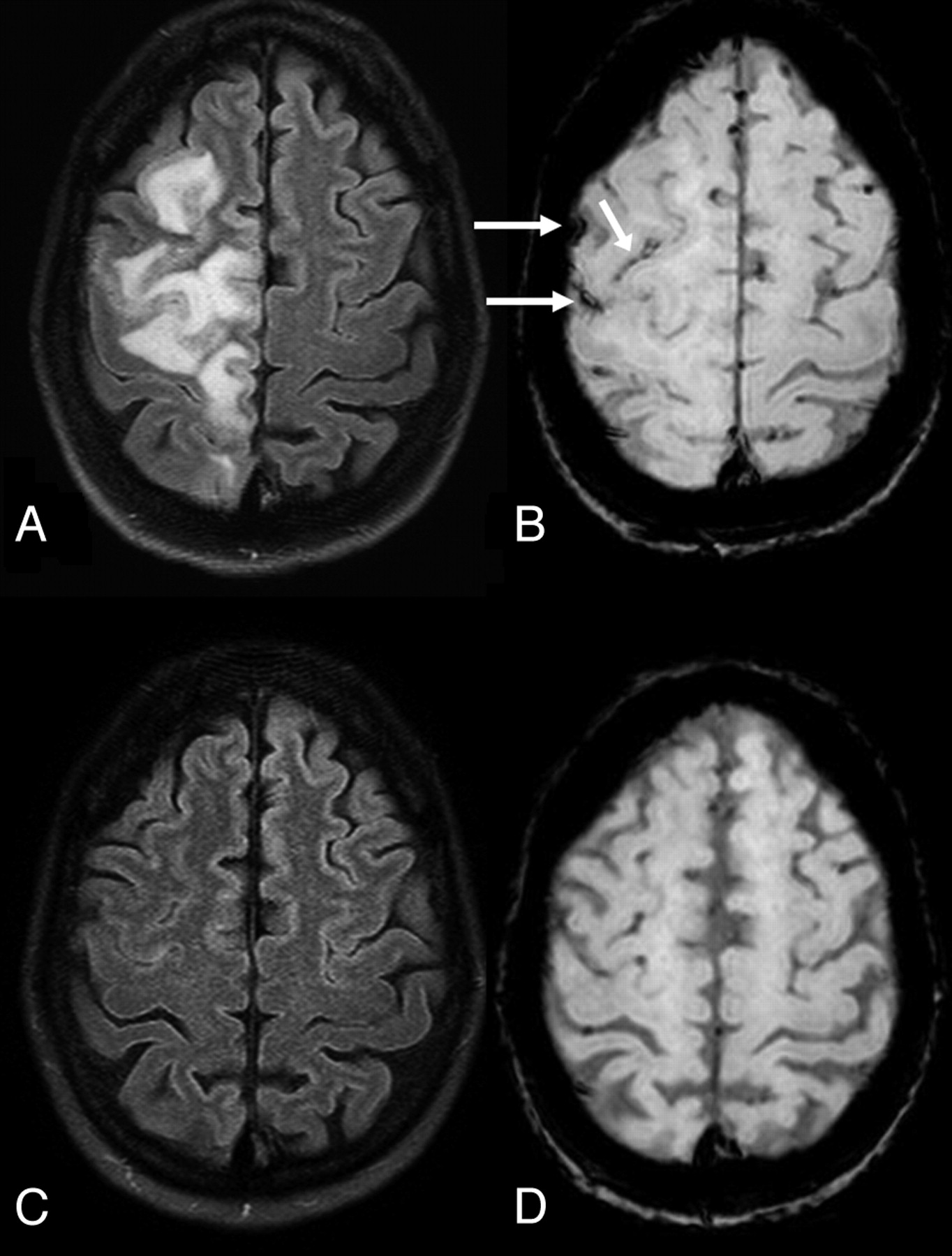
    A 48-year-old woman with severe extent of PRES on 1.5T FLAIR images (A and B), based on involvement of the cerebellum (arrows, A), basal ganglia (arrows, B), and brain stem (not shown) and because the cerebral edema extends from the ventricular margin to the cortex (B). Although the severity was denoted, SWI on that presenting MR image (C) did not demonstrate any MH. Follow-up FLAIR image (D) obtained on the same magnet 22 days later had nearly normal findings, and no MHs were noted on the follow-up SWI images either (not shown).

  • Fig 5.
    • Download figure
    • Open in new tab
    • Download powerpoint
    Fig 5.

    A 52-year-old patient with cyclosporine toxicity. Eleven days before the seizure, a pretransplant surveillance 3T MR imaging with FLAIR (A), SWI (B), and postcontrast T1WI (C) had normal findings. At presentation for seizure with mild PRES, 1.5T FLAIR (D) showed vasogenic edema, with a new punctate MH on SWI (E) and cortical and leptomeningeal contrast enhancement on postcontrast T1WI (F). After the episode of PRES clinically resolved, a 5-month follow-up MR image at 3T showed no edema on FLAIR (G), while the MH remained on SWI (H). The MH also persisted on a 1.5T SWI at 9 months (I).

  • Fig 6.
    • Download figure
    • Open in new tab
    • Download powerpoint
    Fig 6.

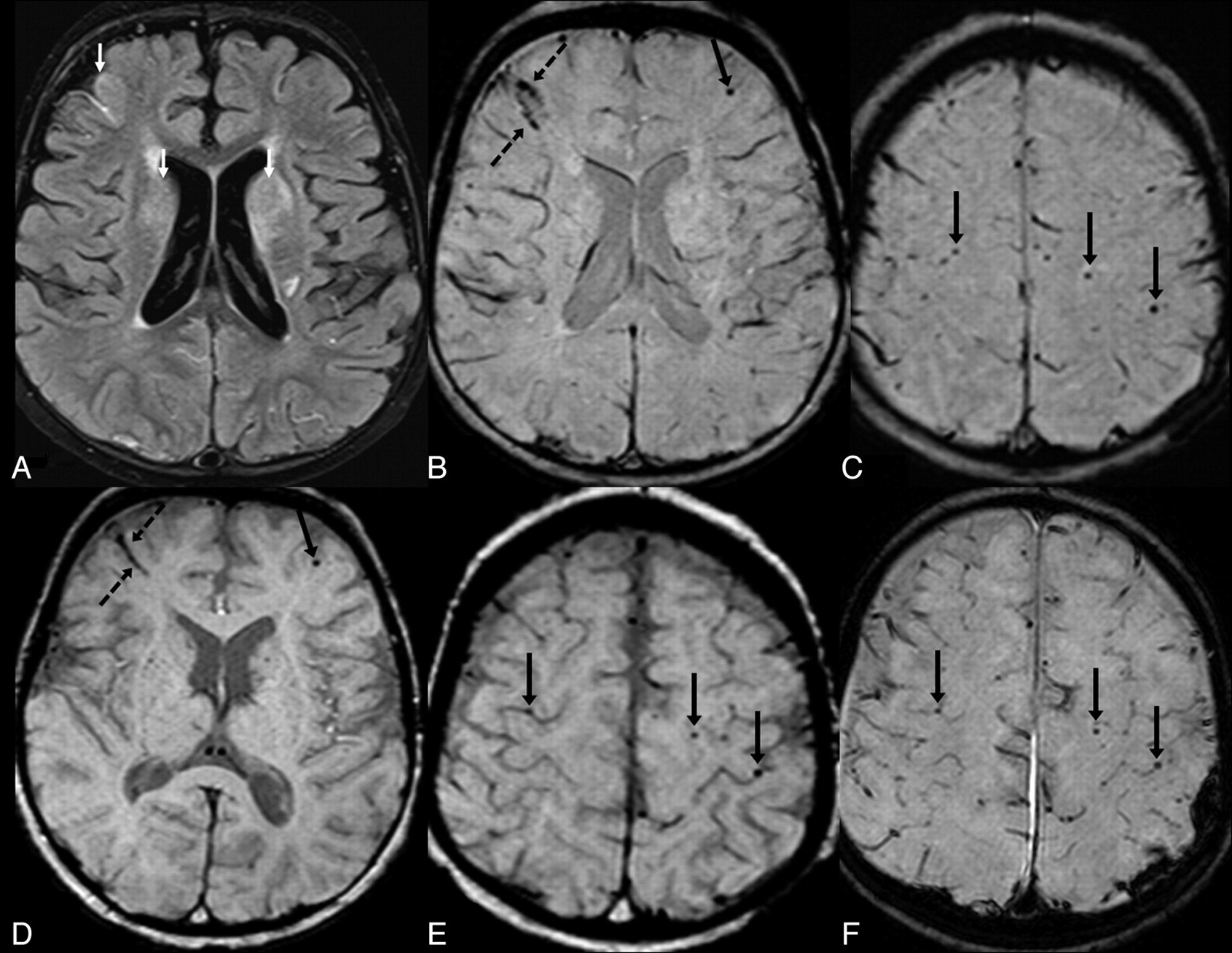
    A 3-year-old child with a seizure from tacrolimus toxicity post–heart transplantation. Thirty-five days before the seizure, a pretransplantation surveillance MR imaging at 1.5T had FLAIR images that appeared to have normal findings (not shown). A, At presentation with PRES, a 1.5T FLAIR image demonstrates edema of the caudate nuclei and frontal lobes (arrows) as well as bilateral parieto-occipital edema (not shown), considered moderate severity. There was dark SAH (dashed arrows, B) and multiple frontal, parietal, and occipital cortical/subcortical MHs (arrows) on SWI (B and C) at presentation, which totaled >20 MHs. On the follow-up 3T MR imaging 8 days later, the regions of vasogenic edema are nearly resolved on FLAIR images (not shown), while SWI demonstrates improved but persistent SAH (dashed arrows, D); the MHs also persist on SWI (D and E). F, Review of SWI from the 35-day pretransplantation 1.5T MR imaging reveals that each of the >20 MHs (arrows) were present before the onset of PRES.

PreviousNext
Back to top

In this issue

American Journal of Neuroradiology: 33 (5)
American Journal of Neuroradiology
Vol. 33, Issue 5
1 May 2012
  • Table of Contents
  • Index by author
Advertisement
Print
Download PDF
Email Article

Thank you for your interest in spreading the word on American Journal of Neuroradiology.

NOTE: We only request your email address so that the person you are recommending the page to knows that you wanted them to see it, and that it is not junk mail. We do not capture any email address.

Enter multiple addresses on separate lines or separate them with commas.
Detection of Microhemorrhage in Posterior Reversible Encephalopathy Syndrome Using Susceptibility-Weighted Imaging
(Your Name) has sent you a message from American Journal of Neuroradiology
(Your Name) thought you would like to see the American Journal of Neuroradiology web site.
CAPTCHA
This question is for testing whether or not you are a human visitor and to prevent automated spam submissions.
Cite this article
A.M. McKinney, B. Sarikaya, C. Gustafson, C.L. Truwit
Detection of Microhemorrhage in Posterior Reversible Encephalopathy Syndrome Using Susceptibility-Weighted Imaging
American Journal of Neuroradiology May 2012, 33 (5) 896-903; DOI: 10.3174/ajnr.A2886

Citation Manager Formats

  • BibTeX
  • Bookends
  • EasyBib
  • EndNote (tagged)
  • EndNote 8 (xml)
  • Medlars
  • Mendeley
  • Papers
  • RefWorks Tagged
  • Ref Manager
  • RIS
  • Zotero
0 Responses
Respond to this article
Share
Bookmark this article
Detection of Microhemorrhage in Posterior Reversible Encephalopathy Syndrome Using Susceptibility-Weighted Imaging
A.M. McKinney, B. Sarikaya, C. Gustafson, C.L. Truwit
American Journal of Neuroradiology May 2012, 33 (5) 896-903; DOI: 10.3174/ajnr.A2886
del.icio.us logo Twitter logo Facebook logo Mendeley logo
  • Tweet Widget
  • Facebook Like
  • Google Plus One
Purchase

Jump to section

  • Article
    • Abstract
    • ABBREVIATIONS:
    • Materials and Methods
    • Results
    • Discussion
    • Conclusions
    • Footnotes
    • References
  • Figures & Data
  • Info & Metrics
  • Responses
  • References
  • PDF

Related Articles

  • No related articles found.
  • PubMed
  • Google Scholar

Cited By...

  • Posterior reversible encephalopathy syndrome (PRES): diagnosis and management
  • Transcellular blood-brain barrier disruption in malaria-induced reversible brain edema
  • Reversible brain edema in experimental cerebral malaria is associated with transcellular blood-brain barrier disruption and delayed microhemorrhages
  • Serial Imaging of Virus-Associated Necrotizing Disseminated Acute Leukoencephalopathy (VANDAL) in COVID-19
  • Potentially Reversible and Recognizable Acute Encephalopathic Syndromes: Disease Categorization and MRI Appearances
  • Controversy of posterior reversible encephalopathy syndrome: what have we learnt in the last 20 years?
  • Emergency management of autonomic dysreflexia with neurologic complications
  • Reply:
  • Utility and Significance of Gadolinium-Based Contrast Enhancement in Posterior Reversible Encephalopathy Syndrome
  • Cytotoxic Edema in Posterior Reversible Encephalopathy Syndrome: Correlation of MRI Features with Serum Albumin Levels
  • Neuroimaging Features and Predictors of Outcome in Eclamptic Encephalopathy: A Prospective Observational Study
  • Crossref (118)
  • Google Scholar

This article has been cited by the following articles in journals that are participating in Crossref Cited-by Linking.

  • Posterior reversible encephalopathy syndrome: clinical and radiological manifestations, pathophysiology, and outstanding questions
    Jennifer E Fugate, Alejandro A Rabinstein
    The Lancet Neurology 2015 14 9
  • Posterior reversible encephalopathy syndrome
    Marlene Fischer, Erich Schmutzhard
    Journal of Neurology 2017 264 8
  • Brain MRI Findings in Patients in the Intensive Care Unit with COVID-19 Infection
    Sedat G. Kandemirli, Lerzan Dogan, Zeynep T. Sarikaya, Simay Kara, Canan Akinci, Dilaver Kaya, Yildiz Kaya, Duzgun Yildirim, Filiz Tuzuner, Mustafa S. Yildirim, Enes Ozluk, Bulent Gucyetmez, Ercan Karaarslan, Isil Koyluoglu, Hande S. Demirel Kaya, Orkhan Mammadov, Ilkay Kisa Ozdemir, Nazire Afsar, Beyza Citci Yalcinkaya, Sevdinaz Rasimoglu, Duygu E. Guduk, Ararso Kedir Jima, Aylin Ilksoz, Vildan Ersoz, Meltem Yonca Eren, Nilufer Celtik, Serdar Arslan, Bora Korkmazer, Saban S. Dincer, Elif Gulek, Ibrahim Dikmen, Murathan Yazici, Serkan Unsal, Taner Ljama, Ismail Demirel, Aykut Ayyildiz, Isil Kesimci, Sahika Bolsoy Deveci, Melih Tutuncu, Osman Kizilkilic, Lutfi Telci, Rehile Zengin, Alp Dincer, Ibrahim O. Akinci, Naci Kocer
    Radiology 2020 297 1
  • Posterior Reversible Encephalopathy Syndrome: Clinical Features and Outcome
    Archana Hinduja
    Frontiers in Neurology 2020 11
  • Central-Variant Posterior Reversible Encephalopathy Syndrome: Brainstem or Basal Ganglia Involvement Lacking Cortical or Subcortical Cerebral Edema
    Alexander M. McKinney, Bharathi D. Jagadeesan, Charles L. Truwit
    American Journal of Roentgenology 2013 201 3
  • Controversy of posterior reversible encephalopathy syndrome: what have we learnt in the last 20 years?
    Bo Gao, Cui Lyu, Alexander Lerner, Alexander M McKinney
    Journal of Neurology, Neurosurgery & Psychiatry 2018 89 1
  • Posterior Reversible Encephalopathy Syndrome (PRES): Pathophysiology and Neuro-Imaging
    Redmond-Craig Anderson, Vishal Patel, Nasim Sheikh-Bahaei, Chia Shang J. Liu, Anandh G. Rajamohan, Mark S. Shiroishi, Paul E. Kim, John L. Go, Alexander Lerner, Jay Acharya
    Frontiers in Neurology 2020 11
  • Posterior Reversible Encephalopathy Syndrome
    Alexandra N. Gewirtz, Virginia Gao, Sarah C. Parauda, Matthew S. Robbins
    Current Pain and Headache Reports 2021 25 3
  • A multi-disciplinary model of risk factors for fatal outcome in posterior reversible encephalopathy syndrome
    Lea M. Alhilali, Arich R. Reynolds, Saeed Fakhran
    Journal of the Neurological Sciences 2014 347 1-2
  • Utility and Significance of Gadolinium-Based Contrast Enhancement in Posterior Reversible Encephalopathy Syndrome
    S.J. Karia, J.B. Rykken, Z.J. McKinney, L. Zhang, A.M. McKinney
    American Journal of Neuroradiology 2016 37 3

More in this TOC Section

  • SWI or T2*: Which MRI Sequence to Use in the Detection of Cerebral Microbleeds? The Karolinska Imaging Dementia Study
  • Progression of Microstructural Damage in Spinocerebellar Ataxia Type 2: A Longitudinal DTI Study
  • Statin Therapy Does Not Affect the Radiographic and Clinical Profile of Patients with TIA and Minor Stroke
Show more BRAIN

Similar Articles

Advertisement

Indexed Content

  • Current Issue
  • Accepted Manuscripts
  • Article Preview
  • Past Issues
  • Editorials
  • Editor's Choice
  • Fellows' Journal Club
  • Letters to the Editor
  • Video Articles

Cases

  • Case Collection
  • Archive - Case of the Week
  • Archive - Case of the Month
  • Archive - Classic Case

Special Collections

  • AJNR Awards
  • ASNR Foundation Special Collection
  • Most Impactful AJNR Articles
  • Photon-Counting CT
  • Spinal CSF Leak Articles (Jan 2020-June 2024)

More from AJNR

  • Trainee Corner
  • Imaging Protocols
  • MRI Safety Corner

Multimedia

  • AJNR Podcasts
  • AJNR Scantastics

Resources

  • Turnaround Time
  • Submit a Manuscript
  • Submit a Video Article
  • Submit an eLetter to the Editor/Response
  • Manuscript Submission Guidelines
  • Statistical Tips
  • Fast Publishing of Accepted Manuscripts
  • Graphical Abstract Preparation
  • Imaging Protocol Submission
  • Evidence-Based Medicine Level Guide
  • Publishing Checklists
  • Author Policies
  • Become a Reviewer/Academy of Reviewers
  • News and Updates

About Us

  • About AJNR
  • Editorial Board
  • Editorial Board Alumni
  • Alerts
  • Permissions
  • Not an AJNR Subscriber? Join Now
  • Advertise with Us
  • Librarian Resources
  • Feedback
  • Terms and Conditions
  • AJNR Editorial Board Alumni

American Society of Neuroradiology

  • Not an ASNR Member? Join Now

© 2025 by the American Society of Neuroradiology All rights, including for text and data mining, AI training, and similar technologies, are reserved.
Print ISSN: 0195-6108 Online ISSN: 1936-959X

Powered by HighWire