Advertisement

Main menu

  • Home
  • Content
    • Current Issue
    • Accepted Manuscripts
    • Article Preview
    • Past Issue Archive
    • Video Articles
    • AJNR Case Collection
    • Case of the Week Archive
    • Case of the Month Archive
    • Classic Case Archive
  • Special Collections
    • AJNR Awards
    • ASNR Foundation Special Collection
    • Most Impactful AJNR Articles
    • Photon-Counting CT
    • Spinal CSF Leak Articles (Jan 2020-June 2024)
  • Multimedia
    • AJNR Podcasts
    • AJNR SCANtastic
    • Trainee Corner
    • MRI Safety Corner
    • Imaging Protocols
  • For Authors
    • Submit a Manuscript
    • Submit a Video Article
    • Submit an eLetter to the Editor/Response
    • Manuscript Submission Guidelines
    • Statistical Tips
    • Fast Publishing of Accepted Manuscripts
    • Graphical Abstract Preparation
    • Imaging Protocol Submission
    • Author Policies
  • About Us
    • About AJNR
    • Editorial Board
    • Editorial Board Alumni
  • More
    • Become a Reviewer/Academy of Reviewers
    • Subscribers
    • Permissions
    • Alerts
    • Feedback
    • Advertisers
    • ASNR Home

User menu

  • Alerts
  • Log in

Search

  • Advanced search
American Journal of Neuroradiology

American Journal of Neuroradiology

ASHNR American Society of Functional Neuroradiology ASHNR American Society of Pediatric Neuroradiology ASSR
  • Alerts
  • Log in

Advanced Search

  • Home
  • Content
    • Current Issue
    • Accepted Manuscripts
    • Article Preview
    • Past Issue Archive
    • Video Articles
    • AJNR Case Collection
    • Case of the Week Archive
    • Case of the Month Archive
    • Classic Case Archive
  • Special Collections
    • AJNR Awards
    • ASNR Foundation Special Collection
    • Most Impactful AJNR Articles
    • Photon-Counting CT
    • Spinal CSF Leak Articles (Jan 2020-June 2024)
  • Multimedia
    • AJNR Podcasts
    • AJNR SCANtastic
    • Trainee Corner
    • MRI Safety Corner
    • Imaging Protocols
  • For Authors
    • Submit a Manuscript
    • Submit a Video Article
    • Submit an eLetter to the Editor/Response
    • Manuscript Submission Guidelines
    • Statistical Tips
    • Fast Publishing of Accepted Manuscripts
    • Graphical Abstract Preparation
    • Imaging Protocol Submission
    • Author Policies
  • About Us
    • About AJNR
    • Editorial Board
    • Editorial Board Alumni
  • More
    • Become a Reviewer/Academy of Reviewers
    • Subscribers
    • Permissions
    • Alerts
    • Feedback
    • Advertisers
    • ASNR Home
  • Follow AJNR on Twitter
  • Visit AJNR on Facebook
  • Follow AJNR on Instagram
  • Join AJNR on LinkedIn
  • RSS Feeds

AJNR is seeking candidates for the AJNR Podcast Editor. Read the position description.

Research ArticleHead & Neck

Diffusion-Weighted MRI: Distinction of Skull Base Chordoma from Chondrosarcoma

K.W. Yeom, R.M. Lober, B.C. Mobley, G. Harsh, H. Vogel, R. Allagio, M. Pearson, M.S.B. Edwards and N.J. Fischbein
American Journal of Neuroradiology May 2013, 34 (5) 1056-1061; DOI: https://doi.org/10.3174/ajnr.A3333
K.W. Yeom
aFrom the Departments of Radiology (K.W.Y., N.J.F.)
  • Find this author on Google Scholar
  • Find this author on PubMed
  • Search for this author on this site
R.M. Lober
bNeurosurgery (R.M.L., G.H., M.S.B.E.)
  • Find this author on Google Scholar
  • Find this author on PubMed
  • Search for this author on this site
B.C. Mobley
dDepartments of Pathology (B.C.M.)
  • Find this author on Google Scholar
  • Find this author on PubMed
  • Search for this author on this site
G. Harsh
bNeurosurgery (R.M.L., G.H., M.S.B.E.)
  • Find this author on Google Scholar
  • Find this author on PubMed
  • Search for this author on this site
H. Vogel
cPathology (H.V.), Stanford University, Palo Alto, California
  • Find this author on Google Scholar
  • Find this author on PubMed
  • Search for this author on this site
R. Allagio
fDepartment of Pathology (R.A.), University of Padova, Padova, Italy.
  • Find this author on Google Scholar
  • Find this author on PubMed
  • Search for this author on this site
M. Pearson
eNeurosurgery (M.P.), Vanderbilt University Medical Center, Nashville, Tennessee
  • Find this author on Google Scholar
  • Find this author on PubMed
  • Search for this author on this site
M.S.B. Edwards
bNeurosurgery (R.M.L., G.H., M.S.B.E.)
  • Find this author on Google Scholar
  • Find this author on PubMed
  • Search for this author on this site
N.J. Fischbein
aFrom the Departments of Radiology (K.W.Y., N.J.F.)
  • Find this author on Google Scholar
  • Find this author on PubMed
  • Search for this author on this site
  • Article
  • Figures & Data
  • Supplemental
  • Info & Metrics
  • Responses
  • References
  • PDF
Loading

Article Figures & Data

Figures

  • Tables
  • Fig 1.
    • Download figure
    • Open in new tab
    • Download powerpoint
    Fig 1.

    ADC measurement. In this 22-month-old girl, poorly differentiated chordoma is seen arising from the clivus (A), which shows minimal or no enhancement (B) and relatively low T2 intensity (C). Regions of interest were drawn at all available axial planes of the tumor on the ADC map (D) and along the entire boundary of the tumor soft tissue, avoiding areas of hemorrhage, necrosis, cyst, or calcification.

  • Fig 2.
    • Download figure
    • Open in new tab
    • Download powerpoint
    Fig 2.

    Sample ADC measurements in various tumors. Chondrosarcoma (A and D), classic chordoma (B), and poorly differentiated chordoma (C).
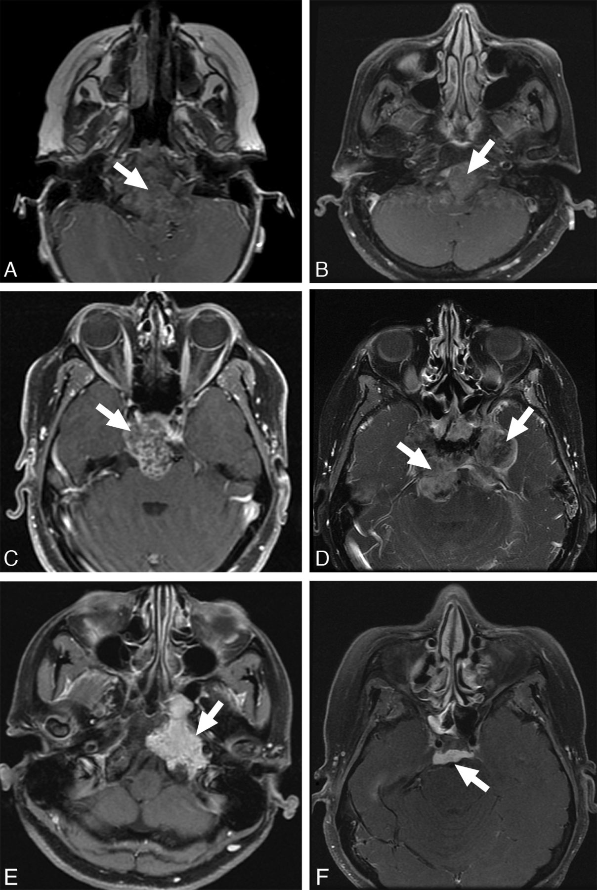
  • Fig 3.
    • Download figure
    • Open in new tab
    • Download powerpoint
    Fig 3.

    Enhancement patterns. Examples of enhancement patterns, mild to none (top), heterogeneous (middle), and solid (bottom) are shown in poorly differentiated chordomas (A and D), classic chordoma (B), and chondrosarcoma (C, E, and F).
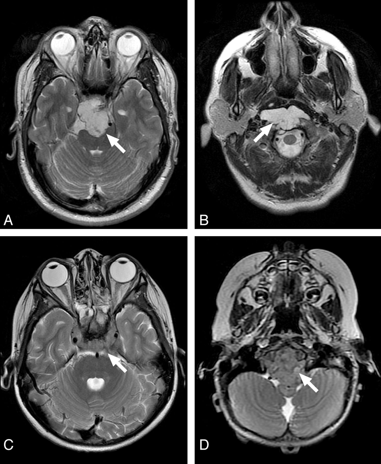
  • Fig 4.
    • Download figure
    • Open in new tab
    • Download powerpoint
    Fig 4.

    Examples of T2 intensity features of the clival tumors. High T2 intensity of chondrosarcoma (A) and classic chordoma (B) are easily recognized. Contrast this with the low T2 intensity of poorly differentiated chordoma (C and D).

Tables

  • Figures
    • View popup
    Table 1:

    Patient characteristics and clinical data (n = 19)

    Entire Cohort (n = 19)Chondrosarcoma (n = 9)Classic Chordoma (n = 7)Poorly Differentiated Chordoma (n = 3)
    Age at diagnosis (yr)
        Median3637367
        Range2–8428–842–642–8
    Sex
        Male8 (42%)3 (33%)3 (43%)2 (67%)
        Female11 (58%)6 (67%)4 (57%)1 (33%)
    Tumor location
        Clival11 (58%)4 (44%)5 (71%)3 (100%)
        Petroclival7 (37%)4 (44%)2 (29%)0 (0%)
        Pterygoid1 (5%)1 (11%)0 (0%)0 (0%)
    New or recurrent/residual
        New16 (84%)8 (89%)5 (71%)3 (100%)
        Recurrent/residual3 (16%)1 (11%)2 (29%)0 (0%)
    • View popup
    Table 2:

    ADC values for each tumor type (10−6 mm2/s)

    TumorMean ADC (Median)Minimum ADC (Median)Maximum ADC (Median)
    Chondrosarcoma (n = 9)2051 ± 262 (1977)1488 ± 360 (1352)2503 ± 512 (2392)
    Classic Chordoma (n = 7)1474 ± 117 (1460)905 ± 118 (860)2199 ± 255 (2217)
    Poorly Differentiated Chordoma (n = 3)875 ± 100 (871)491 ± 210 (469)1503 ± 127 (1557)
    • View popup
    Table 3:

    MRI features for each tumor type

    TumorT2 Hypointensity>90% EnhancementClival Location
    Chondrosarcoma (n = 9)0 (0%)5 (56%)4 (44%)
    Classic chordoma (n = 7)0 (0%)1 (14%)4 (57%)
    Poorly differentiated chordoma (n = 3)3 (100%)0 (0%)3 (100%)
PreviousNext
Back to top

In this issue

American Journal of Neuroradiology: 34 (5)
American Journal of Neuroradiology
Vol. 34, Issue 5
1 May 2013
  • Table of Contents
  • Index by author
Advertisement
Print
Download PDF
Email Article

Thank you for your interest in spreading the word on American Journal of Neuroradiology.

NOTE: We only request your email address so that the person you are recommending the page to knows that you wanted them to see it, and that it is not junk mail. We do not capture any email address.

Enter multiple addresses on separate lines or separate them with commas.
Diffusion-Weighted MRI: Distinction of Skull Base Chordoma from Chondrosarcoma
(Your Name) has sent you a message from American Journal of Neuroradiology
(Your Name) thought you would like to see the American Journal of Neuroradiology web site.
CAPTCHA
This question is for testing whether or not you are a human visitor and to prevent automated spam submissions.
Cite this article
K.W. Yeom, R.M. Lober, B.C. Mobley, G. Harsh, H. Vogel, R. Allagio, M. Pearson, M.S.B. Edwards, N.J. Fischbein
Diffusion-Weighted MRI: Distinction of Skull Base Chordoma from Chondrosarcoma
American Journal of Neuroradiology May 2013, 34 (5) 1056-1061; DOI: 10.3174/ajnr.A3333

Citation Manager Formats

  • BibTeX
  • Bookends
  • EasyBib
  • EndNote (tagged)
  • EndNote 8 (xml)
  • Medlars
  • Mendeley
  • Papers
  • RefWorks Tagged
  • Ref Manager
  • RIS
  • Zotero
0 Responses
Respond to this article
Share
Bookmark this article
Diffusion-Weighted MRI: Distinction of Skull Base Chordoma from Chondrosarcoma
K.W. Yeom, R.M. Lober, B.C. Mobley, G. Harsh, H. Vogel, R. Allagio, M. Pearson, M.S.B. Edwards, N.J. Fischbein
American Journal of Neuroradiology May 2013, 34 (5) 1056-1061; DOI: 10.3174/ajnr.A3333
del.icio.us logo Twitter logo Facebook logo Mendeley logo
  • Tweet Widget
  • Facebook Like
  • Google Plus One
Purchase

Jump to section

  • Article
    • Abstract
    • ABBREVIATION:
    • Materials and Methods
    • Results
    • Discussion
    • Conclusions
    • Acknowledgments
    • Footnotes
    • References
  • Figures & Data
  • Supplemental
  • Info & Metrics
  • Responses
  • References
  • PDF

Related Articles

  • PubMed
  • Google Scholar

Cited By...

  • Time-Saving 3D MR Imaging Protocols with Millimeter and Submillimeter Isotropic Spatial Resolution for Face and Neck Imaging as Implemented at a Single-Site Major Referral Center
  • Differentiation of Skull Base Chondrosarcomas, Chordomas, and Metastases: Utility of DWI and Dynamic Contrast-Enhanced Perfusion MR Imaging
  • Exophytic Lumbar Vertebral Body Mass in an Adult with Back Pain
  • MRI Signal Intensity and Electron Ultrastructure Classification Predict the Long-Term Outcome of Skull Base Chordomas
  • Preoperative MR Imaging to Differentiate Chordoid Meningiomas from Other Meningioma Histologic Subtypes
  • Prognostic Implications of Gadolinium Enhancement of Skull Base Chordomas
  • Role of the Apparent Diffusion Coefficient as a Predictor of Tumor Progression in Patients with Chordoma
  • Chordoid Meningioma: Differentiating a Rare World Health Organization Grade II Tumor from Other Meningioma Histologic Subtypes Using MRI
  • Differentiation of Skull Base Chordomas from Chondrosarcomas by Diffusion-Weighted MRI
  • Crossref (129)
  • Google Scholar

This article has been cited by the following articles in journals that are participating in Crossref Cited-by Linking.

  • Building a global consensus approach to chordoma: a position paper from the medical and patient community
    Silvia Stacchiotti, Josh Sommer
    The Lancet Oncology 2015 16 2
  • Surgery of the lateral skull base: a 50-year endeavour
    E. Zanoletti, A. Mazzoni, A. Martini, R. V. Abbritti, R. Albertini, E. Alexandre, V. Baro, S. Bartolini, D. Bernardeschi, R. Bivona, M. Bonali, I. Borghesi, D. Borsetto, R. Bovo, M. Breun, F. Calbucci, M. L. Carlson, A. Caruso, P. Cayé-Thomasen, D. Cazzador, P. -O. Champagne, R. Colangeli, G. Conte, D. D’Avella, G. Danesi, L. Deantonio, L. Denaro, F. Di Berardino, R. Draghi, F. H. Ebner, N. Favaretto, G. Ferri, A. Fioravanti, S. Froelich, A. Giannuzzi, L. Girasoli, B. R. Grossardt, M. Guidi, R. Hagen, S. Hanakita, D. G. Hardy, V. C. Iglesias, S. Jefferies, H. Jia, M. Kalamarides, I. N. Kanaan, M. Krengli, A. Landi, L. Lauda, D. Lepera, S. Lieber, S. L. K. Lloyd, A. Lovato, F. Maccarrone, R. Macfarlane, J. Magnan, L. Magnoni, D. Marchioni, J. P. Marinelli, G. Marioni, V. Mastronardi, C. Matthies, D. A. Moffat, S. Munari, M. Nardone, R. Pareschi, C. Pavone, E. Piccirillo, G. Piras, L. Presutti, G. Restivo, M. Reznitsky, E. Roca, A. Russo, M. Sanna, L. Sartori, M. Scheich, W. Shehata-Dieler, D. Soloperto, F. Sorrentino, O. Sterkers, A. Taibah, M. Tatagiba, G. Tealdo, D. Vlad, H. Wu, D. Zanetti
    Acta Otorhinolaryngologica Italica 2019 39 3, SUPPLEMENT 1
  • Skull Base Chordomas and Chondrosarcomas
    Natalia Kremenevski, Sven-Martin Schlaffer, Roland Coras, Thomas Mehari Kinfe, Thomas Graillon, Michael Buchfelder
    Neuroendocrinology 2020 110 9-10
  • Diffusion-weighted MRI derived apparent diffusion coefficient identifies prognostically distinct subgroups of pediatric diffuse intrinsic pontine glioma
    Robert M. Lober, Yoon-Jae Cho, Yujie Tang, Patrick D. Barnes, Michael S. Edwards, Hannes Vogel, Paul G. Fisher, Michelle Monje, Kristen W. Yeom
    Journal of Neuro-Oncology 2014 117 1
  • Radiomic analysis of multiparametric magnetic resonance imaging for differentiating skull base chordoma and chondrosarcoma
    Longfei Li, Ke Wang, Xiujian Ma, Zhenyu Liu, Shuo Wang, Jiang Du, Kaibing Tian, Xuezhi Zhou, Wei wei, Kai Sun, Yusong Lin, Zhen Wu, Jie Tian
    European Journal of Radiology 2019 118
  • State-of-the-Art Imaging in Human Chordoma of the Skull Base
    Rene G. C. Santegoeds, Yasin Temel, Jan C. Beckervordersandforth, Jacobus J. Van Overbeeke, Christianne M. Hoeberigs
    Current Radiology Reports 2018 6 5
  • Sellar and Parasellar Imaging
    Carlos Zamora, Mauricio Castillo
    Neurosurgery 2017 80 1
  • Primary Skull Base Chondrosarcomas: A Systematic Review
    Paolo Palmisciano, Ali S. Haider, Mohammadmahdi Sabahi, Chibueze D. Nwagwu, Othman Bin Alamer, Gianluca Scalia, Giuseppe E. Umana, Aaron A. Cohen-Gadol, Tarek Y. El Ahmadieh, Kenny Yu, Omar N. Pathmanaban
    Cancers 2021 13 23
  • Benign and malignant diseases of the clivus
    A. Neelakantan, A.K. Rana
    Clinical Radiology 2014 69 12
  • Is there a role for conventional MRI and MR diffusion-weighted imaging for distinction of skull base chordoma and chondrosarcoma?
    Uta Müller, Rahel A Kubik-Huch, Carmen Ares, Eugen B Hug, Roland Löw, Antonios Valavanis, Frank J Ahlhelm
    Acta Radiologica 2016 57 2

More in this TOC Section

  • Chondrosarcoma vs Synovial Chondromatosis: Imaging
  • WHO Classification Update: Nasal&Skull Base Tumors
  • Peritumoral Signal in Vestibular Schwannomas
Show more HEAD & NECK

Similar Articles

Advertisement

Indexed Content

  • Current Issue
  • Accepted Manuscripts
  • Article Preview
  • Past Issues
  • Editorials
  • Editor's Choice
  • Fellows' Journal Club
  • Letters to the Editor
  • Video Articles

Cases

  • Case Collection
  • Archive - Case of the Week
  • Archive - Case of the Month
  • Archive - Classic Case

Special Collections

  • AJNR Awards
  • ASNR Foundation Special Collection
  • Most Impactful AJNR Articles
  • Photon-Counting CT
  • Spinal CSF Leak Articles (Jan 2020-June 2024)

More from AJNR

  • Trainee Corner
  • Imaging Protocols
  • MRI Safety Corner

Multimedia

  • AJNR Podcasts
  • AJNR Scantastics

Resources

  • Turnaround Time
  • Submit a Manuscript
  • Submit a Video Article
  • Submit an eLetter to the Editor/Response
  • Manuscript Submission Guidelines
  • Statistical Tips
  • Fast Publishing of Accepted Manuscripts
  • Graphical Abstract Preparation
  • Imaging Protocol Submission
  • Evidence-Based Medicine Level Guide
  • Publishing Checklists
  • Author Policies
  • Become a Reviewer/Academy of Reviewers
  • News and Updates

About Us

  • About AJNR
  • Editorial Board
  • Editorial Board Alumni
  • Alerts
  • Permissions
  • Not an AJNR Subscriber? Join Now
  • Advertise with Us
  • Librarian Resources
  • Feedback
  • Terms and Conditions
  • AJNR Editorial Board Alumni

American Society of Neuroradiology

  • Not an ASNR Member? Join Now

© 2025 by the American Society of Neuroradiology All rights, including for text and data mining, AI training, and similar technologies, are reserved.
Print ISSN: 0195-6108 Online ISSN: 1936-959X

Powered by HighWire