Advertisement

Main menu

  • Home
  • Content
    • Current Issue
    • Accepted Manuscripts
    • Article Preview
    • Past Issue Archive
    • Video Articles
    • AJNR Case Collection
    • Case of the Week Archive
    • Case of the Month Archive
    • Classic Case Archive
  • Special Collections
    • AJNR Awards
    • ASNR Foundation Special Collection
    • Most Impactful AJNR Articles
    • Photon-Counting CT
    • Spinal CSF Leak Articles (Jan 2020-June 2024)
  • Multimedia
    • AJNR Podcasts
    • AJNR SCANtastic
    • Trainee Corner
    • MRI Safety Corner
    • Imaging Protocols
  • For Authors
    • Submit a Manuscript
    • Submit a Video Article
    • Submit an eLetter to the Editor/Response
    • Manuscript Submission Guidelines
    • Statistical Tips
    • Fast Publishing of Accepted Manuscripts
    • Graphical Abstract Preparation
    • Imaging Protocol Submission
    • Author Policies
  • About Us
    • About AJNR
    • Editorial Board
    • Editorial Board Alumni
  • More
    • Become a Reviewer/Academy of Reviewers
    • Subscribers
    • Permissions
    • Alerts
    • Feedback
    • Advertisers
    • ASNR Home

User menu

  • Alerts
  • Log in

Search

  • Advanced search
American Journal of Neuroradiology

American Journal of Neuroradiology

ASHNR American Society of Functional Neuroradiology ASHNR American Society of Pediatric Neuroradiology ASSR
  • Alerts
  • Log in

Advanced Search

  • Home
  • Content
    • Current Issue
    • Accepted Manuscripts
    • Article Preview
    • Past Issue Archive
    • Video Articles
    • AJNR Case Collection
    • Case of the Week Archive
    • Case of the Month Archive
    • Classic Case Archive
  • Special Collections
    • AJNR Awards
    • ASNR Foundation Special Collection
    • Most Impactful AJNR Articles
    • Photon-Counting CT
    • Spinal CSF Leak Articles (Jan 2020-June 2024)
  • Multimedia
    • AJNR Podcasts
    • AJNR SCANtastic
    • Trainee Corner
    • MRI Safety Corner
    • Imaging Protocols
  • For Authors
    • Submit a Manuscript
    • Submit a Video Article
    • Submit an eLetter to the Editor/Response
    • Manuscript Submission Guidelines
    • Statistical Tips
    • Fast Publishing of Accepted Manuscripts
    • Graphical Abstract Preparation
    • Imaging Protocol Submission
    • Author Policies
  • About Us
    • About AJNR
    • Editorial Board
    • Editorial Board Alumni
  • More
    • Become a Reviewer/Academy of Reviewers
    • Subscribers
    • Permissions
    • Alerts
    • Feedback
    • Advertisers
    • ASNR Home
  • Follow AJNR on Twitter
  • Visit AJNR on Facebook
  • Follow AJNR on Instagram
  • Join AJNR on LinkedIn
  • RSS Feeds

AJNR is seeking candidates for the AJNR Podcast Editor. Read the position description.

Research ArticleFunctional
Open Access

Effects of the Apolipoprotein E ε4 Allele on Functional MRI during n-Back Working Memory Tasks in Healthy Middle-Aged Adults

C.-J. Chen, C.-C. Chen, D. Wu, N.-F. Chi, P.-C. Chen, Y.-P. Liao, H.-W. Chiu and C.-J. Hu
American Journal of Neuroradiology June 2013, 34 (6) 1197-1202; DOI: https://doi.org/10.3174/ajnr.A3369
C.-J. Chen
aFrom the Departments of Radiology (C.-J.C., Y.-P.L.)
  • Find this author on Google Scholar
  • Find this author on PubMed
  • Search for this author on this site
C.-C. Chen
bNeurology (C.-C.C., D.W., N.-F.C., P.-C.C., C.-J.H.), Taipei Medical University-Shuang Ho Hospital, New Taipei City, Taiwan
cGraduate Institute of Biomedical Informatics (C.-C.C., H.-W.C.)
  • Find this author on Google Scholar
  • Find this author on PubMed
  • Search for this author on this site
D. Wu
bNeurology (C.-C.C., D.W., N.-F.C., P.-C.C., C.-J.H.), Taipei Medical University-Shuang Ho Hospital, New Taipei City, Taiwan
  • Find this author on Google Scholar
  • Find this author on PubMed
  • Search for this author on this site
N.-F. Chi
bNeurology (C.-C.C., D.W., N.-F.C., P.-C.C., C.-J.H.), Taipei Medical University-Shuang Ho Hospital, New Taipei City, Taiwan
  • Find this author on Google Scholar
  • Find this author on PubMed
  • Search for this author on this site
P.-C. Chen
bNeurology (C.-C.C., D.W., N.-F.C., P.-C.C., C.-J.H.), Taipei Medical University-Shuang Ho Hospital, New Taipei City, Taiwan
  • Find this author on Google Scholar
  • Find this author on PubMed
  • Search for this author on this site
Y.-P. Liao
aFrom the Departments of Radiology (C.-J.C., Y.-P.L.)
  • Find this author on Google Scholar
  • Find this author on PubMed
  • Search for this author on this site
H.-W. Chiu
cGraduate Institute of Biomedical Informatics (C.-C.C., H.-W.C.)
  • Find this author on Google Scholar
  • Find this author on PubMed
  • Search for this author on this site
C.-J. Hu
bNeurology (C.-C.C., D.W., N.-F.C., P.-C.C., C.-J.H.), Taipei Medical University-Shuang Ho Hospital, New Taipei City, Taiwan
dDepartment of Neurology (C-.J.H.), Taipei Medical University, Taipei, Taiwan.
  • Find this author on Google Scholar
  • Find this author on PubMed
  • Search for this author on this site
  • Article
  • Figures & Data
  • Info & Metrics
  • Responses
  • References
  • PDF
Loading

Abstract

BACKGROUND AND PURPOSE: APOE4 is the best-documented genetic risk factor for sporadic AD. Previous research showed that APOE4 is associated with increased risk of occurrence and earlier onset of AD in a gene dose–dependent manner. However, the specific role of APOE4 in processing of brain functions requires further investigation. Investigators have used fMRI to measure brain activity on the basis of the blood oxygen level–dependent contrast. This study investigates the effects of APOE4 on fMRI during n-back WM tasks in healthy middle-aged adults.

MATERIALS AND METHODS: From 110 participants, 81 individuals without objective or subjective cognitive impairment underwent APOE genotyping. Nine APOE4 carriers and 9 age- and sex-matched non-APOE4 controls were recruited for fMRI examinations during WM tasks.

RESULTS: Both groups displayed increased brain activation in response to increases in WM loads. During low-WM-load tasks, the APOE4 carriers recruited significantly greater additional processing resources than the non-APOE4 carriers. During moderate- and high-WM-load tasks, the APOE4 carrier group displayed fewer increases in activation than the non-APOE4 carrier group.

CONCLUSIONS: APOE genetic polymorphisms may affect brain functioning in subjects without dementia. The patterns of brain activation during different levels of WM load suggest possible subclinical impairment of WM capacity in APOE4 carriers (ClinicalTrials.gov registration: NCT01287819).

ABBREVIATIONS:

AD
Alzheimer disease
APOE4
Apolipoprotein E ε4 allele
BOLD
blood oxygen level–dependent
MMSE
Mini-Mental State Examination
WM
working memory

AD is the most common cause of dementia in the world. Although the specific pathophysiology of AD remains unclear, compelling evidence has shown that genetic factors play an important role in its occurrence.1 There are 3 genes linked to familial AD, including amyloid precursor protein, presenilin-1, and presenilin-2.2 APOE4 is the best-documented genetic risk factor for sporadic AD.3 The genetic polymorphisms for the APOE gene include 3 alleles: ε2, 3, and 4. Previous research identified APOE4 as associated with increased risk of occurrence and earlier onset of AD in a gene dose–dependent manner.4,5 APOE4 carriers present with more severe memory impairment and greater reductions in regional brain volume and metabolism, in comparison with non-APOE4 carriers.6⇓⇓–9 These findings indicate that APOE4 is important for brain functioning, though the mechanisms by which it exerts its effects need further clarification. Genetic background can thus confer changes in brain structure, metabolism, and functioning, or even behaviors.

Investigators extensively use fMRI to evaluate brain function. Most studies use an indirect-measurement method based on the blood oxygen level–dependent contrast.10,11 Under normal physiologic conditions, the amplitude of the BOLD signal is linearly connected to neural activity and can provide task-specific information concerning neural functions and networks.12⇓–14

WM has the ability to actively keep information for further use by prioritizing, modifying, and protecting it from interference.15 Therefore, the operations of WM consist of memory itself and retention of active information in a stable yet flexible manner.16 Most patients with AD present with progressive impairment of episodic memory, and such dysfunction constitutes the core diagnostic criterion of probable AD.17 However, several findings in the literature also unveil deficits in WM and executive function in patients with AD and mild cognitive impairment.18⇓–20 Previous studies of fMRI documented that WM emerges from interactions among higher sensory, attentional, and mnemonic functions, with separable neural bases.21⇓⇓⇓–25 WM has a fundamental cognitive function, which can be evaluated by fMRI, and thus provides a good domain for testing the differences among individuals with various genetic backgrounds. fMRI studies of APOE4 carriers have shown decreased neural activity in the medial temporal lobe and increased (probable compensatory) neural activity in prefrontal and parietal regions under tasks of encoding and retrieval of episodic memory.26,27 Neural activity alteration in WM in subjects at risk of AD is not conclusive.28 An initial study by Burggren et al29 by using a digit-span task showed no difference in activation patterns between elderly APOE4 carriers and non-APOE4 carriers. They concluded that additional cognitive effort in persons at genetic risk for AD is specific to episodic encoding. Later studies by using an n-back WM paradigm showed no difference in activation patterns between APOE4 carriers and non-APOE4 carriers in young adults but increased activation in the frontal and parietal lobe in elderly APOE4 carriers.30,31 To the best of our knowledge, there is no publication to date about neural activity alteration in WM in middle-aged subjects at risk of AD, especially with different WM loads.

Materials and Methods

Participants

One hundred ten subjects who underwent physical examinations at Taipei Medical University–Shuang Ho Hospital participated in this study. All participants provided written informed consent, and this study was approved by the hospital institutional review board. The participants self-completed the Alzheimer's Disease 8 (AD8) questionnaire for subjective cognitive impairment and were assessed with the MMSE for objective cognitive impairment by a neuropsychologist.32,33 Participants with AD8 >2 or MMSE <26 were excluded. Eighty-one participants without subjective or objective cognitive impairment were selected for further genetic analysis, from which 12 APOE4 carriers, all ε3/4 heterozygous, and 69 non-APOE4 carriers were found. Nine APOE4 carriers underwent fMRI analysis. Nine age-and sex-matched controls were randomly selected from the non-APOE4 carriers.

Working Memory Task

WM capacity can be tested by using a variety of tasks. A commonly used measure in fMRI is the n-back task. The participant is required to monitor a series of stimuli and to respond whenever a stimulus is presented that is the same as the one presented n trials previously. Parametric designs, comparing n = 1, n = 2, and n = 3 trials are often used. Some studies have also used a 0-back control condition. In this study, n-back WM tasks were presented by using E-Prime, Version 2.0 (http://www.pstnet.com/eprime.cfm). At the beginning, a task guide describing the rules of the study was shown. The n-back tasks comprised 4 conditions, 0-, 1-, 2-, and 3-back. The 0-back control condition had a minimal WM load, and individuals were asked to decide whether the current letter matched a single target letter that was specified before the condition began. During the 1-back condition, they were asked to decide whether the current letter matched the previous one. During the 2-back and 3-back conditions, the participants were asked to decide whether the number currently presented matched the number that had been presented 2 and 3 trials back in the sequence. Each condition was conducted in a single run, which consisted of 3 epochs. Each epoch contained 30 seconds of presentation with numbers and 30 seconds of fixation on a crosshair. Twelve numbers were presented in the 30-second period, and each consisted of a 0.5-second appearance of a black number on the screen against a white background and 2 seconds of a fixation cross. All stimuli were projected onto an overhead screen, which participants could view through a mirror located on the scanner's head coil. Participants were instructed to press a response pad with the right index finger when the currently presented number was a target or to press the middle finger when the currently presented number was not a target. The number of correct and incorrect responses was recorded.

Conditions were counterbalanced among participants: Each condition was equally often preceded and followed by each of the other conditions. Before the scans, each participant was instructed on the entire experimental procedure and practiced the tasks outside the scanner to reduce anxiety. To further reduce any group effect independent of WM processes, all participants were trained before scanning to ensure that they could perform the 1-back condition to an accuracy criterion of 70%.

Imaging Methods

Imaging was performed on a 3T MR imaging system (Discovery MR750; GE Healthcare, Milwaukee, Wisconsin). An 8-channel head coil was used for signal reception. Care was taken to minimize the effects of movement by instructing participants to remain still and by placing foam padding around the head. Functional data were collected by using an EPI sequence (TR = 3000 ms, TE = 35 ms, flip angle = 90°, FOV = 230 mm2, matrix = 64 × 64, 40 sections, section thickness = 3 mm, and intersection gap = 1 mm) for a 3.5-minute period resulting in 70 volumes. Two dummy scans, which enabled the MR signal to achieve a steady state, were applied in each scan. A T1-weighted anatomic dataset was obtained from each subject by using brain-volume imaging (TR = 8.2 ms, TE = 3.2 ms, TI = 450 ms, flip angle = 12°, FOV = 240 mm2, matrix = 256 × 256, 160 sections, voxel size = 0.9375 × 0.9375 × 1 mm3). T2-weighted images were acquired by using fast recovery fast spin-echo (TR = 5700 ms, TE = 100 ms, FOV = 230 mm2, matrix = 416 × 416, 20 sections, section thickness = 5 mm, intersection gap = 2 mm, and NEX = 1).

Data Analysis

fMRI data were preprocessed by using SPM5 (Wellcome Department of Imaging Neuroscience, London, UK) implemented in Matlab, Version 7.9 (MathWorks, Natick, Massachusetts). To reduce motion artifacts, we realigned each participant's functional images with the first volume of the series by using the rigid-body transformation procedure and resectioned them by using fourth-degree B-spline function interpolation while adjusting for residual motion-related signal changes. All motion-corrected images were spatially normalized to the EPI template of the Montreal Neurological Institute. The normalization process involved minimizing the residual sum of squared differences among the images to be normalized. Before modeling the fMRI signal changes, normalized EPI images were spatially smoothed by convolution with a 3D Gaussian filter of 6 × 6 × 6 mm3 full width at half maximum to increase the signal-to-noise ratio, validate the statistical analysis, and reduce anatomic variations among participants.

Statistical Analysis

Demographic and neuropsychological test data for both groups were compared by using 2-tailed t tests or χ2 tests. For each n-back condition, the datasets were analyzed by modeling the experimental conditions by using boxcar functions convolved with a hemodynamic response function in the context of the general linear model used by SPM5. To obtain the brain-activation patterns for each group, we entered smoothed normalized scans for all participants into this general linear model and created contrast images for between 0- and 1-back (1 > 0), 1- and 2-back (2 > 1), and 2- and 3-back (3 > 2) for each group. A 1-sample t test was applied for within-group analyses. The probability threshold value was set at .01 uncorrected, with a minimum cluster extent of 3 contiguous voxels. The generated contrast images for 1- > 0-back, 2- > 1-back, and 3- > 2-back were then used for the second-level multiparticipant/between-group random effects analyses, to obtain the activation differences between APOE4 carriers and noncarriers. The 2-sample t test was applied for between-group analyses. The probability threshold value was set at .01 uncorrected, with a minimum cluster extent of 3 contiguous voxels.

Results

The Table describes the basic demographic characteristics of both groups. The groups did not differ significantly regarding sex, age, and years of education. Group accuracies for the n-back task did not differ among APOE4 carriers and non-APOE4 carriers. There was, however, a significant trend of increasing WM load associated with declines in performance accuracy in both groups (P < .001).

View this table:
  • View inline
  • View popup

Demographic information, cognitive function, and accuracy rates of n-back WM in participants

Figure 1 shows activation maps, during the n-back task, of APOE4 carriers and controls in a surface-rendered projection displayed on a standardized brain atlas (display threshold, P < .01; extent, 3 voxels). Both groups displayed increased activation in bilateral frontal and parietal regions, consistent with activation of WM circuitry.

Fig 1.
  • Download figure
  • Open in new tab
  • Download powerpoint
Fig 1.

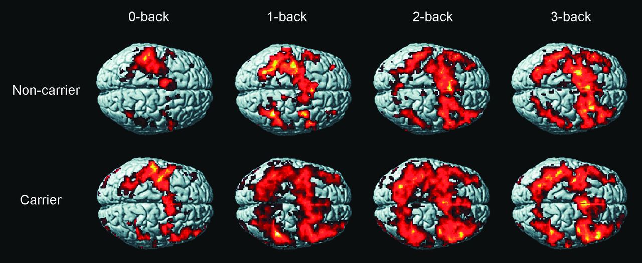
Activation maps of n-back task of APOE4 carriers and non-APOE4 carriers in a surface-rendered projection displayed on a standardized brain atlas (display threshold, P < .01; extent, 3 voxels). Increased activation in bilateral frontal and parietal regions is noted, consistent with activation of WM circuitry in both groups.

Figure 2 displays activation maps, during 1-back > 0-back, 2-back > 1-back, and 3-back > 2-back conditions, of APOE4 carriers and non-APOE4 carriers. Both groups displayed increased brain activation in response to each increase in WM load. The degree of increase in brain activation differed among the groups and was greatest in the 1-back > 0-back conditions in the APOE4 carriers and in the 2-back > 1-back conditions in the non-APOE4 carriers. Visual comparison of the between-group differences in the 1-back > 0-back conditions shows lesser activation in the non-APOE4 carrier group. In 2-back > 1-back and 3-back > 2-back conditions, the APOE4 carrier group demonstrated fewer increases in activation than the non-APOE4 carriers.

Fig 2.
  • Download figure
  • Open in new tab
  • Download powerpoint
Fig 2.

Activation maps of 1-back > 0-back, 2-back > 1-back, and 3-back > 2-back conditions of APOE4 carriers and non-APOE4 carriers in a surface-rendered projection displayed on a standardized brain atlas (display threshold, P < .01; extent, 3 voxels). In both groups, an increase in brain activation occurred in response to each increase in WM load. The degree of increased brain activation was different in both groups. It was greatest in 2-back > 1-back conditions in non-APOE4 carriers, and greatest in 1-back > 0-back conditions in APOE4 carriers. A visual comparison of the between-group difference in 1-back > 0-back conditions shows less extent of activation in the noncarrier group. In 2-back 1-back and 3-back > 2-back conditions, less increase in activation is seen in the carrier group.

Discussion

APOE4 carriers displayed more rapid memory decline and mesial temporal lobe atrophy than non-APOE4 carriers.34⇓⇓⇓⇓⇓⇓–41 The present study recruited individuals without subjective or objective cognitive impairment, hence subjects with normal cognition, to test the effects of APOE4 on the brain functioning of WM by using BOLD fMRI. Performance of n-back WM was not different between groups (P = .41–.85), similar to the findings of a previous publication.42 fMRI analysis showed that during the low-WM-load task, functional activation significantly increased to a greater extent in the APOE4 carriers than in the non-APOE4 carriers. As the WM load increased, the non-APOE4 carriers maintained their ability to further increase activation. In contrast, the APOE4 carriers showed fewer increases in activation during the moderate- and high-WM-load conditions. One possible explanation for this phenomenon is mild impairment of WM capacity in the APOE4 carriers. In the low-WM load (1- back > 0-back conditions), the APOE4 carriers recruited additional processing resources to compensate for processing inefficiencies. In contrast, the non-APOE4 carriers were not impaired or challenged by this low-WM load and, therefore, required relatively fewer increases in processing resources to perform the task. As the WM load increased, the non-APOE4 carriers continued to show increasing activation, presumably because they still had processing reserves available to be drawn on with increasing WM demand. The APOE4 carriers, however, had already recruited most of their (assuming a finite amount) available resources, and thus little additional activation occurred.

In this study, the accuracy rate of 0-back, 2-back, and 3-back WM tasks appeared insignificantly better in the APOE4 carrier group. Among the APOE4 carriers, the accuracy rates did not dramatically decline along with the increase of memory loads when the brain activation recruitment had reached the plateau. It is speculated that there is a ceiling effect of working memory in this study. Continuing increases in memory load might enable the detection of the decline of performance in APOE4 carriers. Dickerson et al43 demonstrated increased hippocampal activation in mild cognitive impairment compared with normal aging and AD under fMRI scanning during face-name associative encoding tasks. They hypothesized that there is a phase of increased medial temporal lobe activation early in the course of prodromal Alzheimer disease. Our findings of increased functional activation under low WM load in APOE4 carriers may agree with their hypothesis and may be applied to the cognitively normal high-risk subjects. Explanations of such functional alteration include a compensatory mechanism for regional neuronal loss and disruption of the intrinsic connectivity of memory networks.44,45 Filbey et al46 also found compensatory neural activation in the medial frontal and parahippocampal gyrus in young and elderly APOE4 carriers during a 9-square-grid visual WM task, similar to our findings in low-WM load.

The relationship between the WM fMRI findings and underlying pathology is not clear at present. It could reflect a hereditary pattern in APOE4 carriers or could be a consequence of functional decline of the subclinical brain. Using resting-state fMRI, Sheline et al47 showed that APOE4 alters the neural network in individuals with normal cognition without amyloid burden, as detected by using Pittsburgh compound B–amyloid PET. However, the long-term effects of these types of brain functional changes remain unknown.

There are a few limitations to this study. First, amyloid pathology was not completely excluded. Numerous studies have proved that amyloid burden can occur many years before objective or subjective cognitive declines, especially among APOE4 carriers.8,48,49 In the present study, amyloid pathology, which results in brain subclinical dysfunction, might potentially have contributed to the increase in BOLD. Second, the sample size of this study was small. APOE4 carrier rates are low in Asian populations relative to whites. This study recruited only 9 APOE4 carriers and 9 non-APOE4 carriers and was able to achieve statistical significance. Third, the insignificantly better performance of some n-back WM tasks in the APOE4 carrier group was unexpected and was not perfectly compatible with the fMRI presentation. Future large-scale research investigations, by using amyloid imaging and long-term follow-ups, are, therefore, warranted.

Conclusions

In summary, fMRI BOLD significantly increased in the APOE4 carriers during low-WM-load tasks but did not increase during moderate- and high-WM loads, which occurred in non-APOE4 carriers. Based on previous research and our findings, these differences could have a hereditary basis. The phenomenon could reflect an impaired WM processing resource associated with subclinical brain dysfunction. This study, therefore, also provided further evidence to confirm the effects of genetic background on brain functioning. Further studies are required to fully elucidate the underlying mechanisms.

Footnotes

  • C.-J. Chen and C.-C. Chen contributed equally to this study.

  • Disclosures: Chih-Chung Chen—RELATED: Grant: Center of Excellence for Clinical Trial and Research in Neurology and Neurosurgery.* Dean Wu—RELATED: Grant: Center of Excellence for Clinical Trial and Research in Neurology and Neurosurgery.* Po-Chih Chen—RELATED: Grant: Center of Excellence for Clinical Trial and Research in Neurology and Neurosurgery.* Hung-Wen Chiu—RELATED: Grant: Center of Excellence for Clinical Trial and Research in Neurology and Neurosurgery.* Chaur-Jong Hu—RELATED: Grant: Taiwan government,* Comments: for sponsoring this study. *Money paid to the institution.

  • This work was supported by the Center of Excellence for Clinical Trial and Research in Neurology and Neurosurgery.

Indicates open access to non-subscribers at www.ajnr.org

REFERENCES

  1. 1.↵
    1. van Duijn CM,
    2. Clayton D,
    3. Chandra V,
    4. et al
    . Familial aggregation of Alzheimer's disease and related disorders: a collaborative re-analysis of case-control studies. Int J Epidemiol 1991;20(suppl 2):S13–20
    PubMed
  2. 2.↵
    1. Waring SC,
    2. Rosenberg RN
    . Genome-wide association studies in Alzheimer disease. Arch Neurol 2008;65:329–34
    CrossRefPubMed
  3. 3.↵
    1. Strittmatter WJ,
    2. Saunders AM,
    3. Schmechel D,
    4. et al
    . Apolipoprotein E: high-avidity binding to beta-amyloid and increased frequency of type 4 allele in late-onset familial Alzheimer disease. Proc Natl Acad Sci U S A 1993;90:1977–81
    Abstract/FREE Full Text
  4. 4.↵
    1. Small GW
    . Neuroimaging and genetic assessment for early diagnosis of Alzheimer's disease. J Clin Psychiatry 1996;57(suppl 14):9–13
  5. 5.↵
    1. Chen K,
    2. Reiman EM,
    3. Alexander GE,
    4. et al
    . Correlations between apolipoprotein E epsilon4 gene dose and whole brain atrophy rates. Am J Psychiatry 2007;164:916–21
    CrossRefPubMed
  6. 6.↵
    1. Caselli RJ,
    2. Graff-Radford NR,
    3. Reiman EM,
    4. et al
    . Preclinical memory decline in cognitively normal apolipoprotein E-epsilon4 homozygotes. Neurology 1999;53:201–07
    CrossRef
  7. 7.↵
    1. Pievani M,
    2. Galluzzi S,
    3. Thompson PM,
    4. et al
    . APOE4 is associated with greater atrophy of the hippocampal formation in Alzheimer's disease. Neuroimage 2011;55:909–19
    CrossRefPubMed
  8. 8.↵
    1. Donix M,
    2. Burggren AC,
    3. Suthana NA,
    4. et al
    . Longitudinal changes in medial temporal cortical thickness in normal subjects with the APOE-4 polymorphism. Neuroimage 2010;53:37–43
    CrossRefPubMed
  9. 9.↵
    1. Reiman EM,
    2. Chen K,
    3. Alexander GE,
    4. et al
    . Correlations between apolipoprotein E epsilon4 gene dose and brain-imaging measurements of regional hypometabolism. Proc Natl Acad Sci U S A 2005;102:8299–302
    Abstract/FREE Full Text
  10. 10.↵
    1. Ogawa S,
    2. Lee TM,
    3. Nayak AS,
    4. et al
    . Oxygenation-sensitive contrast in magnetic resonance image of rodent brain at high magnetic fields. Magn Reson Med 1990;14:68–78
    CrossRefPubMed
  11. 11.↵
    1. Ogawa S,
    2. Lee TM,
    3. Kay AR,
    4. et al
    . Brain magnetic resonance imaging with contrast dependent on blood oxygenation. Proc Natl Acad Sci U S A 1990;87:9868–72
    Abstract/FREE Full Text
  12. 12.↵
    1. Logothetis NK,
    2. Pauls J,
    3. Augath M,
    4. et al
    . Neurophysiological investigation of the basis of the fMRI signal. Nature 2001;412:150–57
    CrossRefPubMed
  13. 13.↵
    1. Ogawa S,
    2. Lee TM,
    3. Stepnoski R,
    4. et al
    . An approach to probe some neural systems interaction by functional MRI at neural time scale down to milliseconds. Proc Natl Acad Sci U S A 2000;97:11026–31
    Abstract/FREE Full Text
  14. 14.↵
    1. Rees G,
    2. Friston K,
    3. Koch C
    . A direct quantitative relationship between the functional properties of human and macaque V5. Nat Neurosci 2000;3:716–23
    CrossRefPubMed
  15. 15.↵
    1. Baddeley A
    . Working memory. Science 1992;255:556–59
    Abstract/FREE Full Text
  16. 16.↵
    1. Bledowski C,
    2. Kaiser J,
    3. Rahm B
    . Basic operations in working memory: contributions from functional imaging studies. Behav Brain Res 2010;214:172–79
    CrossRefPubMed
  17. 17.↵
    1. Dubois B,
    2. Feldman HH,
    3. Jacova C,
    4. et al
    . Research criteria for the diagnosis of Alzheimer's disease: revising the NINCDS-ADRDA criteria. Lancet Neurol 2007;6:734–46
    CrossRefPubMed
  18. 18.↵
    1. Rosen VM,
    2. Bergeson JL,
    3. Putnam K,
    4. et al
    . Working memory and apolipoprotein E: what's the connection? Neuropsychologia 2002;40:2226–33
    CrossRefPubMed
  19. 19.↵
    1. Belleville S,
    2. Chertkow H,
    3. Gauthier S
    . Working memory and control of attention in persons with Alzheimer's disease and mild cognitive impairment. Neuropsychology 2007;21:458–69
    CrossRefPubMed
  20. 20.↵
    1. Belleville S,
    2. Sylvain-Roy S,
    3. de Boysson C,
    4. et al
    . Characterizing the memory changes in persons with mild cognitive impairment. Prog Brain Res 2008;169:365–75
    CrossRefPubMed
  21. 21.↵
    1. D'Esposito M,
    2. Postle BR,
    3. Jonides J,
    4. et al
    . The neural substrate and temporal dynamics of interference effects in working memory as revealed by event-related functional MRI. Proc Natl Acad Sci U S A 1999;96:7514–19
    Abstract/FREE Full Text
  22. 22.↵
    1. Rypma B,
    2. D'Esposito M
    . Isolating the neural mechanisms of age-related changes in human working memory. Nat Neurosci 2000;3:509–15
    CrossRefPubMed
  23. 23.↵
    1. Ranganath C,
    2. Johnson MK,
    3. D'Esposito M
    . Prefrontal activity associated with working memory and episodic long-term memory. Neuropsychologia 2003;41:378–89
    CrossRefPubMed
  24. 24.↵
    1. Narayanan NS,
    2. Prabhakaran V,
    3. Bunge SA,
    4. et al
    . The role of the prefrontal cortex in the maintenance of verbal working memory: an event-related FMRI analysis. Neuropsychology 2005;19:223–32
    CrossRefPubMed
  25. 25.↵
    1. Crone EA,
    2. Wendelken C,
    3. Donohue S,
    4. et al
    . Neurocognitive development of the ability to manipulate information in working memory. Proc Natl Acad Sci U S A 2006;103:9315–20
    Abstract/FREE Full Text
  26. 26.↵
    1. Bondi MW,
    2. Houston WS,
    3. Eyler LT,
    4. et al
    . fMRI evidence of compensatory mechanisms in older adults at genetic risk for Alzheimer disease. Neurology 2005;64:501–08
    CrossRef
  27. 27.↵
    1. Kukolja J,
    2. Thiel CM,
    3. Eggermann T,
    4. et al
    . Medial temporal lobe dysfunction during encoding and retrieval of episodic memory in non-demented APOE epsilon4 carriers. Neuroscience 2010;168:487–97
    CrossRefPubMed
  28. 28.↵
    1. Trachtenberg AJ,
    2. Filippini N,
    3. Mackay CE
    . The effects of APOE-ε4 on the BOLD response. Neurobiol Aging 2012;33:323–34
    CrossRefPubMed
  29. 29.↵
    1. Burggren AC,
    2. Small GW,
    3. Sabb FW,
    4. et al
    . Specificity of brain activation patterns in people at genetic risk for Alzheimer disease. Am J Geriatr Psychiatry 2002;10:44–51
    CrossRefPubMed
  30. 30.↵
    1. Mondadori CR,
    2. de Quervain DJ,
    3. Buchmann A,
    4. et al
    . Better memory and neural efficiency in young apolipoprotein E epsilon4 carriers. Cereb Cortex 2007;17:1934–47
    Abstract/FREE Full Text
  31. 31.↵
    1. Wishart HA,
    2. Saykin AJ,
    3. Rabin LA,
    4. et al
    . Increased brain activation during working memory in cognitively intact adults with the APOE epsilon4 allele. Am J Psychiatry 2006;163:1603–10
    CrossRefPubMed
  32. 32.↵
    1. Galvin JE,
    2. Roe CM,
    3. Powlishta KK,
    4. et al
    . The AD8: a brief informant interview to detect dementia. Neurology 2005;65:559–64
    CrossRef
  33. 33.↵
    1. Folstein MF,
    2. Folstein SE,
    3. McHugh PR
    . “Mini-mental state”: a practical method for grading the cognitive state of patients for the clinician. J Psychiatr Res 1975;12:189–98
    CrossRefPubMed
  34. 34.↵
    1. Dik MG,
    2. Jonker C,
    3. Comijs HC,
    4. et al
    . Memory complaints and APOE-epsilon4 accelerate cognitive decline in cognitively normal elderly. Neurology 2001;57:2217–22
    CrossRef
  35. 35.↵
    1. Caselli RJ,
    2. Reiman EM,
    3. Osborne D,
    4. et al
    . Longitudinal changes in cognition and behavior in asymptomatic carriers of the APOE e4 allele. Neurology 2004;62:1990–95
    CrossRef
  36. 36.↵
    1. Caselli RJ,
    2. Reiman EM,
    3. Locke DEC,
    4. et al
    . Cognitive domain decline in healthy apolipoprotein E epsilon4 homozygotes before the diagnosis of mild cognitive impairment. Arch Neurol 2007;64:1306–11
    CrossRefPubMed
  37. 37.↵
    1. Caselli RJ,
    2. Dueck AC,
    3. Osborne D,
    4. et al
    . Longitudinal modeling of age-related memory decline and the APOE epsilon4 effect. N Engl J Med 2009;361:255–63
    CrossRefPubMed
  38. 38.↵
    1. Thomann PA,
    2. Roth AS,
    3. Dos Santos V,
    4. et al
    . Apolipoprotein E polymorphism and brain morphology in mild cognitive impairment. Dement Geriatr Cogn Disord 2008;26:300–05
    CrossRefPubMed
  39. 39.↵
    1. Honea RA,
    2. Vidoni E,
    3. Harsha A,
    4. et al
    . Impact of APOE on the healthy aging brain: a voxel-based MRI and DTI study. J Alzheimers Dis 2009;18:553–64
    PubMed
  40. 40.↵
    1. Spampinato MV,
    2. Rumboldt Z,
    3. Hosker RJ,
    4. et al
    . Apolipoprotein E and gray matter volume loss in patients with mild cognitive impairment and Alzheimer disease. Radiology 2011;258:843–52
    CrossRefPubMed
  41. 41.↵
    1. Lu PH,
    2. Thompson PM,
    3. Leow A,
    4. et al
    . Apolipoprotein E genotype is associated with temporal and hippocampal atrophy rates in healthy elderly adults: a tensor-based morphometry study. J Alzheimers Dis 2011;23:433–42
    PubMed
  42. 42.↵
    1. Greenwood PM,
    2. Lambert C,
    3. Sunderland T,
    4. et al
    . Effects of apolipoprotein E genotype on spatial attention, working memory, and their interaction in healthy, middle-aged adults: results from the National Institute of Mental Health's BIOCARD study. Neuropsychology 2005;19:199–211
    CrossRefPubMed
  43. 43.↵
    1. Dickerson BC,
    2. Salat DH,
    3. Greve DN,
    4. et al
    . Increased hippocampal activation in mild cognitive impairment compared to normal aging and AD. Neurology 2005;65:404–11
    CrossRefPubMed
  44. 44.↵
    1. Hämäläinen A,
    2. Pihlajamäki M,
    3. Tanila H,
    4. et al
    . Increased fMRI responses during encoding in mild cognitive impairment. Neurobiol Aging 2007;28:1889–903
    CrossRefPubMed
  45. 45.↵
    1. Sperling RA,
    2. Dickerson BC,
    3. Pihlajamaki M,
    4. et al
    . Functional alterations in memory networks in early Alzheimer's disease. Neuromolecular Med 2010;12:27–43
    CrossRefPubMed
  46. 46.↵
    1. Filbey FM,
    2. Chen G,
    3. Sunderland T,
    4. et al
    . Failing compensatory mechanisms during working memory in older apolipoprotein E-epsilon4 healthy adults. Brain Imaging Behav 2010;4:177–88
    CrossRefPubMed
  47. 47.↵
    1. Sheline YI,
    2. Morris JC,
    3. Snyder AZ,
    4. et al
    . APOE4 allele disrupts resting state fMRI connectivity in the absence of amyloid plaques or decreased CSF Aβ42. J Neurosci 2010;30:17035–40
    Abstract/FREE Full Text
  48. 48.↵
    1. Pike KE,
    2. Savage G,
    3. Villemagne VL,
    4. et al
    . Beta-amyloid imaging and memory in non-demented individuals: evidence for preclinical Alzheimer's disease. Brain 2007;130(pt 11):2837–44
    Abstract/FREE Full Text
  49. 49.↵
    1. Reiman EM,
    2. Chen K,
    3. Liu X,
    4. et al
    . Fibrillar amyloid-beta burden in cognitively normal people at 3 levels of genetic risk for Alzheimer's disease. Proc Natl Acad Sci U S A 2009;106:6820–25
    Abstract/FREE Full Text
  • Received July 6, 2012.
  • Accepted after revision August 26, 2012.
  • © 2013 by American Journal of Neuroradiology
View Abstract
PreviousNext
Back to top

In this issue

American Journal of Neuroradiology: 34 (6)
American Journal of Neuroradiology
Vol. 34, Issue 6
1 Jun 2013
  • Table of Contents
  • Index by author
Advertisement
Print
Download PDF
Email Article

Thank you for your interest in spreading the word on American Journal of Neuroradiology.

NOTE: We only request your email address so that the person you are recommending the page to knows that you wanted them to see it, and that it is not junk mail. We do not capture any email address.

Enter multiple addresses on separate lines or separate them with commas.
Effects of the Apolipoprotein E ε4 Allele on Functional MRI during n-Back Working Memory Tasks in Healthy Middle-Aged Adults
(Your Name) has sent you a message from American Journal of Neuroradiology
(Your Name) thought you would like to see the American Journal of Neuroradiology web site.
CAPTCHA
This question is for testing whether or not you are a human visitor and to prevent automated spam submissions.
Cite this article
C.-J. Chen, C.-C. Chen, D. Wu, N.-F. Chi, P.-C. Chen, Y.-P. Liao, H.-W. Chiu, C.-J. Hu
Effects of the Apolipoprotein E ε4 Allele on Functional MRI during n-Back Working Memory Tasks in Healthy Middle-Aged Adults
American Journal of Neuroradiology Jun 2013, 34 (6) 1197-1202; DOI: 10.3174/ajnr.A3369

Citation Manager Formats

  • BibTeX
  • Bookends
  • EasyBib
  • EndNote (tagged)
  • EndNote 8 (xml)
  • Medlars
  • Mendeley
  • Papers
  • RefWorks Tagged
  • Ref Manager
  • RIS
  • Zotero
0 Responses
Respond to this article
Share
Bookmark this article
Effects of the Apolipoprotein E ε4 Allele on Functional MRI during n-Back Working Memory Tasks in Healthy Middle-Aged Adults
C.-J. Chen, C.-C. Chen, D. Wu, N.-F. Chi, P.-C. Chen, Y.-P. Liao, H.-W. Chiu, C.-J. Hu
American Journal of Neuroradiology Jun 2013, 34 (6) 1197-1202; DOI: 10.3174/ajnr.A3369
del.icio.us logo Twitter logo Facebook logo Mendeley logo
  • Tweet Widget
  • Facebook Like
  • Google Plus One
Purchase

Jump to section

  • Article
    • Abstract
    • ABBREVIATIONS:
    • Materials and Methods
    • Results
    • Discussion
    • Conclusions
    • Footnotes
    • REFERENCES
  • Figures & Data
  • Info & Metrics
  • Responses
  • References
  • PDF

Related Articles

  • No related articles found.
  • PubMed
  • Google Scholar

Cited By...

  • Differential Aging Trajectories of Modulation of Activation to Cognitive Challenge in APOE {varepsilon}4 Groups: Reduced Modulation Predicts Poorer Cognitive Performance
  • {beta}-Amyloid Deposition Is Associated with Decreased Right Prefrontal Activation during Task Switching among Cognitively Normal Elderly
  • Altered Brain Activation Patterns Under Different Working Memory Loads in Patients With Type 2 Diabetes
  • Crossref (26)
  • Google Scholar

This article has been cited by the following articles in journals that are participating in Crossref Cited-by Linking.

  • Neuroimaging Modalities in Alzheimer’s Disease: Diagnosis and Clinical Features
    JunHyun Kim, Minhong Jeong, Wesley R. Stiles, Hak Soo Choi
    International Journal of Molecular Sciences 2022 23 11
  • Altered Brain Activation Patterns Under Different Working Memory Loads in Patients With Type 2 Diabetes
    Yaojing Chen, Zhen Liu, Junying Zhang, Kai Xu, Sisi Zhang, Dongfeng Wei, Zhanjun Zhang
    Diabetes Care 2014 37 12
  • The Contribution of Genetic Factors to Cognitive Impairment and Dementia: Apolipoprotein E Gene, Gene Interactions, and Polygenic Risk
    Jialing Fan, Wuhai Tao, Xin Li, He Li, Junying Zhang, Dongfeng Wei, Yaojing Chen, Zhanjun Zhang
    International Journal of Molecular Sciences 2019 20 5
  • Functional neuroimaging findings in healthy middle-aged adults at risk of Alzheimer’s disease
    Mirette Habib, Elijah Mak, Silvy Gabel, Li Su, Guy Williams, Adam Waldman, Katie Wells, Karen Ritchie, Craig Ritchie, John T. O’Brien
    Ageing Research Reviews 2017 36
  • Bridging Integrator 1 (BIN1) Genotype Effects on Working Memory, Hippocampal Volume, and Functional Connectivity in Young Healthy Individuals
    Xiaolong Zhang, Jin-Tai Yu, Jin Li, Chao Wang, Lan Tan, Bing Liu, Tianzi Jiang
    Neuropsychopharmacology 2015 40 7
  • Aβ-related hyperactivation in frontoparietal control regions in cognitively normal elderly
    Hwamee Oh, Jason Steffener, Qolamreza R. Razlighi, Christian Habeck, Dan Liu, Yunglin Gazes, Sarah Janicki, Yaakov Stern
    Neurobiology of Aging 2015 36 12
  • Brain Aging and APOE ε4 Interact to Reveal Potential Neuronal Compensation in Healthy Older Adults
    Elisa Scheller, Lena V. Schumacher, Jessica Peter, Jacob Lahr, Julius Wehrle, Christoph P. Kaller, Christian Gaser, Stefan Klöppel
    Frontiers in Aging Neuroscience 2018 10
  • The Elusive Nature of APOE ε4 in Mid-adulthood: Understanding the Cognitive Profile
    Claire Lancaster, Naji Tabet, Jennifer Rusted
    Journal of the International Neuropsychological Society 2017 23 3
  • β-Amyloid Deposition Is Associated with Decreased Right Prefrontal Activation during Task Switching among Cognitively Normal Elderly
    Hwamee Oh, Jason Steffener, Qolamreza R. Razlighi, Christian Habeck, Yaakov Stern
    The Journal of Neuroscience 2016 36 6
  • Adverse Effects of the Apolipoprotein E ε4 Allele on Episodic Memory, Task Switching and Gray Matter Volume in Healthy Young Adults
    Jianfei Nao, Hongzan Sun, Qiushi Wang, Shuang Ma, Shuo Zhang, Xiaoyu Dong, Ying Ma, Xiaoming Wang, Dongming Zheng
    Frontiers in Human Neuroscience 2017 11

More in this TOC Section

  • Kurtosis and Epileptogenic Tubers: A Pilot Study
  • Glutaric Aciduria Type 1: DK vs. Conventional MRI
  • Predicting Outcomes in Tuberous Sclerosis Epilepsy
Show more Functional

Similar Articles

Advertisement

Indexed Content

  • Current Issue
  • Accepted Manuscripts
  • Article Preview
  • Past Issues
  • Editorials
  • Editor's Choice
  • Fellows' Journal Club
  • Letters to the Editor
  • Video Articles

Cases

  • Case Collection
  • Archive - Case of the Week
  • Archive - Case of the Month
  • Archive - Classic Case

Special Collections

  • AJNR Awards
  • ASNR Foundation Special Collection
  • Most Impactful AJNR Articles
  • Photon-Counting CT
  • Spinal CSF Leak Articles (Jan 2020-June 2024)

More from AJNR

  • Trainee Corner
  • Imaging Protocols
  • MRI Safety Corner

Multimedia

  • AJNR Podcasts
  • AJNR Scantastics

Resources

  • Turnaround Time
  • Submit a Manuscript
  • Submit a Video Article
  • Submit an eLetter to the Editor/Response
  • Manuscript Submission Guidelines
  • Statistical Tips
  • Fast Publishing of Accepted Manuscripts
  • Graphical Abstract Preparation
  • Imaging Protocol Submission
  • Evidence-Based Medicine Level Guide
  • Publishing Checklists
  • Author Policies
  • Become a Reviewer/Academy of Reviewers
  • News and Updates

About Us

  • About AJNR
  • Editorial Board
  • Editorial Board Alumni
  • Alerts
  • Permissions
  • Not an AJNR Subscriber? Join Now
  • Advertise with Us
  • Librarian Resources
  • Feedback
  • Terms and Conditions
  • AJNR Editorial Board Alumni

American Society of Neuroradiology

  • Not an ASNR Member? Join Now

© 2025 by the American Society of Neuroradiology All rights, including for text and data mining, AI training, and similar technologies, are reserved.
Print ISSN: 0195-6108 Online ISSN: 1936-959X

Powered by HighWire