
Article Figures & Data
Figures
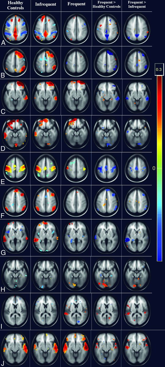
- Fig 1.
Whole-brain cluster-correlation maps of seed-to-voxel–based resting-state functional connectivity (FDR-corrected P < .001) with seed regions in the medial prefrontal cortex (A and B), right anterior prefrontal cortex (C), left anterior prefrontal cortex (D), left primary somatosensory cortex (E), right middle temporal gyrus (F), left angular gyrus (G), left precuneus (H), left posterior entorhinal cortex (I), and right medial temporal gyrus (J). The columns represents the healthy controls (column 1), the infrequent-seizure group (column 2), the frequent-seizure group (column 3), the frequent-seizure group versus healthy controls (column 4), and the frequent-seizure group versus the infrequent-seizure group (column 5). The colors represent the significance of connectivity; red indicates an increase in connectivity, and blue indicates a decrease in connectivity.
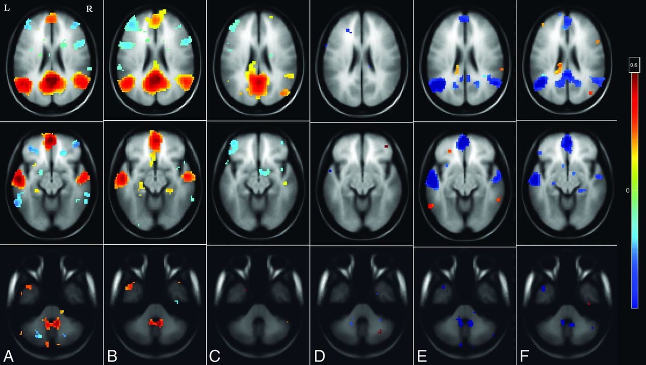
- Fig 2.
Whole-brain cluster-correlation maps of seed-to-voxel–based resting-state functional connectivity for the PCC seed region (FDR-corrected P < .001). Shown is DMN connectivity using PCC seed at 3 different axial levels: at the level of ventricles in the top row, midbrain in the middle row, and the cerebellum in the bottom row for healthy controls (A), the infrequent-seizure group (B), the frequent-seizure group (C), the infrequent-seizure group versus healthy controls (D), the frequent-seizure group versus healthy controls (E), and the infrequent-seizure group versus the frequent-seizure group (F). The colors represent the significance of connectivity; red indicates an increase in connectivity, and blue indicates a decrease in connectivity.

Tables
- Table 1:
Demographic and clinical features of 2 groups (with frequent or infrequent seizures) of patients with HWE
Clinical Feature Frequent-Seizure Group (>2/mo) Infrequent-Seizure Group (≤2/mo) P Value Male/female ratio, n 14:4 13:5 .700 Mean (±SD) age at evaluation, y 29.06 ± 9.86 28.67 ± 10.78 .910 Mean (±SD) age at onset, y 22.61 ± 8.39 20.69 ± 10.36 .546 Mean (±SD) duration of illness, y 6.56 ± 6.48 7.39 ± 9.76 .546 HWE attacks per month <.0001 Mean (±SD) 2.61 ± 0.69 0.62 ± 0.52 Median (range) 2.5 (2–4) 0.43 (0.02–1.66) <.0001 1:1 episodes, n (%)a 8 (44.4) 1 (5.6) .717 Family history of any type of epilepsy, n (%) 6 (33) 5 (27) .463 History of febrile convulsion, n 1 0 Family history of HWE, n (%) 4 (22) 1 (5.6) Self-induction phenomena, n (%) 2 (11.1) 2 (11.1) Abnormal EEG, n 0 2 (11.1) Focal abnormalities in EEG, n 0 2 Mean (±SD) time between last seizure and fMRI, days 10.6 ± 7.5 9.38 ± 6.9 .735 Complex partial seizures, n 12 10 Generalized tonic-clonic seizures, n 6 8 ↵a Patients who were having seizures every time they took a hot-water bath.
- Table 2:
Seed-to-voxel–based connectivity results in the frequent-seizure and healthy control groups
Seed Region Connectivity Region P Value (FDR Corrected) Cluster Size (No. of Voxels) β Valuea T Valueb Posterior cingulate cortex L angular gyrus (decreased) .0003 381 −0.21 6.84 L temporopolar region (decreased) .0009 325 −0.24 7.11 Medial prefrontal cortex (decreased) .0009 247 −0.15 6.14 L lateral parietal cortex (decreased) .002 92 −0.15 4.96 L inferior parietal cortex (decreased) .002 52 −0.13 4.64 L superior temporal cortex (decreased) .005 37 −0.21 5.22 L primary somatosensory cortex R primary motor cortex (decreased) .001 336 −0.22 8.70 L primary motor cortex (decreased) .001 187 −0.17 6.51 L superior temporal gyrus L precuneus (decreased) .002 112 −0.11 5.79 R dorsal frontal cortex (decreased) .005 77 −0.11 5.82 L primary auditory cortex R premotor cortex (decreased) .005 94 −0.17 6.87 R dorsal frontal cortex R and L superior temporal gyrus (decreased) .005 117 −0.12 5.86 L lateral parietal cortex R cerebellar tonsil (decreased) .005 43 −0.09 5.13 Medial prefrontal cortex L and R dorsal posterior cingulate cortex (decreased) .005 166 −0.11 5.52 L primary somatosensory cortex (decreased) .005 137 −0.11 6.19 R medial temporal gyrus R temporopolar region (increased) .005 146 0.17 7.96 R posterior entorhinal cortex (increased) .005 108 0.17 6.28 L precuneus L secondary visual cortex (increased) .001 121 0.15 7.78 L primary visual cortex (increased) .003 83 0.13 5.49 - Table 3:
Seed-to-voxel–based connectivity results in the infrequent-seizure and healthy control groups
Seed Region Connectivity Region P Value (FDR Corrected) Cluster Size (No. of Voxels) β Valuea T Valueb L posterior entorhinal cortex L fusiform gyrus (decreased) .004 140 −0.15 6.73 L anterior prefrontal cortex L ventral posterior cingulate cortex (increased) .003 136 0.12 7.34 L dorsal posterior cingulate cortex (increased) .005 74 0.10 6.17 R anterior cingulate cortex R posterior superior temporal gyrus (increased) .002 172 0.10 6.82 - Table 4:
Seed-to-voxel–based connectivity results in the frequent- and infrequent-seizure groups
Seed Region Connectivity Region P Value (FDR Corrected) Cluster Size (No. of Voxels) β Valuea T Valueb Medial prefrontal cortex Precuneus (decreased) .00004 279 −0.19 9.76 Posterior cingulate cortex (decreased) .00004 347 −0.25 5.72 R anterior prefrontal cortex R dorsal frontal cortex (decreased) .005 284 −0.27 9.17 R middle temporal gyrus (decreased) .005 147 −0.18 6.13 L anterior prefrontal cortex L middle temporal gyrus (decreased) .005 92 −0.11 5.57 Posterior cingulate cortex (decreased) .005 110 −0.10 5.43 L pyramis (decreased) .005 122 −0.14 6.02 L primary somatosensory cortex R premotor cortex (decreased) .0001 192 −0.26 9.31 L premotor cortex (decreased) .002 148 −0.18 8.66 L angular gyrus L superior temporal gyrus (decreased) .002 108 −0.11 6.63 Anterior cingulate L piriform cortex (decreased) .005 56 −0.11 5.39 L lateral parietal cortices Precuneus (decreased) .0006 309 −0.18 8.17 L somatosensory association area (decreased) .003 164 −0.13 7.96 Posterior cingulate cortex L angular gyrus (decreased) .00006 321 −0.17 7.84 L inferior parietal cortex (decreased) .0009 191 −0.16 6.52 L thalamus R somatosensory association cortex (decreased) .009 78 −0.11 4.67 L posterior and anterior entorhinal cortex R primary auditory cortex (increased) .005 112 0.10 5.34 L primary auditory cortex (increased) .005 93 0.11 5.71


Jump to section
Related Articles
Cited By...
- Improved Seizure Onset-Zone Lateralization in Temporal Lobe Epilepsy using 7T Resting-State fMRI: A Direct Comparison with 3T
- Atypical intrinsic neural timescales in temporal lobe epilepsy
- Functional Network Connectivity Imprint In Febrile Seizures
- Thalamic deep brain stimulation as a paradigm to reduce consciousness: implications for cortico-striatal dynamics, absence epilepsy and consciousness studies