
Article Figures & Data
Figures
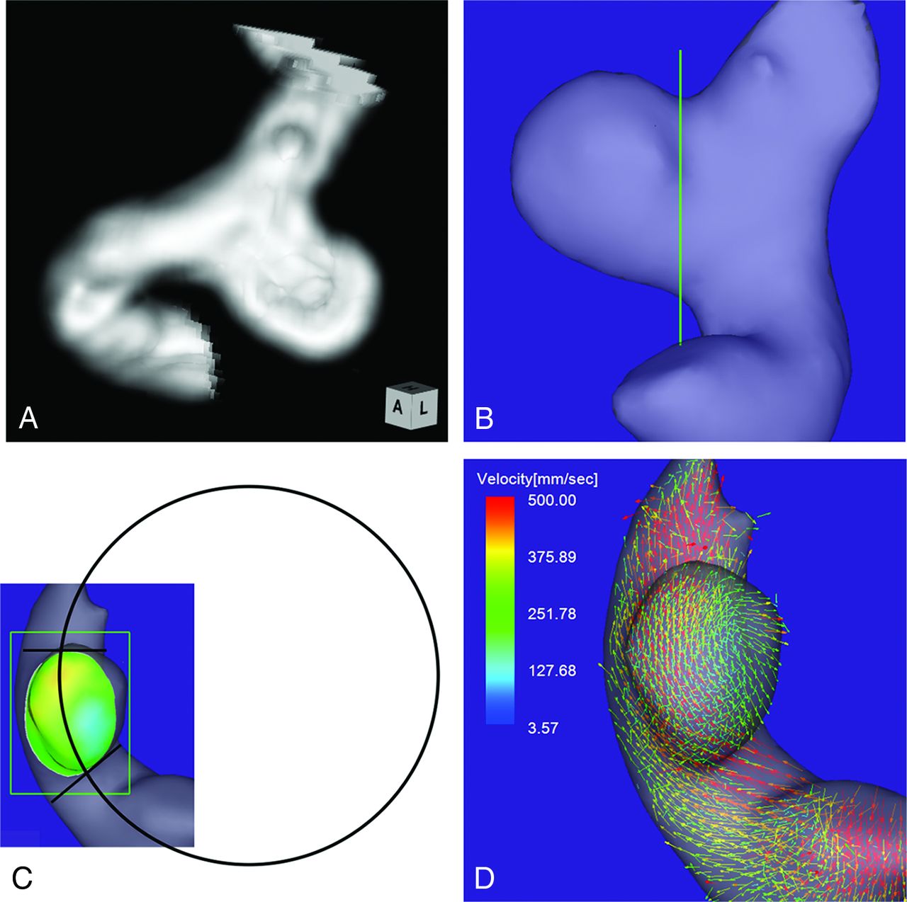
- Fig 1.
Case 1. A 64-year-old woman had an unruptured aneurysm at the medial C2 segment of the right ICA. A, 3D TOF MRA image. B, Image shows the section plane corresponding to the aneurysm orifice. C, En face image of the section plane. A 4D flow MR image demonstrates the inflow zone (orange) located on the distal neck and a circle in 2D fitting the central axis of the arterial part developing the aneurysm. The radius is 7.1 mm. D, A 4D flow MR image demonstrates a velocity vector map at peak systole.
- Fig 2.
The distribution of the radius of the parent artery curvature measured in 2D on an en face image of the section plane corresponding to the orifice in group 1, 2, and 3 aneurysms. Group 1, 2, and 3 aneurysms have inflow zone locations on the distal neck, lateral side of the neck, and proximal neck, respectively. The radius in group 1 is significantly larger than that in group 2 (asterisk 1: 8.3 mm, median; interquartile range, 4.5 mm; versus 4.5 mm, median; IQR, 1.9 mm; P < .0001) or those in groups 2 and 3 (asterisk 2: 8.3 mm, median; IQR, 4.5 mm; versus 5.0 mm, median; IQR, 1.4 mm; P < .0001).
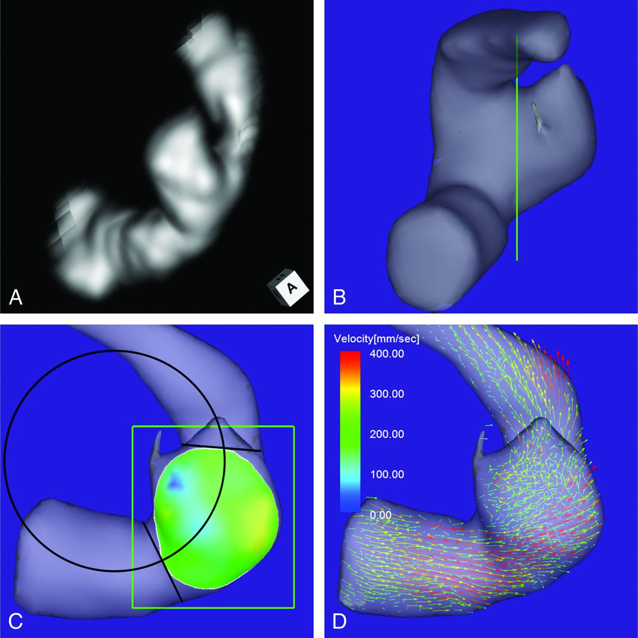
- Fig 3.
Case 2. An 80-year-old man had an unruptured aneurysm at the lateral C3 segment of the right ICA. A, A 3D TOF MRA image. B, Image shows the section plane corresponding to the aneurysm orifice. C, En face image of the section plane. A 4D flow MR image demonstrates the inflow zone (bright yellow) located on the lateral side of the neck and a circle in 2D fitting the central axis of the arterial part developing the aneurysm. The radius is 3.8 mm. D, A 4D flow MR image demonstrates a velocity vector map at peak systole, revealing that high-velocity vector components continue along the external side of the parent artery curvature.
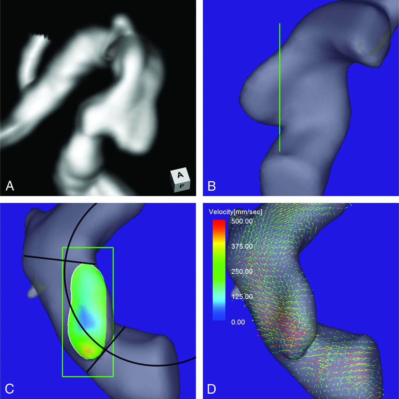
- Fig 4.
Case 3. A 73-year-old man had an unruptured aneurysm at the medial C2 segment of the right ICA. A, A 3D TOF MRA image. B, Image shows the section plane corresponding to the aneurysm orifice. C, En face image of the section plane. A 4D flow MR image demonstrates the inflow zone (orange) located in a part of the proximal neck extending beyond the central axis of the parent artery and a circle in 2D fitting the central axis of the arterial part developing the aneurysm. The radius is 4.9 mm. D, A 4D flow MR image demonstrates a velocity vector map at peak systole revealing that high-velocity vector components continue along the external side of the parent artery.


Tables
Aneurysm parameters: correlations between groups 1 and 2 and between group 1 and the other 2 groupsa
Parameters Group 1 (n = 10) Group 2 (n = 19) Group 3 (n = 3) P Value Group 1 vs 2 Group 1 vs Others MD 4.6 (1.7) 5.1 (2.5) 3.7 (0.5) .491 (NS) .714 (NS) ND 4.3 (1.2) 4.2 (1.5) 4.0 (0.7) .818 (NS) .855 (NS) MD/ND ratio 1.1 (0.1) 1.2 (0.4) 0.9 (0.0) .383 (NS) .745 (NS) Maximum perpendicular height 3.4 (0.7) 4.3 (2.6) 3.4 (0.3) .190 (NS) .318 (NS) Aspect ratio 1.1 (0.1) 1.2 (0.4) 0.7 (0.2) .136 (NS) .255 (NS) Maximum height 3.9 (0.9) 4.4 (2.5) 3.4 (0.3) .291 (NS) .555 (NS) Size ratio 1.1 (0.1) 1.4 (0.6) 0.8 (0.3) .598 (NS) .440 (NS) Neck width 3.4 (0.6) 3.5 (1.5) 2.3 (0.3) .335 (NS) .699 (NS) Maximum-orifice ratio 1.1 (0.3) 1.2 (0.6) 1.2 (0.2) .568 (NS) .502 (NS) Width-orifice ratio 0.8 (0.2) 1.0 (0.4) 0.6 (0.3) .215 (NS) .350 (NS) Radius of curvature 8.3 (4.5) 4.5 (1.9) 5.0 (0.3) <.0001 (S) <.0001 (S) Note:—S indicates significant; NS, not significant by the comparison test adjusted for the P value; MD, maximum diameter; ND, neck diameter.
↵a Data are median (interquartile range). Units of parameters except for ratios are mm.


