Advertisement

Main menu

  • Home
  • Content
    • Current Issue
    • Accepted Manuscripts
    • Article Preview
    • Past Issue Archive
    • Video Articles
    • AJNR Case Collection
    • Case of the Week Archive
    • Case of the Month Archive
    • Classic Case Archive
  • Special Collections
    • AJNR Awards
    • ASNR Foundation Special Collection
    • Most Impactful AJNR Articles
    • Photon-Counting CT
    • Spinal CSF Leak Articles (Jan 2020-June 2024)
  • Multimedia
    • AJNR Podcasts
    • AJNR SCANtastic
    • Trainee Corner
    • MRI Safety Corner
    • Imaging Protocols
  • For Authors
    • Submit a Manuscript
    • Submit a Video Article
    • Submit an eLetter to the Editor/Response
    • Manuscript Submission Guidelines
    • Statistical Tips
    • Fast Publishing of Accepted Manuscripts
    • Graphical Abstract Preparation
    • Imaging Protocol Submission
    • Author Policies
  • About Us
    • About AJNR
    • Editorial Board
    • Editorial Board Alumni
  • More
    • Become a Reviewer/Academy of Reviewers
    • Subscribers
    • Permissions
    • Alerts
    • Feedback
    • Advertisers
    • ASNR Home

User menu

  • Alerts
  • Log in

Search

  • Advanced search
American Journal of Neuroradiology

American Journal of Neuroradiology

ASHNR American Society of Functional Neuroradiology ASHNR American Society of Pediatric Neuroradiology ASSR
  • Alerts
  • Log in

Advanced Search

  • Home
  • Content
    • Current Issue
    • Accepted Manuscripts
    • Article Preview
    • Past Issue Archive
    • Video Articles
    • AJNR Case Collection
    • Case of the Week Archive
    • Case of the Month Archive
    • Classic Case Archive
  • Special Collections
    • AJNR Awards
    • ASNR Foundation Special Collection
    • Most Impactful AJNR Articles
    • Photon-Counting CT
    • Spinal CSF Leak Articles (Jan 2020-June 2024)
  • Multimedia
    • AJNR Podcasts
    • AJNR SCANtastic
    • Trainee Corner
    • MRI Safety Corner
    • Imaging Protocols
  • For Authors
    • Submit a Manuscript
    • Submit a Video Article
    • Submit an eLetter to the Editor/Response
    • Manuscript Submission Guidelines
    • Statistical Tips
    • Fast Publishing of Accepted Manuscripts
    • Graphical Abstract Preparation
    • Imaging Protocol Submission
    • Author Policies
  • About Us
    • About AJNR
    • Editorial Board
    • Editorial Board Alumni
  • More
    • Become a Reviewer/Academy of Reviewers
    • Subscribers
    • Permissions
    • Alerts
    • Feedback
    • Advertisers
    • ASNR Home
  • Follow AJNR on Twitter
  • Visit AJNR on Facebook
  • Follow AJNR on Instagram
  • Join AJNR on LinkedIn
  • RSS Feeds

AJNR is seeking candidates for the AJNR Podcast Editor. Read the position description.

Research ArticleAdult Brain

Constrained Source Space MR Spectroscopy: Multiple Voxels, No Gradient Readout

K. Landheer, A. Sahgal, S. Das and S.J. Graham
American Journal of Neuroradiology August 2015, 36 (8) 1436-1443; DOI: https://doi.org/10.3174/ajnr.A4319
K. Landheer
aFrom the Departments of Medical Biophysics (K.L., S.J.G.)
dPhysical Sciences (K.L., S.J.G.), Sunnybrook Research Institute, Toronto, Ontario, Canada
  • Find this author on Google Scholar
  • Find this author on PubMed
  • Search for this author on this site
A. Sahgal
cRadiation Oncology, Sunnybrook Odette Cancer Centre (A.S.), University of Toronto, Ontario, Canada
  • Find this author on Google Scholar
  • Find this author on PubMed
  • Search for this author on this site
S. Das
bSurgery (S.D.)
eKeenan Research Centre (S.D.), St. Michael's Hospital, Toronto, Ontario, Canada.
  • Find this author on Google Scholar
  • Find this author on PubMed
  • Search for this author on this site
S.J. Graham
aFrom the Departments of Medical Biophysics (K.L., S.J.G.)
dPhysical Sciences (K.L., S.J.G.), Sunnybrook Research Institute, Toronto, Ontario, Canada
  • Find this author on Google Scholar
  • Find this author on PubMed
  • Search for this author on this site
  • Article
  • Figures & Data
  • Info & Metrics
  • Responses
  • References
  • PDF
Loading

Article Figures & Data

Figures

  • Tables
  • Fig 1.
    • Download figure
    • Open in new tab
    • Download powerpoint
    Fig 1.

    A, Pulse diagram for CSSMRS. The first radiofrequency pulse has a flip angle of α (where α is <90°) and is cosine modulated, such that the subsequent spin echo after the third radiofrequency pulse excites 2 voxels. Shaded gradients are crusher gradients. The section-select rephasing lobe for the y gradient is added directly to the first crusher. The gradient-echo readout in the dotted box is optional for voxel localization verification. RF indicates radiofrequency; DAQ, data acquisition. B, An anatomic T1-weighted image of patient 6 with the nominal voxel locations overlaid and a brain tumor evident in the left middle temporal gyrus. The 2 spectra for this patient are displayed in the bottom row of Fig 2.

  • Fig 2.
    • Download figure
    • Open in new tab
    • Download powerpoint
    Fig 2.

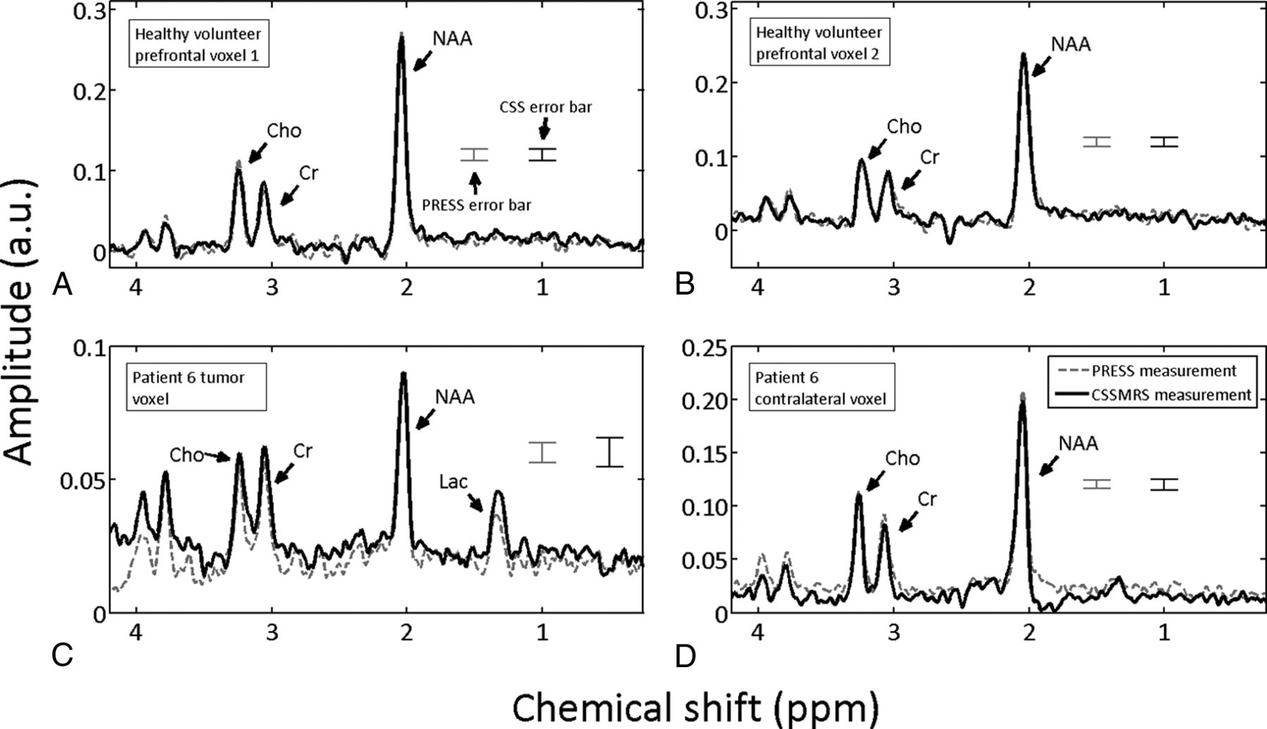
    Spectra from a healthy volunteer (A and B) and a patient with brain cancer (C and D) measured with both CSSMRS and PRESS. Spectra from patient 6 are shown, because this patient exhibited the median g-factor, typifying CSSMRS reconstruction quality. Errors represent the standard deviation over 128 excitations. a.u. indicates arbitrary units.

  • Fig 3.
    • Download figure
    • Open in new tab
    • Download powerpoint
    Fig 3.

    A, Spectra from a healthy volunteer at 30-millisecond echo time, obtained by using both CSSMRS and PRESS. The labeled metabolites are myo-inositol (mI), Cho, Cr, Glx, and NAA. B, The unapodized spectrum obtained from CSSMRS from patient 1 (highest g-factor) along with the automated quantitation of short echo time MR spectroscopy spectra (AQSES) fit.

  • Fig 4.
    • Download figure
    • Open in new tab
    • Download powerpoint
    Fig 4.

    Measured and simulated differences between the CSSMRS and PRESS measurement for 6 different voxel separations for the 3 main metabolites within a healthy adult brain: NAA, Cho, and Cr. The signal from the CSSMRS voxel that was kept in a fixed position was reconstructed and compared with the PRESS measurement obtained from the same location. The black and gray lines represent the measured and simulated values, respectively. The g factors are also displayed for reference above the top x-axis, though there is a nonlinear relationship between g factor and voxel separation. Error bars represent Cramer-Rao bounds.

  • Fig 5.
    • Download figure
    • Open in new tab
    • Download powerpoint
    Fig 5.

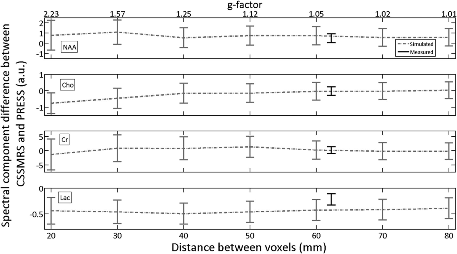
    Simulated metabolite quantification values for 7 different voxel separations for the 4 main metabolites within the tumor spectra for patient 6: NAA, Cho, Cr, and lactate (Lac). The quantified values were from the stationary voxel placed within the tumor and are plotted in gray. The black data points located at 62 cm in each plot are the experimental results for this patient, corresponding to the first difference column values listed in Tables 2–5 for patient 6. The estimated g factors are also displayed above the top x-axis for reference, though there is a nonlinear relationship between the g factor and voxel separation.

Tables

  • Figures
    • View popup
    Table 1:

    Summary of patients with brain tumor studied in experiment 2

    Patient No.Age (yr)SexDiseaseRadiation Treatment StatusTumor LocationTumor Size (vs Voxel Size)
    136FGrade II oligodendrogliomaNoneRight cingulate gyrusLarger
    284MGrade IV glioblastomaCurrently undergoing focused radiationLeft middle temporal gyrusComparable
    379MGrade IV glioblastomaCurrently undergoing focused radiationLeft superior temporal gyrusLarger
    461FBrain metastases from breast cancer60 days since completion of focused radiationLeft middle temporal gyrusSmaller
    579MBrain metastases from colon cancer70 days since completion of focused radiationRight superior temporal gyrusSmaller
    661MGrade IV glioblastomaCurrently undergoing focused radiationLeft middle temporal gyrusLarger
    • Note:—F indicates female; M, male.

    • View popup
    Table 2:

    Quantified NAA values from PRESS and CSSMRS for both voxelsa

    Patient No.CSSMRS Tumor Voxel (a.u.)PRESS Tumor Voxel (a.u.)Difference (a.u.)CSSMRS Healthy Voxel (a.u.)PRESS Healthy Voxel (a.u.)Difference (a.u.)
    15.70 ± 0.654.86 ± 0.580.83 ± 0.879.73 ± 0.289.88 ± 0.20−0.15 ± 0.35
    22.70 ± 0.442.08 ± 0.400.62 ± 0.599.34 ± 0.358.84 ± 0.350.50 ± 0.49
    31.70 ± 0.191.60 ± 0.200.10 ± 0.289.14 ± 0.129.42 ± 0.14−0.28 ± 0.18
    43.33 ± 0.540.14 ± 0.253.19 ± 0.598.09 ± 0.388.89 ± 0.36−0.81 ± 0.52
    55.27 ± 0.294.22 ± 0.211.05 ± 0.358.87 ± 0.3810.93 ± 0.55−2.06 ± 0.66
    65.89 ± 0.275.41 ± 0.330.48 ± 0.436.81 ± 0.216.98 ± 0.20−0.17 ± 0.29
    • Note:—a.u. indicates arbitrary units.

    • ↵a Shown are means and standard deviations (Cramer-Rao bounds).

    • View popup
    Table 3:

    Quantified Cho values from PRESS and CSSMRS for both voxelsa

    Patient No.CSSMRS Tumor Voxel (a.u.)PRESS Tumor Voxel (a.u.)Difference (a.u.)CSSMRS Healthy Voxel (a.u.)PRESS Healthy Voxel (a.u.)Difference (a.u.)
    12.93 ± 0.392.89 ± 0.240.04 ± 0.451.72 ± 0.181.73 ± 0.13−0.01 ± 0.22
    21.86 ± 0.271.67 ± 0.170.19 ± 0.321.89 ± 0.202.22 ± 0.22−0.33 ± 0.30
    30.67 ± 0.120.96 ± 0.17−0.28 ± 0.212.06 ± 0.072.31 ± 0.08−0.25 ± 0.11
    40.97 ± 0.930.83 ± 0.330.15 ± 0.991.36 ± 0.182.05 ± 0.21−0.69 ± 0.75
    51.66 ± 0.121.81 ± 0.11−0.15 ± 0.161.56 ± 0.263.02 ± 0.66−1.46 ± 0.71
    61.56 ± 0.171.59 ± 0.20−0.02 ± 0.261.75 ± 0.131.80 ± 0.12−0.05 ± 0.17
    • Note:—a.u. indicates arbitrary units.

    • ↵a Shown are means and standard deviations (Cramer-Rao bounds).

    • View popup
    Table 4:

    Quantified Cr values from PRESS and CSSMRS for both voxelsa

    Patient No.CSSMRS Tumor Voxel (a.u.)PRESS Tumor Voxel (a.u.)Difference (a.u.)CSSMRS Healthy Voxel (a.u.)PRESS Healthy Voxel (a.u.)Difference (a.u.)
    110.62 ± 2.447.77 ± 1.072.85 ± 2.666.34 ± 0.776.50 ± 0.61−0.16 ± 0.98
    23.82 ± 1.681.66 ± 0.552.16 ± 1.779.09 ± 0.8910.40 ± 1.07−1.32 ± 1.39
    32.41 ± 0.771.56 ± 0.670.85 ± 1.028.73 ± 0.328.37 ± 0.350.36 ± 0.48
    44.66 ± 4.170.42 ± 0.634.24 ± 4.228.94 ± 1.038.60 ± 0.95−0.25 ± 1.40
    56.19 ± 0.615.98 ± 0.480.21 ± 0.7710.44 ± 1.7610.58 ± 2.97−0.15 ± 3.45
    68.21 ± 0.748.00 ± 0.880.22 ± 1.156.13 ± 0.566.19 ± 0.52−0.05 ± 0.77
    • Note:—a.u. indicates arbitrary units.

    • ↵a Shown are means and standard deviations (Cramer-Rao bounds).

    • View popup
    Table 5:

    Quantified lactate values from PRESS and CSSMRS for both voxelsa

    Patient No.CSSMRS Tumor Voxel (a.u.)PRESS Tumor Voxel (a.u.)Difference (a.u.)CSSMRS Healthy Voxel (a.u.)PRESS Healthy Voxel (a.u.)Difference (a.u.)
    11.31 ± 0.091.46 ± 0.08−0.16 ± 0.120.03 ± 0.040.23 ± 0.07−0.20 ± 0.08
    23.36 ± 0.113.16 ± 0.100.20 ± 0.150.60 ± 0.110.51 ± 0.110.09 ± 0.16
    32.83 ± 0.042.77 ± 0.040.06 ± 0.050.25 ± 0.040.28 ± 0.050.03 ± 0.06
    4b3.22 ± 0.142.76 ± 0.130.46 ± 0.191.84 ± 0.101.59 ± 0.100.25 ± 0.14
    52.26 ± 0.082.32 ± 0.07−0.06 ± 0.100.73 ± 0.130.87 ± 0.17−0.14 ± 0.21
    60.54 ± 0.070.76 ± 0.08−0.22 ± 0.110.22 ± 0.050.29 ± 0.05−0.08 ± 0.07
    • Note:—a.u. indicates arbitrary units.

    • ↵a Shown are means and standard deviations (Cramer-Rao bounds).

    • ↵b Lipid contamination from scalp mislabeled as lactate in healthy voxel.

PreviousNext
Back to top

In this issue

American Journal of Neuroradiology: 36 (8)
American Journal of Neuroradiology
Vol. 36, Issue 8
1 Aug 2015
  • Table of Contents
  • Index by author
  • Complete Issue (PDF)
Advertisement
Print
Download PDF
Email Article

Thank you for your interest in spreading the word on American Journal of Neuroradiology.

NOTE: We only request your email address so that the person you are recommending the page to knows that you wanted them to see it, and that it is not junk mail. We do not capture any email address.

Enter multiple addresses on separate lines or separate them with commas.
Constrained Source Space MR Spectroscopy: Multiple Voxels, No Gradient Readout
(Your Name) has sent you a message from American Journal of Neuroradiology
(Your Name) thought you would like to see the American Journal of Neuroradiology web site.
CAPTCHA
This question is for testing whether or not you are a human visitor and to prevent automated spam submissions.
Cite this article
K. Landheer, A. Sahgal, S. Das, S.J. Graham
Constrained Source Space MR Spectroscopy: Multiple Voxels, No Gradient Readout
American Journal of Neuroradiology Aug 2015, 36 (8) 1436-1443; DOI: 10.3174/ajnr.A4319

Citation Manager Formats

  • BibTeX
  • Bookends
  • EasyBib
  • EndNote (tagged)
  • EndNote 8 (xml)
  • Medlars
  • Mendeley
  • Papers
  • RefWorks Tagged
  • Ref Manager
  • RIS
  • Zotero
0 Responses
Respond to this article
Share
Bookmark this article
Constrained Source Space MR Spectroscopy: Multiple Voxels, No Gradient Readout
K. Landheer, A. Sahgal, S. Das, S.J. Graham
American Journal of Neuroradiology Aug 2015, 36 (8) 1436-1443; DOI: 10.3174/ajnr.A4319
del.icio.us logo Twitter logo Facebook logo Mendeley logo
  • Tweet Widget
  • Facebook Like
  • Google Plus One
Purchase

Jump to section

  • Article
    • Abstract
    • ABBREVIATIONS:
    • Materials and Methods
    • Results
    • Discussion
    • Conclusions
    • Footnotes
    • References
  • Figures & Data
  • Info & Metrics
  • Responses
  • References
  • PDF

Related Articles

  • No related articles found.
  • PubMed
  • Google Scholar

Cited By...

  • No citing articles found.
  • Crossref (1)
  • Google Scholar

This article has been cited by the following articles in journals that are participating in Crossref Cited-by Linking.

  • Fourier‐based decomposition for simultaneous 2‐voxel MRS acquisition with 2SPECIAL
    Layla Tabea Riemann, Christoph Stefan Aigner, Ralf Mekle, Oliver Speck, Georg Rose, Bernd Ittermann, Sebastian Schmitter, Ariane Fillmer
    Magnetic Resonance in Medicine 2022 88 5

More in this TOC Section

  • Diagnostic Neuroradiology of Monoclonal Antibodies
  • Clinical Outcomes After Chiari I Decompression
  • Segmentation of Brain Metastases with BLAST
Show more Adult Brain

Similar Articles

Advertisement

Indexed Content

  • Current Issue
  • Accepted Manuscripts
  • Article Preview
  • Past Issues
  • Editorials
  • Editor's Choice
  • Fellows' Journal Club
  • Letters to the Editor
  • Video Articles

Cases

  • Case Collection
  • Archive - Case of the Week
  • Archive - Case of the Month
  • Archive - Classic Case

Special Collections

  • AJNR Awards
  • ASNR Foundation Special Collection
  • Most Impactful AJNR Articles
  • Photon-Counting CT
  • Spinal CSF Leak Articles (Jan 2020-June 2024)

More from AJNR

  • Trainee Corner
  • Imaging Protocols
  • MRI Safety Corner

Multimedia

  • AJNR Podcasts
  • AJNR Scantastics

Resources

  • Turnaround Time
  • Submit a Manuscript
  • Submit a Video Article
  • Submit an eLetter to the Editor/Response
  • Manuscript Submission Guidelines
  • Statistical Tips
  • Fast Publishing of Accepted Manuscripts
  • Graphical Abstract Preparation
  • Imaging Protocol Submission
  • Evidence-Based Medicine Level Guide
  • Publishing Checklists
  • Author Policies
  • Become a Reviewer/Academy of Reviewers
  • News and Updates

About Us

  • About AJNR
  • Editorial Board
  • Editorial Board Alumni
  • Alerts
  • Permissions
  • Not an AJNR Subscriber? Join Now
  • Advertise with Us
  • Librarian Resources
  • Feedback
  • Terms and Conditions
  • AJNR Editorial Board Alumni

American Society of Neuroradiology

  • Not an ASNR Member? Join Now

© 2025 by the American Society of Neuroradiology All rights, including for text and data mining, AI training, and similar technologies, are reserved.
Print ISSN: 0195-6108 Online ISSN: 1936-959X

Powered by HighWire