
Article Figures & Data
Figures
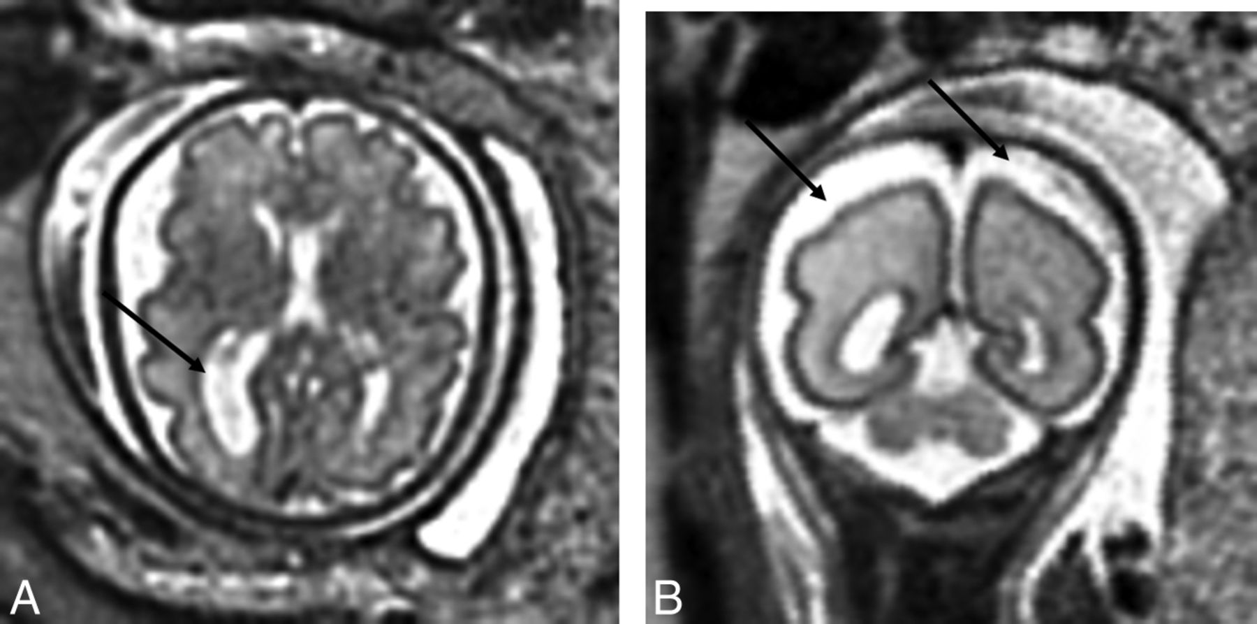
- Fig 1.
A, T2-weighted axial view of the brain of a 32-week-old fetus with CHD with unilateral ventriculomegaly. B, T2-weighted axial view of the brain of a 27.29-week-old fetus with CHD with extra-axial spaces. Reprinted from Brossard-Racine et al.10
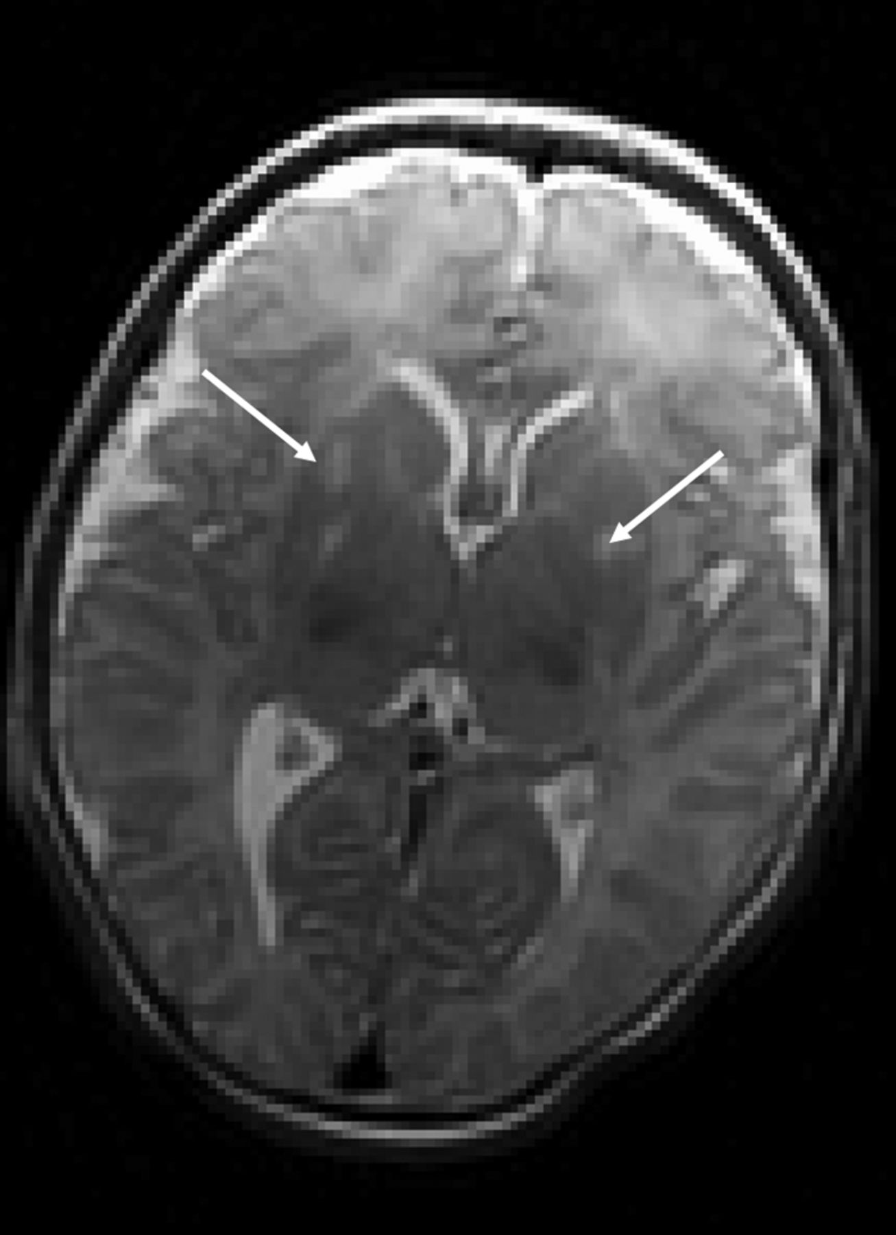
- Fig 2.
T2-weighted axial view of a neonate with CHD with punctate white matter injury.
- Fig 3.
T2-weighted axial view of a neonate with CHD with bilateral deep gray matter infarcts.
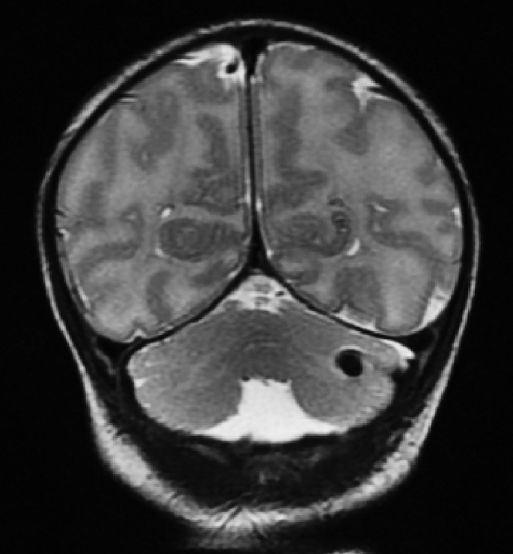
- Fig 4.
T2-weighted coronal view of a neonate with CHD with unilateral cerebellar hemorrhage.

Tables
Diagnosis Distribution (%) Abnormal Findings on Fetal MRI (%) Abnormal Findings on Preop MRI (%) Dextro-transposition of the great arteries 31 (30.1) 5 (29.4) 9 (27.3) Hypoplastic left-heart syndrome 20 (19.4) 3 (17.6) 9 (27.3) Double-outlet right ventricle 13 (12.6) 2 (11.8) 2 (6.1) Tetralogy of Fallot 11 (10.7) 2 (11.8) 2 (6.1) Atrioventricular septal defect 8 (7.8) 3 (17.6) 1 (3.0) Tricuspid atresia 4 (3.9) 1 (5.9) 3 (12.1) Ventricular septal defect 4 (3.9) 0 (0.0) 2 (6.1) Pulmonary atresia 3 (2.9) 0 (0.0) 2 (6.1) Ebstein anomaly 2 (1.9) 0 (0.0) 1 (3.0) Pulmonary stenosis 2 (1.9) 0 (0.0) 1 (3.0) Aortic stenosis 1 (1.0) 0 (0.0) 0 (0.0) Severe coarctation of the aorta 2 (1.9) 0 (0.0) 0 (0.0) Hypoplastic right-heart syndrome 1 (1.0) 0 (0.0) 0 (0.0) Truncus arteriosus 1 (1.0) 1 (5.9) 1 (3.0) Total 103 (100) 17 (100) 33 (100) Note:—Preop indicates preoperative.
Antenatal Mean ± SD/Median (Range)/No. (%) Pregnancy-induced diabetes 7 (6.8%) Group B streptococcus 13 (13.3%) Hyperthyroidism 3 (2.9%) Hypothyroidism 8 (7.8%) Pregnancy-induced hypertension 3 (2.9%) Placenta previa 3 (2.9%) Placenta abruption 2 (1.9%) Preterm labor 3 (2.9%) Chorioamnionitis 1 (1.0%) Intrapartum Induced labor 48 (52.7%) Emergency cesarean delivery 22 (21.8%) Gestational age at birth (wk) 38.5 ± 1.3 Birth weight (g) 3173 ± 552 Apgar score 1 min/5 min 8 (1–9)/8(5–9) Intubation at birth 18 (19%) SNAP-II score 5 (0–68) Perinatal (prior to MRI) Lowest pH 7.318 ± 0.062 Lowest pO2 38.63 ± 23.72 Highest pCO2 47.65 ± 12.00 Cardiac catheterization 11 (10.9%) Balloon atrial septostomy 15 (14.6%) Note:—pCO2 indicates carbon dioxide partial pressure; pO2, oxygen partial pressure.
Abnormal Fetal MRI Findings Abnormal Neonatal MRI Findings Predictive Value Yes No Total Yes 9 8 17 Positive, 9/17 = 52.9% No 24 62 86 Negative, 62/86 = 72.1% Total 33 70 n = 103 Sensitivity, 9/33 = 27.3% Specificity, 62/70 = 88.6% Normal pWMI Hemorrhage NHPI Dev. Abn.b Total Normal 62 11 6 3 4 86 Extracerebral space 2 2 0 0 0 4 Ventriculomegaly 3 0 0 2 1 6 WM abn. 1 0 1 1 0 3 Immature brain 2 0 0 0 0 2 Vermis hypoplasia 0 1 0 0 1 2 Total 70 14 7 6 6 103 Note:—Dev. Abn. indicates developmental abnormalities.
↵a Cases with multiple abnormalities were included only once in the table on the basis of the most severe finding.
↵b Developmental anomaly includes congenital malformation, delayed maturation, and cerebral atrophy (extra-axial space and/or ventricular enlargement).
- Table 5:
Clinical characteristics of neonates with CHD with and without newly acquired brain injurya
Neonates without Injury (n = 62) Neonates with New Injury (n = 20) P Value Induced labor 28 (45.2%) 10 (50.0%) .570 Vaginal delivery 29 (46.8%) 15 (65.0%) .082 Gestational age at birth (wk) 38.93 (33.86–40.71) 38.71 (36.00–40.00) .134 Birth weight (g) 3300 (1077–4276) 3085 (2240–4220) .525 Apgar score 1 min/5 min 8 (1–9)/8 (5–9) 8 (5–9)/8 (7–9) .816/.266 Intubation at birth 14 (22.5%) 3 (12.5%) .373 SNAP-II score 5 (0–43) 5 (0–28) .471 Lowest pH prior to MRI, (mean) 7.320 (7.174 ± 7.490) 7.326 (7.220 ± 7.454) .567 Lowest pO2 prior to MRI 34.85 (20–214) 33.75 (22–78) .673 Highest pCO2 prior to MRI 46.50 (22–83) 46.25 (24–61) .794 Cardiac catheterization prior to MRI 3 (4.8%) 6 (30.0%) .006 Balloon atrial sepstostomy prior to MRI 10 (16.1%) 4 (20.0%) .612 Age at MRI (days) 3.0 (1.0–76.0) 3.0 (1.0–12.0) .063 Time between fetal and neonatal MRI (wk) 6.00 (1.28–20.85) 4.86 (1.00–10.43) .011 ↵a Values are median (range) or No. (%).


