
Article Figures & Data
Figures
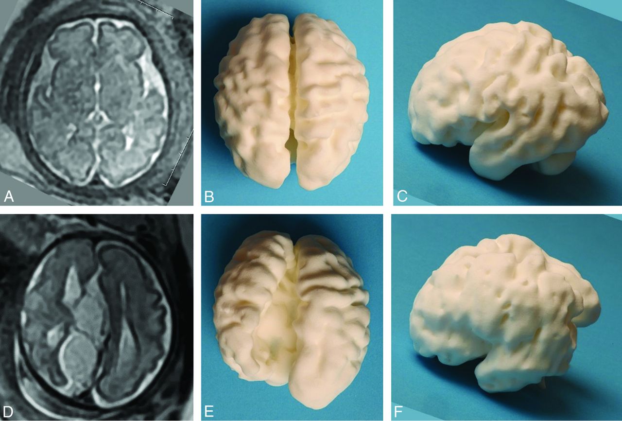
- Fig 1.
Images of the 3D printed model produced via laser sintering from an in utero MR imaging study performed at 30 weeks' gestational age in a fetus with ventriculomegaly and an interhemispheric cyst recognized on ultrasonography, compared with an age-matched fetus with no brain abnormality. A 2D single-shot fast spin-echo image in the axial plane of the normal brain is shown (A), along with superior (B) and left lateral (C) views of the 3D printed model. D–F, The matching images from the fetus with agenesis of the corpus callosum and extra-axial cysts, which do not communicate with the ventricular system (type II cysts of Barkovich et al),7 are shown. Note that the orientation of the 2D image has been altered to match the 3D model for ease of interpretation. The left hemisphere contains widespread heterotopia, a feature that was confirmed at postmortem examination.
- Fig 2.
Images of the 3D printed models produced via laser sintering from 2 in utero MR imaging studies performed at 2 gestational ages in a fetus with lissencephaly compared with an age-matched fetus with no brain abnormality. A 2D single-shot fast spin-echo image in the axial plane of the normal brain at 22 weeks' gestation is shown (A), along with superior (B) and left lateral (C) views of the 3D printed model. The same format is shown for a healthy 30-week fetus (D–F) and the fetus with lissencephaly (G–L).
- Fig 3.
A 3D printed model produced via laser sintering with the internal anatomy of the brain shown from an attached 2D single-shot fast spin-echo image to produce a section of the fetal brain—superior (A) and oblique (B) projections.
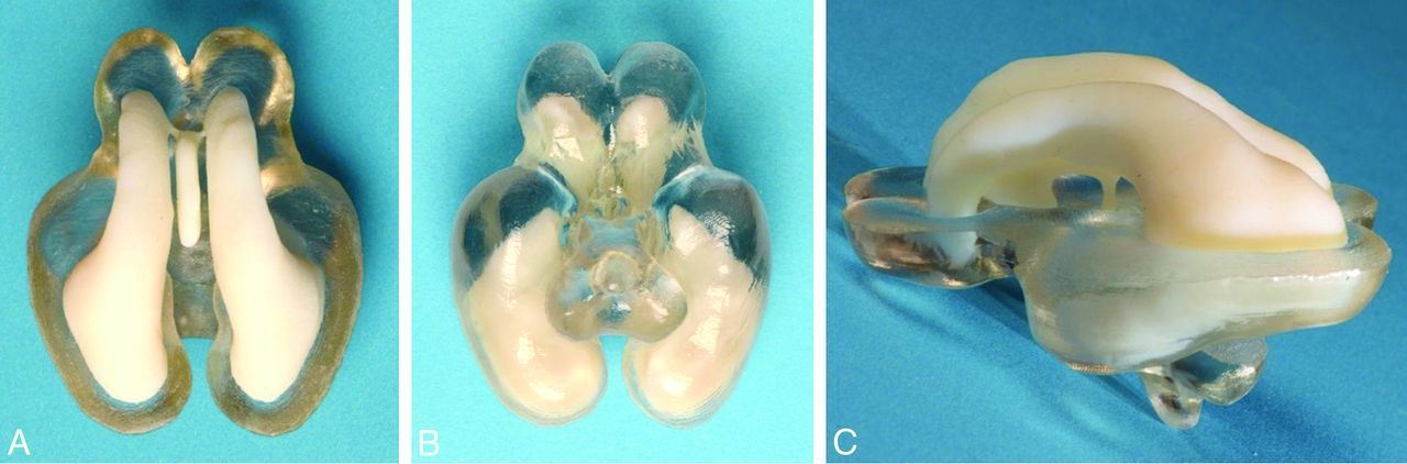
- Fig 4.
Dual-material 3D printed brain produced on an Objet Connex 500 jetting system (manufactured courtesy of Professor Richard Bibb, Loughborough Design School, Loughborough, UK). Separate STereoLithography files were exported from 3D Slicer, one consisting of the segmented entire ventricular system and the other of part of the brain parenchyma. The ventricular system is printed in the same white material as the other brains, while the parenchyma is printed in a clear material. The superior (A), inferior (B), and left lateral (C) views show the relationship between the ventricles and brain to advantage.


Tables
Imaging sequence parameters for the 3D FIESTA acquisition
3D FIESTA Steady-State Balanced Gradient-Echo TR (ms) 4.2 TE (ms) 2.1 Flip angle 60° Bandwidth (Hz) 125 NEX 0.75 Section thickness/gap (mm) 2.2/0 No. of partitions 26 FOV (mm) 340 × 270 Matrix size 320/256 Interpolation phase/secondary phase ZIP 512/ ZIP 2 Scan time (sec) 21 Note:—ZIP indicates interpolation values.


Jump to section
Related Articles
Cited By...
- No citing articles found.