
Article Figures & Data
Figures
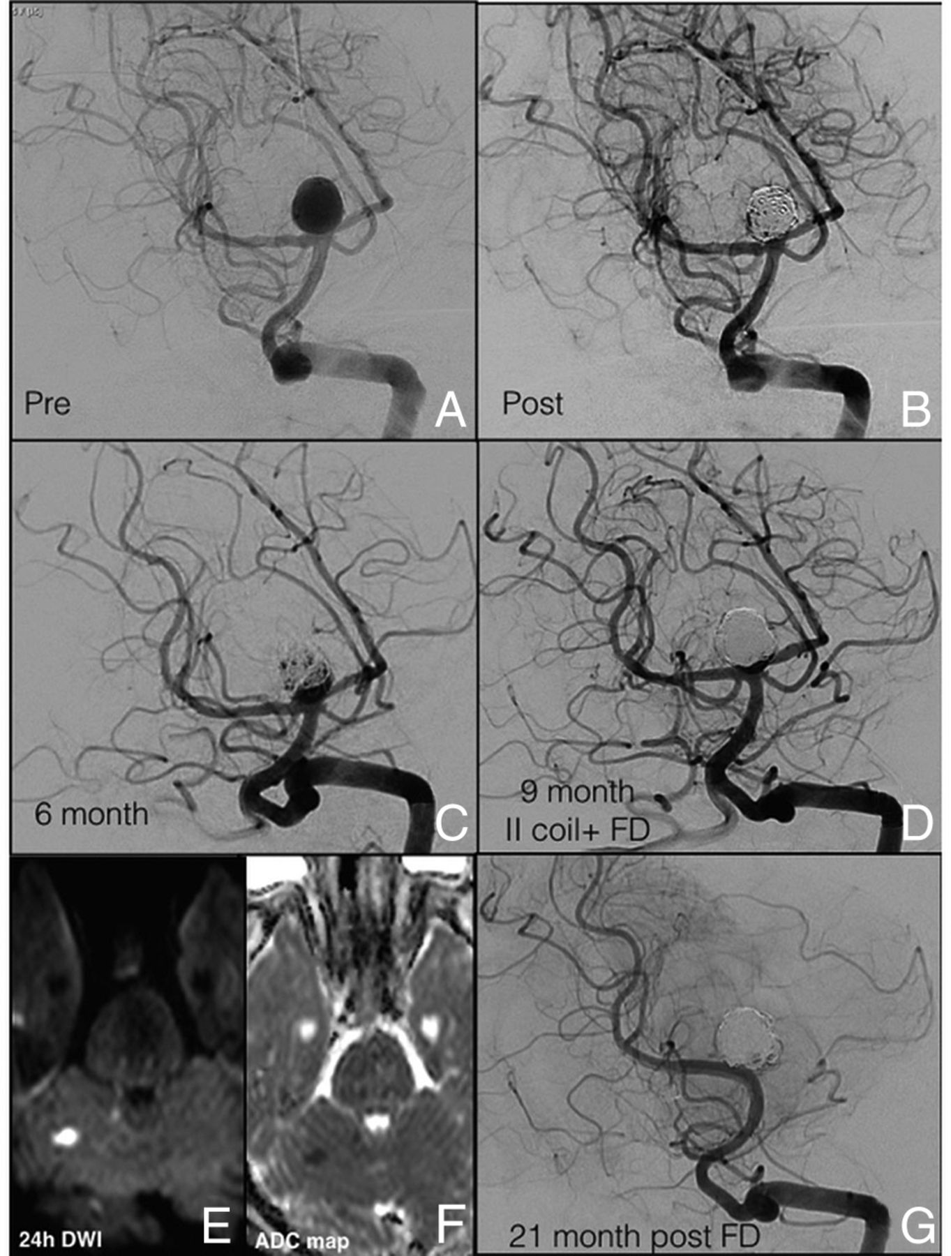
- Fig 1.
A, DSA shows a ruptured large BAA (10 × 10 mm; neck, 4.4 mm). B, Aneurysmal embolization was performed with the balloon-remodeling technique. C, Six-month DSA follow-up shows a significant neck recanalization (5 mm × 6 mm). D, At the 9-month follow-up, a second coiling remodeling technique associated with the deployment of a FRED flow-diverter stent across the right PCA was performed. E and F, The 24-hour postprocedural MR imaging, with DWI and ADC map, shows the presence of a small ischemic right cerebellar lesion. G, At 21-month DSA follow-up, both covered superior cerebellar arteries were not visualized, and the persistence of complete BAA occlusion was confirmed (mRR class I; mRS, 0).
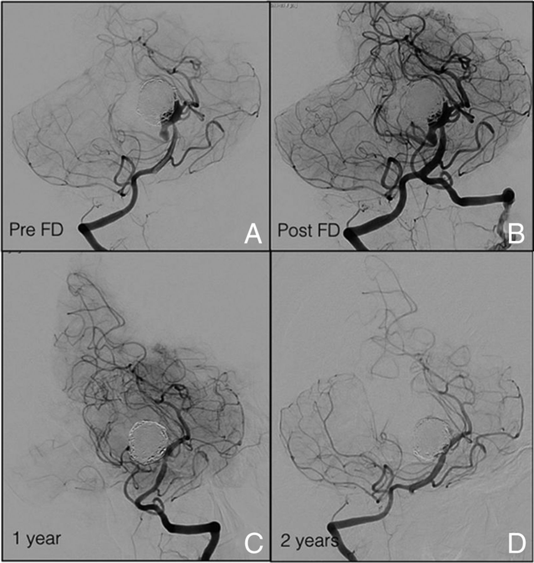
- Fig 2.
A, DSA shows the remnant of a wide-neck, large BAA aneurysm that involved both superior cerebellar arteries and the left P1 segment, with a 5-mm right superior cerebellar artery fusiform aneurysm, and a fetal origin of the right PCA, treated with a simple coiling. B, Six weeks later, the remnant was treated with a Pipeline stent. C, Twelve-month and D, 24-month follow-up DSA reveal complete aneurysmal occlusion (mRR class I).
- Fig 3.
A, DSA shows a wide-neck, large BAA aneurysm (B) treated in a single session with coiling and a Silk flow-diverter stent across the left PCA. C and D, MR imaging shows the presence of a midbrain hematoma 12 hours after the treatment. E, Anteroposterior view DSA shows residual filling at the level of the aneurysmal neck. A second Pipeline flow diverter was then deployed. F, The 2-week DSA follow-up shows complete aneurysmal sac occlusion (mRR class I).
- Fig 4.
A, The initial DSA demonstrates a wide-neck, large BAA involving the origin of both PCAs and superior cerebellar arteries (B) treated with a stent-assisted coiling technique (Neuroform 3.5 mm × 20 mm). C, The 18-month DSA follow-up reveals an aneurysmal recurrence at the level of the neck. D, A Pipeline stent was used to treat the neck remnant with (E) adequate aneurysm occlusion at 15-month DSA follow-up (mRR class IIIa) demonstrated in the working projection. F, The compression test performed confirms the result.
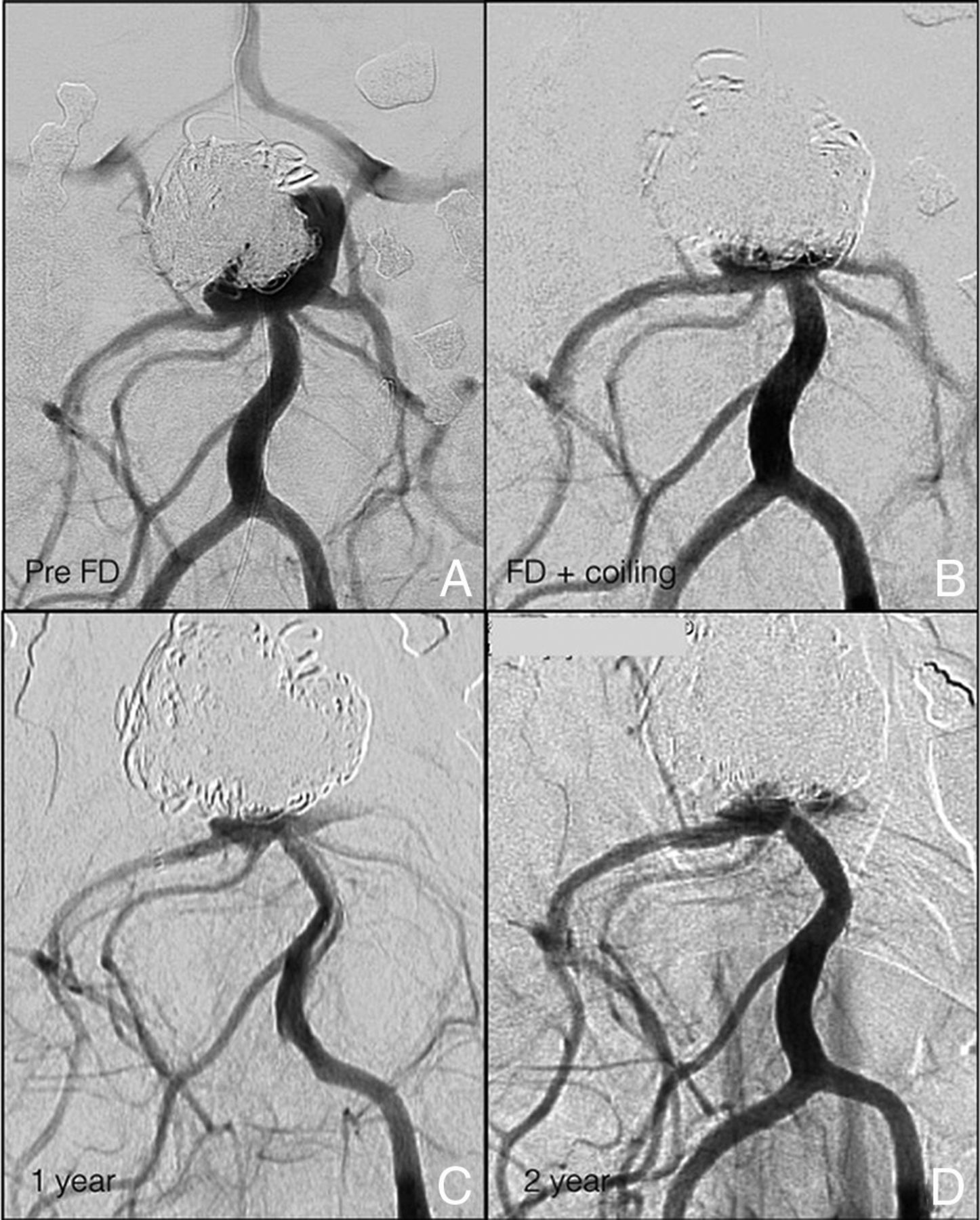
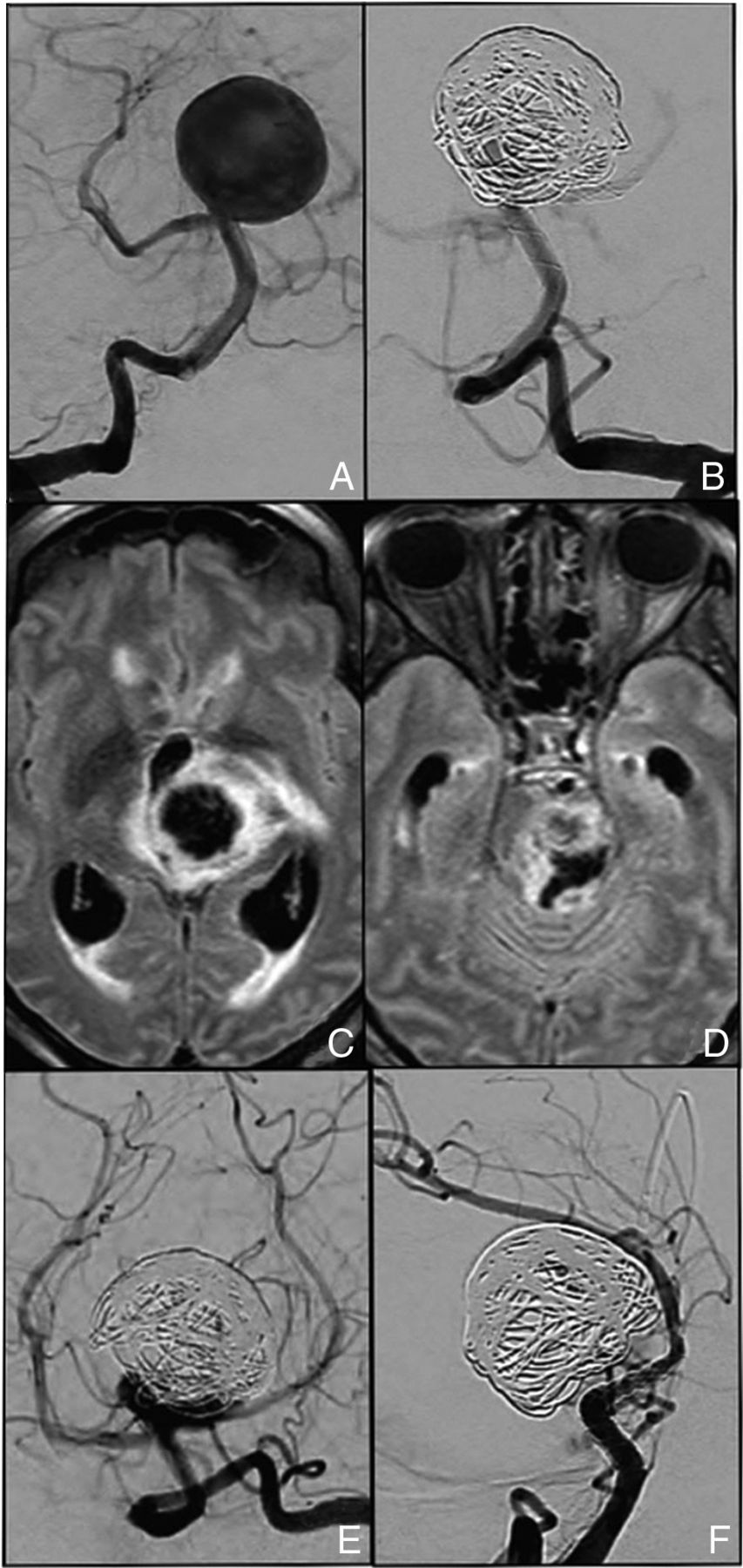
- Fig 5.
A, The preoperative DSA shows a large, wide-neck BAA, twice partially coiled in an emergency setting, responsible for a chiasmal compression syndrome. B, Additional coiling of the sac remnant occurred and a subsequent flow-diverter stent (FRED 3.5 mm × 22–16 mm) was deployed across the right P1 segment. C, Twelve-month and D, 24-month DSA follow-up demonstrate adequate BAA occlusion (mRR class IIIa), and the visual field was completely recovered.


Jump to section
Related Articles
Cited By...
- Flow diversion for the treatment of intracranial bifurcation aneurysms: a systematic review and meta-analysis
- Preliminary experience with the use of low profile visualized intraluminal support device in basilar artery for aneurysm treatment
- The bendy' basilar: progressive aneurysm tilting and arterial deformation can be a delayed outcome after coiling of large basilar apex aneurysms