Advertisement

Main menu

  • Home
  • Content
    • Current Issue
    • Accepted Manuscripts
    • Article Preview
    • Past Issue Archive
    • Video Articles
    • AJNR Case Collection
    • Case of the Week Archive
    • Case of the Month Archive
    • Classic Case Archive
  • Special Collections
    • AJNR Awards
    • ASNR Foundation Special Collection
    • Most Impactful AJNR Articles
    • Photon-Counting CT
    • Spinal CSF Leak Articles (Jan 2020-June 2024)
  • Multimedia
    • AJNR Podcasts
    • AJNR SCANtastic
    • Trainee Corner
    • MRI Safety Corner
    • Imaging Protocols
  • For Authors
    • Submit a Manuscript
    • Submit a Video Article
    • Submit an eLetter to the Editor/Response
    • Manuscript Submission Guidelines
    • Statistical Tips
    • Fast Publishing of Accepted Manuscripts
    • Graphical Abstract Preparation
    • Imaging Protocol Submission
    • Author Policies
  • About Us
    • About AJNR
    • Editorial Board
    • Editorial Board Alumni
  • More
    • Become a Reviewer/Academy of Reviewers
    • Subscribers
    • Permissions
    • Alerts
    • Feedback
    • Advertisers
    • ASNR Home

User menu

  • Alerts
  • Log in

Search

  • Advanced search
American Journal of Neuroradiology

American Journal of Neuroradiology

ASHNR American Society of Functional Neuroradiology ASHNR American Society of Pediatric Neuroradiology ASSR
  • Alerts
  • Log in

Advanced Search

  • Home
  • Content
    • Current Issue
    • Accepted Manuscripts
    • Article Preview
    • Past Issue Archive
    • Video Articles
    • AJNR Case Collection
    • Case of the Week Archive
    • Case of the Month Archive
    • Classic Case Archive
  • Special Collections
    • AJNR Awards
    • ASNR Foundation Special Collection
    • Most Impactful AJNR Articles
    • Photon-Counting CT
    • Spinal CSF Leak Articles (Jan 2020-June 2024)
  • Multimedia
    • AJNR Podcasts
    • AJNR SCANtastic
    • Trainee Corner
    • MRI Safety Corner
    • Imaging Protocols
  • For Authors
    • Submit a Manuscript
    • Submit a Video Article
    • Submit an eLetter to the Editor/Response
    • Manuscript Submission Guidelines
    • Statistical Tips
    • Fast Publishing of Accepted Manuscripts
    • Graphical Abstract Preparation
    • Imaging Protocol Submission
    • Author Policies
  • About Us
    • About AJNR
    • Editorial Board
    • Editorial Board Alumni
  • More
    • Become a Reviewer/Academy of Reviewers
    • Subscribers
    • Permissions
    • Alerts
    • Feedback
    • Advertisers
    • ASNR Home
  • Follow AJNR on Twitter
  • Visit AJNR on Facebook
  • Follow AJNR on Instagram
  • Join AJNR on LinkedIn
  • RSS Feeds

AJNR is seeking candidates for the AJNR Podcast Editor. Read the position description.

Research ArticleAdult Brain
Open Access

Longitudinal Persistence of Meningeal Enhancement on Postcontrast 7T 3D-FLAIR MRI in Multiple Sclerosis

S.N. Jonas, I. Izbudak, A.A. Frazier and D.M. Harrison
American Journal of Neuroradiology October 2018, 39 (10) 1799-1805; DOI: https://doi.org/10.3174/ajnr.A5796
S.N. Jonas
aFrom the Department of Radiology (S.N.J., A.A.F.), University of Maryland Medical Center, Baltimore, Maryland
  • Find this author on Google Scholar
  • Find this author on PubMed
  • Search for this author on this site
  • ORCID record for S.N. Jonas
I. Izbudak
bDepartment of Radiology/Neuroradiology (I.I), Johns Hopkins University, Baltimore, Maryland
  • Find this author on Google Scholar
  • Find this author on PubMed
  • Search for this author on this site
  • ORCID record for I. Izbudak
A.A. Frazier
aFrom the Department of Radiology (S.N.J., A.A.F.), University of Maryland Medical Center, Baltimore, Maryland
  • Find this author on Google Scholar
  • Find this author on PubMed
  • Search for this author on this site
  • ORCID record for A.A. Frazier
D.M. Harrison
cDepartment of Neurology, University of Maryland School of Medicine (D.M.H.), Baltimore, Maryland.
  • Find this author on Google Scholar
  • Find this author on PubMed
  • Search for this author on this site
  • ORCID record for D.M. Harrison
  • Article
  • Figures & Data
  • Supplemental
  • Info & Metrics
  • Responses
  • References
  • PDF
Loading

Article Figures & Data

Figures

  • Tables
  • Fig 1.
    • Download figure
    • Open in new tab
    • Download powerpoint
    Fig 1.

    Original illustration depicting the 4 morphologies of meningeal enhancement seen in this analysis. Subarachnoid spread/fill pattern (represented by green in A) is an amorphous and ill-defined collection of contrast pooling within the cerebral sulci. The subarachnoid nodular pattern (B) is defined as a punctate, discrete site of meningeal enhancement located within the cerebral sulci abutting the pial surface. The vessel wall pattern (C) is characterized by extension of contrast along the outer margin of large meningeal vessels with a preserved internal flow void creating a characteristic tram-track appearance. The dural pattern (D) is a circumscribed, rounded focus of contrast situated along the dural margin without extension into the subarachnoid space. The perivascular, tubular white structures (seen in schematics A, B, and D) represent the recently discovered meningeal lymphatic system. Reaccumulation of leaked contrast from the CSF into these meningeal lymph channels is a potential mechanism for the venous rim pattern (C).
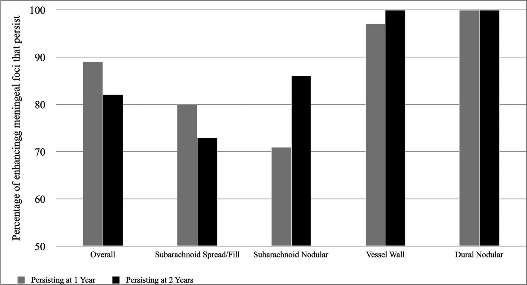
  • Fig 2.
    • Download figure
    • Open in new tab
    • Download powerpoint
    Fig 2.

    Graph displaying the percentages of baseline enhancing meningeal foci that persist 1 year later (gray bars) and 2 years later (black bars). All 31 participants were scanned at baseline and at 1 year, but 2-year data are limited to 15 participants. At 1 year, persistence was noted in 253/284 (89%) overall foci, 91/114 (80%) subarachnoid spread/fill foci, 10/14 (71%) subarachnoid nodular foci, 104/107 (97%) vessel wall foci, and 46/46 (100%) dural nodular foci. At 2 years, persistence was noted in 132/161 (82%) overall foci, 45/62 (73%) subarachnoid spread/fill foci, 6/7 (86%) subarachnoid nodular foci, 55/55 (100%) vessel wall foci, and 34/34 (100%) dural foci.

  • Fig 3.
    • Download figure
    • Open in new tab
    • Download powerpoint
    Fig 3.

    Examples of persisting foci of meningeal enhancement on delayed postcontrast FLAIR at 7T. Sagittal reformatted images show subarachnoid spread/fill enhancement that persists from December 15, 2015 (A), to March 3, 2017 (B), in a 58-year-old woman with relapsing-remitting MS. Axial images show subarachnoid nodular enhancement that persists from October 8, 2014 (C), to March 3, 2017 (D), in a 49-year-old man with relapsing-remitting MS. Axial images show vessel wall enhancement that persists from March 14, 2016 (E), to April 4, 2017 (F), in a 57-year-old man with primary-progressive MS. Axial images show dural enhancement that persists from May 9, 2016 (G), to May 31, 2017 (H), in a 44-year-old woman with secondary-progressive MS. Note that no intrinsic signal was observed in these locations on precontrast acquisitions (not shown).

  • Fig 4.
    • Download figure
    • Open in new tab
    • Download powerpoint
    Fig 4.

    Examples of resolving foci of meningeal enhancement on delayed postcontrast FLAIR at 7T. Coronal reformatted images show subarachnoid spread/fill enhancement that resolves between October 23, 2014 (A), and February 26, 2016 (B), in a 49-year-old woman with relapsing-remitting MS. Coronal reformatted images show subarachnoid nodular enhancement within the cerebellar folia that resolves between October 8, 2014 (C), and February 19, 2016 (D), in a 49-year-old man with relapsing-remitting MS. Sagittal formatted images show vessel wall enhancement that resolves from May 9, 2016 (E), to May 31, 2017 (F), in a 44-year-old woman with secondary-progressive MS. Note that no foci of meningeal enhancement classified as a dural subtype resolved in this study.

Tables

  • Figures
    • View popup
    Table 1:

    MRI sequence parameters

    SequenceResolution (mm)TRTITEParallel ImagingFlip AngleTime (min:sec)
    MP2RAGE0.7 × 0.7 × 0.7TRvolume = 8.25 sTI1 = 1 s1.97 msSENSE = 2 × 2FA1 = 7°9:46
    TRTFE = 6.9 msTI2 = 3.3 sFA2 = 5°
    MPFLAIR0.7 × 0.7 × 0.78000 ms2077 ms400 msSENSE = 2 × 390°10:48
    • Note:—SENSE indicates sensitivity encoding; MPFLAIR, magnetization-prepared FLAIR; FA, flip angle; TFE, turbo field echo.

    • View popup
    Table 2:

    Cohort baseline characteristicsa

    Characteristics
    Age at enrollment (yr)49 (26–61)
    Sex11/31 Men (35%), 20/31 women (65%)
    Disease subtype at enrollment21/31 (68%) RR, 7/31 (23%) SP,
    3/31 (10%) PP
    Disease duration at enrollment (mo)109 (8–461)
    Patients with new relapses in past 30 days1/31 (3%)
    No. of relapses in past year per subject0 (0–3)
    Modified Fatigue Impact Scale score at enrollment43 (0–78)
    Symbol Digit Modality Test at enrollment50 (35–81)
    Expanded Disability Status Scale score at enrollment3 (1–6.5)
    Immunomodulatory treatment status at baseline
        On treatment25/31 (81%)
        Not on treatment6/31 (19%)
    Treatment type at baseline
        Interferon3/25 (12%)
        Glatiramer6/25 (24%)
        Natalizumab2/25 (8%)
        Teriflunomide1/25 (4%)
        Fingolimod4/25 (16%)
        Dimethyl fumarate9/25 (36%)
    No. of subjects who switched between disease-modifying therapies from baseline to follow-up scans10/31
    • Note:—RR indicates relapsing-remitting MS; SP, secondary-progressive MS; PP, primary-progressive MS.

    • ↵a Median values are shown with the range of observed values in parentheses.

    • View popup
    Table 3:

    Anatomic distribution within the brain of enhancing meningeal foci at baseline

    Brain RegionNo. of Foci at BaselinePercentage of Foci at Baseline
    Right frontal6021.1
    Left frontal6422.5
    Right parietal4415.5
    Left parietal4415.5
    Right occipital207.0
    Left occipital248.4
    Right temporal165.6
    Left temporal82.8
    Right cerebellum20.7
    Left cerebellum20.7
    • View popup
    Table 4:

    Wilcoxon rank sum test for longitudinal persistence of meningeal enhancement versus demographic and clinical factorsa

    On Treatment (n = 25)Not on Treatment (n = 6)Progressive MS (n = 10)Nonprogressive MS (n = 21)EDSS Progressor at 1 yrb (n = 7)EDSS Nonprogressor at 1 yrb (n = 24)
    Total No. of overall foci persisting at 1 yr per subject9 (1–24)9 (1–15)8 (1–24)9 (1–15)12 (1–15)7.5 (1–24)c
    Total No. of subarachnoid spread/fill foci persisting at 1 yr per subject2 (0–9)2 (0–6)2 (0–9)2 (0–9)5 (1–9)2 (0–9)
    Total No. of subarachnoid nodular foci persisting at 1 yr per subject0 (0–2)0 (0–1)0 (0–0)0 (0–2)c0 (0–0)0 (0–2)
    Total No. of vessel wall foci persisting at 1 yr per subject3 (0–11)3.5 (1–6)2 (0–6)3 (0–11)3 (0–11)3 (0–7)
    Total No. of dural foci persisting at 1 yr per subject1 (0–9)1 (0–6)1 (0–9)1 (0–6)1 (0–6)1 (0–9)
    • ↵a Median values are listed with the range of observed values in parentheses.

    • ↵b Criteria for EDSS progressor status is listed in the “Materials and Methods” section.

    • ↵c P < .05.

PreviousNext
Back to top

In this issue

American Journal of Neuroradiology: 39 (10)
American Journal of Neuroradiology
Vol. 39, Issue 10
1 Oct 2018
  • Table of Contents
  • Index by author
  • Complete Issue (PDF)
Advertisement
Print
Download PDF
Email Article

Thank you for your interest in spreading the word on American Journal of Neuroradiology.

NOTE: We only request your email address so that the person you are recommending the page to knows that you wanted them to see it, and that it is not junk mail. We do not capture any email address.

Enter multiple addresses on separate lines or separate them with commas.
Longitudinal Persistence of Meningeal Enhancement on Postcontrast 7T 3D-FLAIR MRI in Multiple Sclerosis
(Your Name) has sent you a message from American Journal of Neuroradiology
(Your Name) thought you would like to see the American Journal of Neuroradiology web site.
CAPTCHA
This question is for testing whether or not you are a human visitor and to prevent automated spam submissions.
Cite this article
S.N. Jonas, I. Izbudak, A.A. Frazier, D.M. Harrison
Longitudinal Persistence of Meningeal Enhancement on Postcontrast 7T 3D-FLAIR MRI in Multiple Sclerosis
American Journal of Neuroradiology Oct 2018, 39 (10) 1799-1805; DOI: 10.3174/ajnr.A5796

Citation Manager Formats

  • BibTeX
  • Bookends
  • EasyBib
  • EndNote (tagged)
  • EndNote 8 (xml)
  • Medlars
  • Mendeley
  • Papers
  • RefWorks Tagged
  • Ref Manager
  • RIS
  • Zotero
0 Responses
Respond to this article
Share
Bookmark this article
Longitudinal Persistence of Meningeal Enhancement on Postcontrast 7T 3D-FLAIR MRI in Multiple Sclerosis
S.N. Jonas, I. Izbudak, A.A. Frazier, D.M. Harrison
American Journal of Neuroradiology Oct 2018, 39 (10) 1799-1805; DOI: 10.3174/ajnr.A5796
del.icio.us logo Twitter logo Facebook logo Mendeley logo
  • Tweet Widget
  • Facebook Like
  • Google Plus One
Purchase

Jump to section

  • Article
    • Abstract
    • ABBREVIATIONS:
    • Materials and Methods
    • Results
    • Discussion
    • Conclusions
    • Footnotes
    • References
  • Figures & Data
  • Supplemental
  • Info & Metrics
  • Responses
  • References
  • PDF

Related Articles

  • PubMed
  • Google Scholar

Cited By...

  • Meningeal contrast enhancement in multiple sclerosis: assessment of field strength, acquisition delay, and clinical relevance
  • Leptomeningeal Enhancement in Multiple Sclerosis and Other Neurological Diseases: A Systematic Review and Meta-Analysis
  • Leptomeningeal Contrast Enhancement Is Related to Focal Cortical Thinning in Relapsing-Remitting Multiple Sclerosis: A Cross-Sectional MRI Study
  • Crossref (37)
  • Google Scholar

This article has been cited by the following articles in journals that are participating in Crossref Cited-by Linking.

  • 7T MRI cerebral leptomeningeal enhancement is common in relapsing-remitting multiple sclerosis and is associated with cortical and thalamic lesions
    Jonathan Zurawski, Shahamat Tauhid, Renxin Chu, Fariha Khalid, Brian C Healy, Howard L Weiner, Rohit Bakshi
    Multiple Sclerosis Journal 2020 26 2
  • Leptomeningeal enhancement in multiple sclerosis and other neurological diseases: A systematic review and Meta-Analysis
    Benjamin V. Ineichen, Charidimos Tsagkas, Martina Absinta, Daniel S. Reich
    NeuroImage: Clinical 2022 33
  • No association between cortical lesions and leptomeningeal enhancement on 7-Tesla MRI in multiple sclerosis
    Mehrnaz Ighani, Samuel Jonas, Izlem Izbudak, Seongjin Choi, Alfonso Lema-Dopico, Jun Hua, Erin E O’Connor, Daniel M Harrison
    Multiple Sclerosis Journal 2020 26 2
  • Imaging Mechanisms of Disease Progression in Multiple Sclerosis: Beyond Brain Atrophy
    Francesca Bagnato, Susan A. Gauthier, Cornelia Laule, George R. Wayne Moore, Riley Bove, Zhengxin Cai, Julien Cohen‐Adad, Daniel M. Harrison, Eric C. Klawiter, Sarah A. Morrow, Gülin Öz, William D. Rooney, Seth A. Smith, Peter A. Calabresi, Roland G. Henry, Jiwon Oh, Daniel Ontaneda, Daniel Pelletier, Daniel S. Reich, Russell T. Shinohara, Nancy L. Sicotte
    Journal of Neuroimaging 2020 30 3
  • Determinants and Biomarkers of Progression Independent of Relapses in Multiple Sclerosis
    Massimiliano Calabrese, Paolo Preziosa, Antonio Scalfari, Elisa Colato, Damiano Marastoni, Martina Absinta, Marco Battaglini, Nicola De Stefano, Massimiliano Di Filippo, Simon Hametner, Owain W. Howell, Matilde Inglese, Hans Lassmann, Roland Martin, Richard Nicholas, Richard Reynolds, Maria A. Rocca, Agnese Tamanti, Marco Vercellino, Luisa Maria Villar, Massimo Filippi, Roberta Magliozzi
    Annals of Neurology 2024 96 1
  • Deciphering Multiple Sclerosis Progression
    Virginia Meca-Lallana, Leticia Berenguer-Ruiz, Joan Carreres-Polo, Sara Eichau-Madueño, Jaime Ferrer-Lozano, Lucía Forero, Yolanda Higueras, Nieves Téllez Lara, Angela Vidal-Jordana, Francisco Carlos Pérez-Miralles
    Frontiers in Neurology 2021 12
  • Effect of ocrelizumab on leptomeningeal inflammation and humoral response to Epstein-Barr virus in multiple sclerosis. A pilot study
    Robert Zivadinov, Dejan Jakimovski, Murali Ramanathan, Ralph HB. Benedict, Niels Bergsland, Michael G. Dwyer, Bianca Weinstock-Guttman
    Multiple Sclerosis and Related Disorders 2022 67
  • Use of a Commercial 7-T MRI Scanner for Clinical Brain Imaging: Indications, Protocols, Challenges, and Solutions—A Single-Center Experience
    Can Özütemiz, Matthew White, Wendy Elvendahl, Yigitcan Eryaman, Małgorzata Marjańska, Gregory J. Metzger, Rémi Patriat, Jeramy Kulesa, Noam Harel, Yoichi Watanabe, Andrea Grant, Guglielmo Genovese, Zuzan Cayci
    American Journal of Roentgenology 2023 221 6
  • Leptomeningeal, dura mater and meningeal vessel wall enhancements in multiple sclerosis
    Franziska E. Hildesheim, Deepa P. Ramasamy, Niels Bergsland, Dejan Jakimovski, Michael G. Dwyer, David Hojnacki, Alexis A. Lizarraga, Channa Kolb, Svetlana Eckert, Bianca Weinstock-Guttman, Robert Zivadinov
    Multiple Sclerosis and Related Disorders 2021 47
  • CSF enhancement on post-contrast fluid-attenuated inversion recovery images; a systematic review
    Whitney M. Freeze, Merel van der Thiel, Jeroen de Bresser, Catharina J.M. Klijn, Ellis S. van Etten, Jacobus F.A. Jansen, Louise van der Weerd, Heidi I.L. Jacobs, Walter H. Backes, Susanne J. van Veluw
    NeuroImage: Clinical 2020 28

More in this TOC Section

  • Diagnostic Neuroradiology of Monoclonal Antibodies
  • Clinical Outcomes After Chiari I Decompression
  • Segmentation of Brain Metastases with BLAST
Show more Adult Brain

Similar Articles

Advertisement

Indexed Content

  • Current Issue
  • Accepted Manuscripts
  • Article Preview
  • Past Issues
  • Editorials
  • Editor's Choice
  • Fellows' Journal Club
  • Letters to the Editor
  • Video Articles

Cases

  • Case Collection
  • Archive - Case of the Week
  • Archive - Case of the Month
  • Archive - Classic Case

Special Collections

  • AJNR Awards
  • ASNR Foundation Special Collection
  • Most Impactful AJNR Articles
  • Photon-Counting CT
  • Spinal CSF Leak Articles (Jan 2020-June 2024)

More from AJNR

  • Trainee Corner
  • Imaging Protocols
  • MRI Safety Corner

Multimedia

  • AJNR Podcasts
  • AJNR Scantastics

Resources

  • Turnaround Time
  • Submit a Manuscript
  • Submit a Video Article
  • Submit an eLetter to the Editor/Response
  • Manuscript Submission Guidelines
  • Statistical Tips
  • Fast Publishing of Accepted Manuscripts
  • Graphical Abstract Preparation
  • Imaging Protocol Submission
  • Evidence-Based Medicine Level Guide
  • Publishing Checklists
  • Author Policies
  • Become a Reviewer/Academy of Reviewers
  • News and Updates

About Us

  • About AJNR
  • Editorial Board
  • Editorial Board Alumni
  • Alerts
  • Permissions
  • Not an AJNR Subscriber? Join Now
  • Advertise with Us
  • Librarian Resources
  • Feedback
  • Terms and Conditions
  • AJNR Editorial Board Alumni

American Society of Neuroradiology

  • Not an ASNR Member? Join Now

© 2025 by the American Society of Neuroradiology All rights, including for text and data mining, AI training, and similar technologies, are reserved.
Print ISSN: 0195-6108 Online ISSN: 1936-959X

Powered by HighWire