Advertisement

Main menu

  • Home
  • Content
    • Current Issue
    • Accepted Manuscripts
    • Article Preview
    • Past Issue Archive
    • Video Articles
    • AJNR Case Collection
    • Case of the Week Archive
    • Case of the Month Archive
    • Classic Case Archive
  • Special Collections
    • AJNR Awards
    • ASNR Foundation Special Collection
    • Most Impactful AJNR Articles
    • Photon-Counting CT
    • Spinal CSF Leak Articles (Jan 2020-June 2024)
  • Multimedia
    • AJNR Podcasts
    • AJNR SCANtastic
    • Trainee Corner
    • MRI Safety Corner
    • Imaging Protocols
  • For Authors
    • Submit a Manuscript
    • Submit a Video Article
    • Submit an eLetter to the Editor/Response
    • Manuscript Submission Guidelines
    • Statistical Tips
    • Fast Publishing of Accepted Manuscripts
    • Graphical Abstract Preparation
    • Imaging Protocol Submission
    • Author Policies
  • About Us
    • About AJNR
    • Editorial Board
    • Editorial Board Alumni
  • More
    • Become a Reviewer/Academy of Reviewers
    • Subscribers
    • Permissions
    • Alerts
    • Feedback
    • Advertisers
    • ASNR Home

User menu

  • Alerts
  • Log in

Search

  • Advanced search
American Journal of Neuroradiology

American Journal of Neuroradiology

ASHNR American Society of Functional Neuroradiology ASHNR American Society of Pediatric Neuroradiology ASSR
  • Alerts
  • Log in

Advanced Search

  • Home
  • Content
    • Current Issue
    • Accepted Manuscripts
    • Article Preview
    • Past Issue Archive
    • Video Articles
    • AJNR Case Collection
    • Case of the Week Archive
    • Case of the Month Archive
    • Classic Case Archive
  • Special Collections
    • AJNR Awards
    • ASNR Foundation Special Collection
    • Most Impactful AJNR Articles
    • Photon-Counting CT
    • Spinal CSF Leak Articles (Jan 2020-June 2024)
  • Multimedia
    • AJNR Podcasts
    • AJNR SCANtastic
    • Trainee Corner
    • MRI Safety Corner
    • Imaging Protocols
  • For Authors
    • Submit a Manuscript
    • Submit a Video Article
    • Submit an eLetter to the Editor/Response
    • Manuscript Submission Guidelines
    • Statistical Tips
    • Fast Publishing of Accepted Manuscripts
    • Graphical Abstract Preparation
    • Imaging Protocol Submission
    • Author Policies
  • About Us
    • About AJNR
    • Editorial Board
    • Editorial Board Alumni
  • More
    • Become a Reviewer/Academy of Reviewers
    • Subscribers
    • Permissions
    • Alerts
    • Feedback
    • Advertisers
    • ASNR Home
  • Follow AJNR on Twitter
  • Visit AJNR on Facebook
  • Follow AJNR on Instagram
  • Join AJNR on LinkedIn
  • RSS Feeds

AJNR is seeking candidates for the AJNR Podcast Editor. Read the position description.

Research ArticleAdult Brain
Open Access

Sequential Apparent Diffusion Coefficient for Assessment of Tumor Progression in Patients with Low-Grade Glioma

I.E. Chen, N. Swinburne, N.M. Tsankova, M.M. Hefti, A. Aggarwal, A.H. Doshi, A. Hormigo, B.N. Delman and K. Nael
American Journal of Neuroradiology June 2018, 39 (6) 1039-1046; DOI: https://doi.org/10.3174/ajnr.A5639
I.E. Chen
aFrom the Departments of Radiology (I.E.C., N.S., A.A., A.H.D., B.N.D., K.N.)
  • Find this author on Google Scholar
  • Find this author on PubMed
  • Search for this author on this site
  • ORCID record for I.E. Chen
N. Swinburne
aFrom the Departments of Radiology (I.E.C., N.S., A.A., A.H.D., B.N.D., K.N.)
  • Find this author on Google Scholar
  • Find this author on PubMed
  • Search for this author on this site
  • ORCID record for N. Swinburne
N.M. Tsankova
bPathology (N.M.T., M.M.H.)
  • Find this author on Google Scholar
  • Find this author on PubMed
  • Search for this author on this site
  • ORCID record for N.M. Tsankova
M.M. Hefti
bPathology (N.M.T., M.M.H.)
  • Find this author on Google Scholar
  • Find this author on PubMed
  • Search for this author on this site
  • ORCID record for M.M. Hefti
A. Aggarwal
aFrom the Departments of Radiology (I.E.C., N.S., A.A., A.H.D., B.N.D., K.N.)
  • Find this author on Google Scholar
  • Find this author on PubMed
  • Search for this author on this site
  • ORCID record for A. Aggarwal
A.H. Doshi
aFrom the Departments of Radiology (I.E.C., N.S., A.A., A.H.D., B.N.D., K.N.)
  • Find this author on Google Scholar
  • Find this author on PubMed
  • Search for this author on this site
  • ORCID record for A.H. Doshi
A. Hormigo
cNeurology (A.H.), Icahn School of Medicine at Mount Sinai, New York, New York.
  • Find this author on Google Scholar
  • Find this author on PubMed
  • Search for this author on this site
  • ORCID record for A. Hormigo
B.N. Delman
aFrom the Departments of Radiology (I.E.C., N.S., A.A., A.H.D., B.N.D., K.N.)
  • Find this author on Google Scholar
  • Find this author on PubMed
  • Search for this author on this site
  • ORCID record for B.N. Delman
K. Nael
aFrom the Departments of Radiology (I.E.C., N.S., A.A., A.H.D., B.N.D., K.N.)
  • Find this author on Google Scholar
  • Find this author on PubMed
  • Search for this author on this site
  • ORCID record for K. Nael
  • Article
  • Figures & Data
  • Info & Metrics
  • Responses
  • References
  • PDF
Loading

Article Figures & Data

Figures

  • Tables
  • Fig 1.
    • Download figure
    • Open in new tab
    • Download powerpoint
    Fig 1.

    A VOI was generated using a voxel-based signal intensity threshold method subsuming the entire region of FLAIR hyperintensity. Using coregistered images, we transferred the VOI to ADC for quantitative ADC analysis.
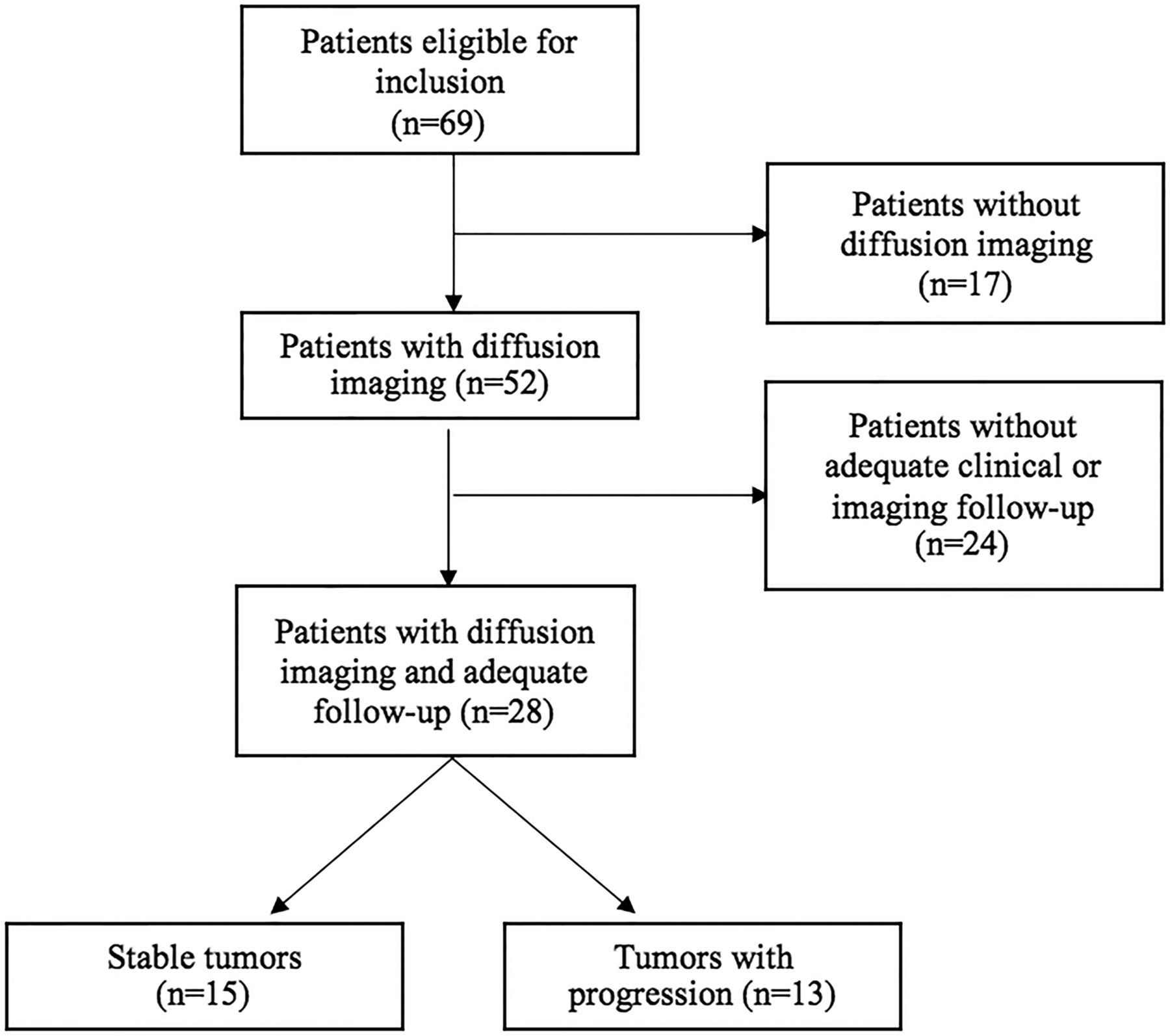
  • Fig 2.
    • Download figure
    • Open in new tab
    • Download powerpoint
    Fig 2.

    Study flow diagram.

  • Fig 3.
    • Download figure
    • Open in new tab
    • Download powerpoint
    Fig 3.

    A 34-year-old woman diagnosed with grade II oligodendroglioma. A, Axial FLAIR/ADC images from 4 sequential MR images are shown (upper row: FLAIR; lower row: ADC; from left to right: first scan after initial diagnosis and at 5, 9, and 14 months). Review of FLAIR images shows stable extent of tumor during the entire follow-up. B, Plotting the normalized ADC10 values demonstrates no significant decrease in ADC10 values, concordant with stability of the tumor.

  • Fig 4.
    • Download figure
    • Open in new tab
    • Download powerpoint
    Fig 4.
    • Download figure
    • Open in new tab
    • Download powerpoint
    Fig 4.

    A 73-year-old woman diagnosed with a grade II oligodendroglioma. A, Axial FLAIR/ADC images from 4 sequential scans are shown (upper row: FLAIR; lower row: ADC; from left to right: first scan after initial diagnosis and at 4, 8, and 28 months). Disease progression was diagnosed at 28 months on conventional imaging and confirmed by surgical pathology. B, Both normalized ADC10 and ADCmean ratios are plotted, demonstrating an interval decrease of ADC10 as early as 4 months following baseline examination, indicating eventual tumor progression. C, Histograms of quantitative normalized ratios of ADC10 and ADCmean for each MR imaging examination.

Tables

  • Figures
    • View popup
    Table 1:

    Baseline and clinical data in patients with and without progression—univariate analysis

    Patient CharacteristicsTotal (n = 28)Progressed (n = 13)Stable (n = 15)P Value
    Age (mean) (SD) (yr)50.4 (16.6)49.1 (19.0)51.6 (14.8).70
    Sex, male/female14:147:67:8.56
    Initial tumor size (mean) (SD) (mL)30.7 (33.8)32.3 (26.3)29.4 (40.1).82
    Tumor type, oligo/astro19/99/410/5.80
    Follow-up time (mean) (range) (mo)40.9 (12–109)42.8 (12–89)39.3 (12–109).75
    No. of MRIs/patient (mean) (SD)8.8 (4.3)9.8 (5.0)7.9 (3.6).26
    Extent of initial tumor resection (No.) (>50% of initial volume)15 (53.5%)8 (61.5%)7 (46.6%).68
    • Note:—oligo indicates oligodendroglioma; astro, astrocytoma.

    • View popup
    Table 2:

    Diagnostic accuracy of ADC interval change—Fisher exact test

    ADCInterval ChangeStable Tumors (n = 15)Progressed (n = 13)P ValueOverall Diagnostic Accuracy
    MeanDecrease7/156/13.850%
    Plateau/increase8/157/13
    10th percentileDecrease3/1512/13<.00186%
    Plateau/increase12/151/13
PreviousNext
Back to top

In this issue

American Journal of Neuroradiology: 39 (6)
American Journal of Neuroradiology
Vol. 39, Issue 6
1 Jun 2018
  • Table of Contents
  • Index by author
  • Complete Issue (PDF)
Advertisement
Print
Download PDF
Email Article

Thank you for your interest in spreading the word on American Journal of Neuroradiology.

NOTE: We only request your email address so that the person you are recommending the page to knows that you wanted them to see it, and that it is not junk mail. We do not capture any email address.

Enter multiple addresses on separate lines or separate them with commas.
Sequential Apparent Diffusion Coefficient for Assessment of Tumor Progression in Patients with Low-Grade Glioma
(Your Name) has sent you a message from American Journal of Neuroradiology
(Your Name) thought you would like to see the American Journal of Neuroradiology web site.
CAPTCHA
This question is for testing whether or not you are a human visitor and to prevent automated spam submissions.
Cite this article
I.E. Chen, N. Swinburne, N.M. Tsankova, M.M. Hefti, A. Aggarwal, A.H. Doshi, A. Hormigo, B.N. Delman, K. Nael
Sequential Apparent Diffusion Coefficient for Assessment of Tumor Progression in Patients with Low-Grade Glioma
American Journal of Neuroradiology Jun 2018, 39 (6) 1039-1046; DOI: 10.3174/ajnr.A5639

Citation Manager Formats

  • BibTeX
  • Bookends
  • EasyBib
  • EndNote (tagged)
  • EndNote 8 (xml)
  • Medlars
  • Mendeley
  • Papers
  • RefWorks Tagged
  • Ref Manager
  • RIS
  • Zotero
0 Responses
Respond to this article
Share
Bookmark this article
Sequential Apparent Diffusion Coefficient for Assessment of Tumor Progression in Patients with Low-Grade Glioma
I.E. Chen, N. Swinburne, N.M. Tsankova, M.M. Hefti, A. Aggarwal, A.H. Doshi, A. Hormigo, B.N. Delman, K. Nael
American Journal of Neuroradiology Jun 2018, 39 (6) 1039-1046; DOI: 10.3174/ajnr.A5639
del.icio.us logo Twitter logo Facebook logo Mendeley logo
  • Tweet Widget
  • Facebook Like
  • Google Plus One
Purchase

Jump to section

  • Article
    • Abstract
    • ABBREVIATIONS:
    • Materials and Methods
    • Results
    • Discussion
    • Conclusions
    • Footnotes
    • REFERENCES
  • Figures & Data
  • Info & Metrics
  • Responses
  • References
  • PDF

Related Articles

  • No related articles found.
  • PubMed
  • Google Scholar

Cited By...

  • No citing articles found.
  • Crossref (8)
  • Google Scholar

This article has been cited by the following articles in journals that are participating in Crossref Cited-by Linking.

  • The Concept of «Peritumoral Zone» in Diffuse Low-Grade Gliomas: Oncological and Functional Implications for a Connectome-Guided Therapeutic Attitude
    Melissa Silva, Catalina Vivancos, Hugues Duffau
    Brain Sciences 2022 12 4
  • Perfusion imaging with arterial spin labeling (ASL)–MRI predicts malignant progression in low‑grade (WHO grade II) gliomas
    Christina M. Flies, Tom J. Snijders, Tom Van Seeters, Marion Smits, Filip Y. F. De Vos, Jeroen Hendrikse, Jan Willem Dankbaar
    Neuroradiology 2021 63 12
  • A Personalized Longitudinal Strategy in Low-Grade Glioma Patients: Predicting Oncological and Neural Interindividual Variability and Its Changes over Years to Think One Step Ahead
    Hugues Duffau
    Journal of Personalized Medicine 2022 12 10
  • Predicting IDH mutation status of lower grade gliomas using standard MR imaging features and apparent diffusion coefficient
    Shuangshuang Song, Jiankun Xu, Leiming Wang, Fan Yu, Yueshan Piao, Miao Zhang, Jie Lu
    Chinese Journal of Academic Radiology 2021 4 3
  • T2–Fluid-Attenuated Inversion Recovery Mismatch Sign in Grade II and III Gliomas: Is There a Coexisting T2–Diffusion-Weighted Imaging Mismatch?
    Niko Radović, Martina Špero, Ana Hrkać Pustahija, Fadi Almahariq, Dubravka Srdoč
    Journal of Computer Assisted Tomography 2022 46 2
  • Congress of Neurological Surgeons systematic review and evidence‑based guidelines on the management of recurrent diffuse low-grade glioma: update
    Kevin Morrow, Andrew Sloan, Jeffrey J. Olson, D. Ryan Ormond
    Journal of Neuro-Oncology 2025 171 1
  • Congress of neurological surgeons systematic review and evidence-based guidelines for the role of imaging in newly diagnosed WHO grade II diffuse glioma in adults: update
    Chaitra Badve, Abraham Nirappel, Simon Lo, Daniel A. Orringer, Jeffrey J. Olson
    Journal of Neuro-Oncology 2025
  • Diagnostic Value/Accuracy of Diffusion-weighted Magnetic Resonance Imaging in Low-grade Glioma: Systematic Review
    Asish Pavanan, Shivali Kashikar, Pratapsingh Hanumansingh Parihar
    Journal of the Scientific Society 2024 51 2

More in this TOC Section

  • Diagnostic Neuroradiology of Monoclonal Antibodies
  • Clinical Outcomes After Chiari I Decompression
  • Segmentation of Brain Metastases with BLAST
Show more Adult Brain

Similar Articles

Advertisement

Indexed Content

  • Current Issue
  • Accepted Manuscripts
  • Article Preview
  • Past Issues
  • Editorials
  • Editor's Choice
  • Fellows' Journal Club
  • Letters to the Editor
  • Video Articles

Cases

  • Case Collection
  • Archive - Case of the Week
  • Archive - Case of the Month
  • Archive - Classic Case

Special Collections

  • AJNR Awards
  • ASNR Foundation Special Collection
  • Most Impactful AJNR Articles
  • Photon-Counting CT
  • Spinal CSF Leak Articles (Jan 2020-June 2024)

More from AJNR

  • Trainee Corner
  • Imaging Protocols
  • MRI Safety Corner

Multimedia

  • AJNR Podcasts
  • AJNR Scantastics

Resources

  • Turnaround Time
  • Submit a Manuscript
  • Submit a Video Article
  • Submit an eLetter to the Editor/Response
  • Manuscript Submission Guidelines
  • Statistical Tips
  • Fast Publishing of Accepted Manuscripts
  • Graphical Abstract Preparation
  • Imaging Protocol Submission
  • Evidence-Based Medicine Level Guide
  • Publishing Checklists
  • Author Policies
  • Become a Reviewer/Academy of Reviewers
  • News and Updates

About Us

  • About AJNR
  • Editorial Board
  • Editorial Board Alumni
  • Alerts
  • Permissions
  • Not an AJNR Subscriber? Join Now
  • Advertise with Us
  • Librarian Resources
  • Feedback
  • Terms and Conditions
  • AJNR Editorial Board Alumni

American Society of Neuroradiology

  • Not an ASNR Member? Join Now

© 2025 by the American Society of Neuroradiology All rights, including for text and data mining, AI training, and similar technologies, are reserved.
Print ISSN: 0195-6108 Online ISSN: 1936-959X

Powered by HighWire