
Article Figures & Data
Figures
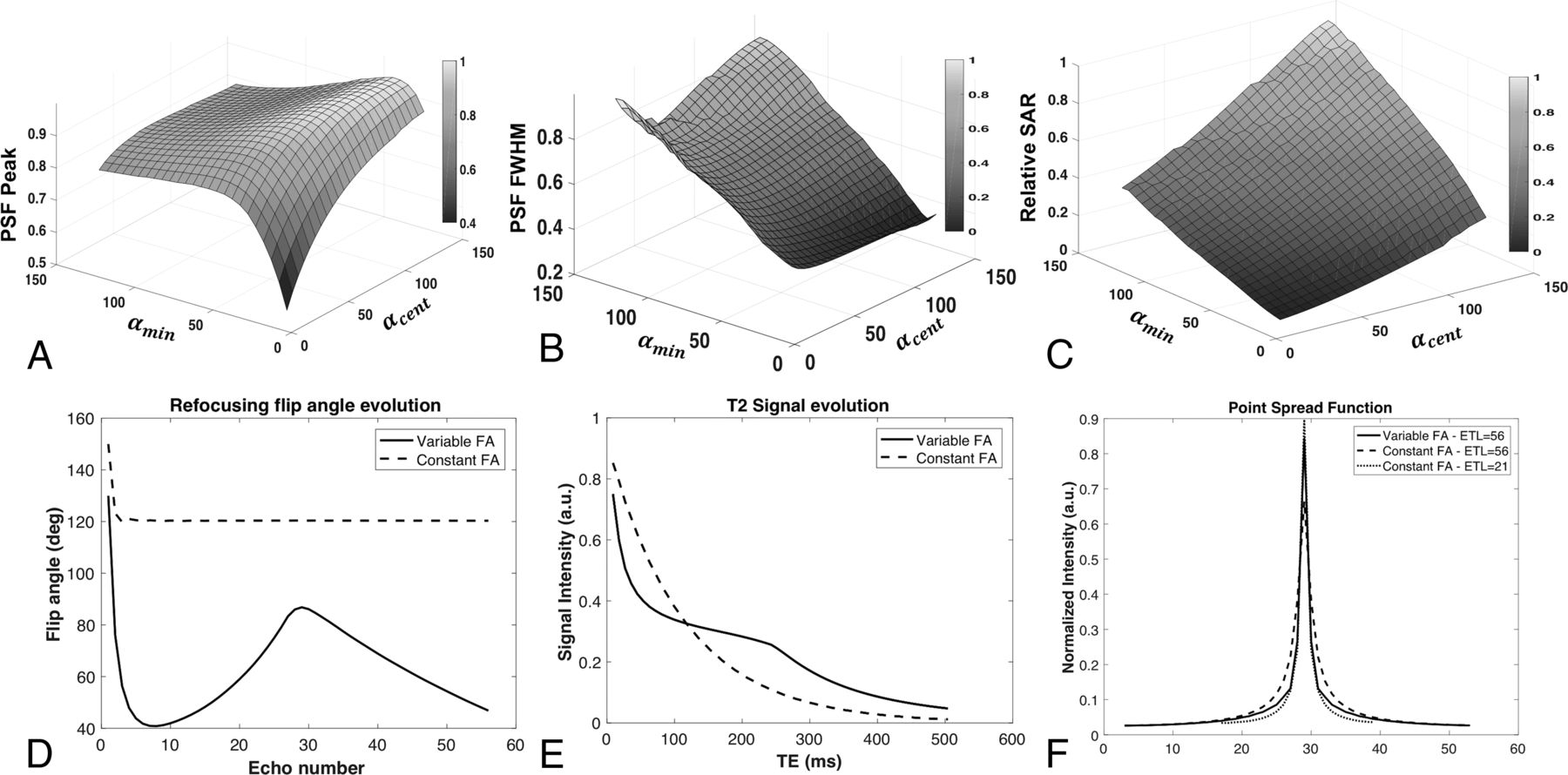
- Fig 1.
Surface plots of the peak and full width at half maximum (FWHM) of the simulated point spread function (PSF) as a function of αmin and αcent are shown in A and B, respectively. C, The relative SAR as a function of the 2 control angles. Note that the computed PSF is maximized at higher values of αcent, however, at the cost of increased SAR. D, The refocusing flip angle modulation scheme for a conventional fast spin-echo and the variable flip angle sequence along with the T2 signal evolution (E). Note that the VFA scheme stabilizes the signal evolution over the echo-train. The point spread functions for the constant and the variable flip angle echo-trains are compared in F. There is a considerable improvement in the PSF with the use of variable refocusing flip angles at longer echo-train lengths, resulting in better spatial resolution and less blurring. FA indicates flip angle; deg, degree; a.u., arbitrary units.
- Fig 2.
Phantom experiments comparing the resolution performance of TSE-VFA. Data were acquired on agarose gel phantoms (A) using the conventional TSE at ETL = 21, ETL = 56, and TSE-VFA at ETL = 56. B, Line plots across the 2 phantoms for the 3 sequences. Note the reduction in ringing when using TSE-VFA at the longer echo-train length of 56. FA indicates flip angle; a.u., arbitrary units.
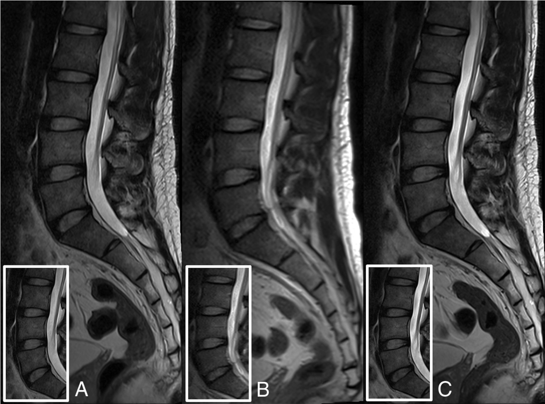
- Fig 3.
Images of the lumbar spine demonstrating better PSF behavior (reduced blurring) with variable flip angle TSE at ETL = 56 (A) compared with conventional TSE at the same ETL (B). C, Image acquired using the TSE sequence at ETL = 21. The TSE-VFA had a lower SAR value of 1.22 compared with the TSE at ETL = 21 (SAR = 1.665). Note that at the effective TE = 105 ms, the contrast between TSE-VFA and the T2-TSE sequence is comparable.
- Fig 4.
Sagittal T2WI TSE (A and C) and TSE-VFA (B and D) images of the lumbar spine for 2 subjects. A and B, The presence of multifocal osseous metastases with a pathologic fracture of L2 (arrowhead). The conventional TSE image (3 min 24 sec scan time) shows aliasing artifacts (dotted arrows) due to motion, which are absent in the TSE-VFA image (1 min 30 sec scan time). C and D, Images are from a patient with multilevel degenerative disc disease and a right subarticular disc protrusion abutting the right L3 nerve root at L2–L3 (white arrow). Note that the small hemangioma within the L1 vertebral body (open arrow) is well-resolved by the TSE-VFA (D). The TSE-VFA image for this subject received a score of 4 for the SNR and the CSF signal compared with a score of 5 for the TSE image.
- Fig 5.
Sagittal STIR TSE (A and C) and sagittal HASTE-VFA (B and D) images of the whole spine for 2 subjects. A and B, Disc protrusions in the lower thoracic spine with several Schmorl nodes in the lumbar spine. C and D, Images from a subject with degenerative disc changes in the lumbar and the lower thoracic spine. Note the increased motion-related artifacts (arrows) with the 9 min 30 sec STIR TSE sequence (A and C) when compared to the single-shot sequence (B and D) with a 1 min 54 sec scan time.


Tables
- Table 1:
Scan parameters used for the conventional TSE and the proposed variable flip angle sequences
Parameters Conventional TSE TSE-VFA HASTE-VFA Resolution (phase × freq) (mm2) 0.81 × 0.73 0.81 × 0.73 1.25 × 1.0 Slice thickness (mm) 3 3 3 Refocusing flip angle 140° αstart = 130° αstart = 130° αmin = 45° αmin = 50° αcent = 110° αcent = 90° αend = 45° αend = 45° Parallel imaging acceleration factor 2 2 1 ETL 21 56 160 TR (ms) 2800 3600 770 Scan time (min) 3 min 25 sec 1 min 28 sec 1 min 57 sec Note:—freq indicates frequency.
Sequence SNR Phantom 1 SNR Phantom 2 SNR Efficiency Phantom 1 SNR Efficiency Phantom 2 Relative Contrast SAR Scan Time (min) Conventional TSE ETL = 21 469.76 248.56 261.38 138.30 0.47 0.24 3 min 13 sec Conventional TSE ETL = 56 412.99 195.29 201.52 95.29 0.46 0.38 4 min 12 sec TSE-VFA ETL = 56 423.54 216.12 338.02 172.48 0.46 0.17 1 min 34 sec Scoring Criteria Mean Score TSE Mean Score TSE-VFA Weighted Gwet AC1 Motion 4.9 ± 0.32 5 ± 0 0.97 Artifacts 5 ± 0 5 ± 0 1 Edge sharpness 5 ± 0 5 ± 0 1 SNR 5 ± 0 4.4 ± 0.52 0.93 Facet joints 5 ± 0 4.8 ± 0.42 1 Endplates 5 ± 0 5 ± 0 1 Nerve roots 5 ± 0 4.8 ± 0.42 1 Spinal cord 5 ± 0 5 ± 0 1 Discs 5 ± 0 5 ± 0 1 Sequence Vertebral Body SNR Vertebral Body SNR Efficiency Vertebrae-Disc Relative Contrast SAR Healthy volunteers TSE 36.08 ± 7.51 19.48 ± 4.05 0.47 ± 0.33 1.69 ± 0.14 TSE-VFA 27.81 ± 4.33 22.94 ± 3.57 0.45 ± 0.32 1.31 ± 0.21 Clinical patients TSE 55.45 ± 18.75 29.93 ± 10.12 0.13 ± 0.69 1.84 ± 0.61 TSE-VFA 45.18 ± 14.96 37.27 ± 12.34 0.12 ± 0.7 1.37 ± 0.34 Scoring Criteria Mean Score TSE Mean Score TSE-VFA P Value of Wilcoxon Testa Weighted Gwet AC1 Motion 4.71 ± 0.59 4.83 ± 0.42 <.001 0.88 Artifacts 4.94 ± 0.23 4.76 ± 0.46 <.001 0.93 Edge sharpness 4.91 ± 0.33 4.79 ± 0.41 <.001 0.93 SNR 4.84 ± 0.40 4.36 ± 0.64b .371b 0.84 Facet joints 4.83 ± 0.40 4.67 ± 0.50 <.001 0.86 Endplates 4.94 ± 0.23 4.83 ± 0.38 <.001 0.94 Nerve roots 4.76 ± 0.49 4.51 ± 0.58 <.001 0.78 Spinal cord 4.73 ± 0.51 4.47 ± 0.61 <.01 0.86 Discs 4.87 ± 0.44 4.80 ± 0.44 <.001 0.91 ↵a The null hypothesis states that the median difference in the image-quality scores between TSE and TSE-VFA is greater than the noninferiority margin Δ, and rejecting the null hypothesis shows noninferiority in performance.
↵b Refers to lack of noninferiority between the TSE-VFA and TSE at a significance level of P < .025.
- Table 6:
Estimate of agreement in diagnostic quality between the conventional TSE and the proposed sequence
Clinical Diagnostic Criteria Lumbar Spine Overall Agreement (%) Positive Agreement (%) Facet joints 100 100 Endplates 100 100 Nerve roots 98.57 98.53 Spinal cord 97.14 97.06 Discs 100 100 Scoring Criteria Mean Score HASTE-VFA Gwet AC1 Interobserver Reliability Motion 4.66 ± 0.48 0.74 Artifacts 4.56 ± 0.50 0.65 Edge sharpness 4.78 ± 0.42 0.94 SNR 4.34 ± 0.75 0.39 Facet joints 4.69 ± 0.47 0.77 Endplates 4.91 ± 0.51 0.98 Nerve roots 4.47 ± 0.57 0.66 Spinal cord 4.72 ± 0.46 0.79 Discs 4.75 ± 0.44 0.91


Jump to section
Related Articles
Cited By...
- No citing articles found.