
Article Figures & Data
Figures
- Fig 1.
Illustration of ROIs on B0 (A–C) and histology (D–F) maps for a representative control and TBI rat. Regions shown are the bilateral cortex, bilateral hippocampus, and corpus callosum.
- Fig 2.
Changes in FA, MD, and MK values for the bilateral cortex (ips, con), bilateral hippocampus (ips, con), and corpus callosum and changes in Da, Dr, Ka, and Kr values for the corpus callosum. The asterisk indicates P < .05, compared with preinjury; hash tag, P < .05, compared with 3 days after TBI; caret, P < .05, compared with 14 days after TBI; ips, ipsilateral; con, contralateral.
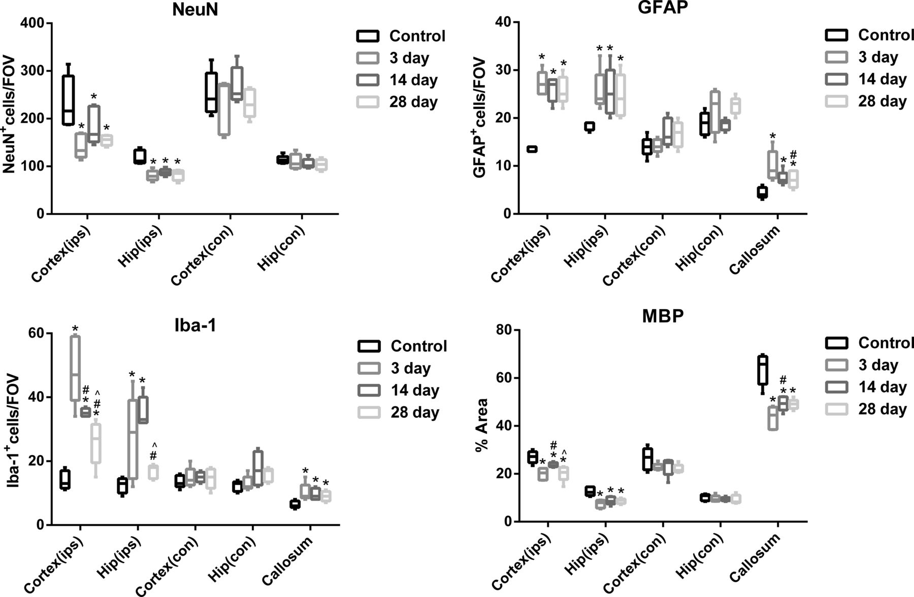
- Fig 3.
Changes in NeuN+, GFAP+, and Iba-1+ cells and MBP area for the bilateral cortex (ips, con), bilateral hippocampus (ips, con), and corpus callosum. Asterisk indicates P < .05, compared with preinjury; hash tag, P < .05, compared with 3 days after TBI; caret, P < .05, compared with 14 days after TBI; ips, ipsilateral; con, contralateral.
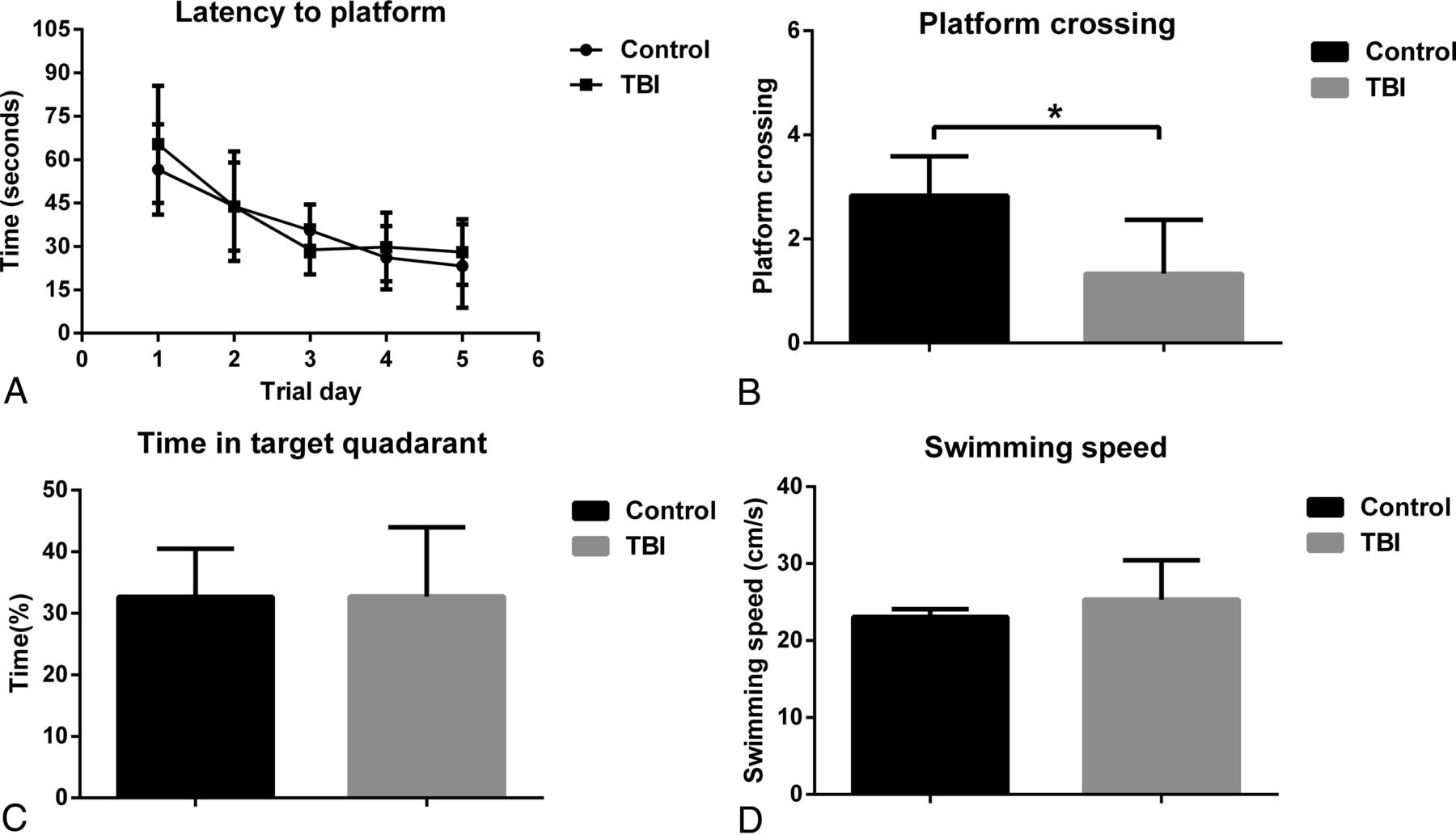
- Fig 4.
The Morris water maze tests results. A, Latency to find the platform. B, Platform-crossing times. C, Time spent in target quadrant. D, The swimming speed. Error bars indicate standard error. Asterisk indicates P < .05.


Jump to section
Related Articles
Cited By...
- A Pilot Study to Establish a Penetrating Traumatic Brain Injury Rat Model for Implantation of a 3D Printed Scaffold
- LONGITUDINAL CHANGES IN WHITE MATTER MICROSTRUCTURAL STATUS FOLLOWING QUANTIFIED HEAD-BALL IMPACTS IN SOCCER: A PRELIMINARY, PROSPECTIVE STUDY
- Neuroanatomical underpinning of diffusion kurtosis measurements in the cerebral cortex of healthy macaque brains
- Longitudinal, Multiparametric MRI Assessment of repetitive mild TBI in rats
- Behavioral and Structural Effects of Single and Repeat Closed-Head Injury