Advertisement

Main menu

  • Home
  • Content
    • Current Issue
    • Accepted Manuscripts
    • Article Preview
    • Past Issue Archive
    • Video Articles
    • AJNR Case Collection
    • Case of the Week Archive
    • Case of the Month Archive
    • Classic Case Archive
  • Special Collections
    • AJNR Awards
    • ASNR Foundation Special Collection
    • Most Impactful AJNR Articles
    • Photon-Counting CT
    • Spinal CSF Leak Articles (Jan 2020-June 2024)
  • Multimedia
    • AJNR Podcasts
    • AJNR SCANtastic
    • Trainee Corner
    • MRI Safety Corner
    • Imaging Protocols
  • For Authors
    • Submit a Manuscript
    • Submit a Video Article
    • Submit an eLetter to the Editor/Response
    • Manuscript Submission Guidelines
    • Statistical Tips
    • Fast Publishing of Accepted Manuscripts
    • Graphical Abstract Preparation
    • Imaging Protocol Submission
    • Author Policies
  • About Us
    • About AJNR
    • Editorial Board
    • Editorial Board Alumni
  • More
    • Become a Reviewer/Academy of Reviewers
    • Subscribers
    • Permissions
    • Alerts
    • Feedback
    • Advertisers
    • ASNR Home

User menu

  • Alerts
  • Log in

Search

  • Advanced search
American Journal of Neuroradiology

American Journal of Neuroradiology

ASHNR American Society of Functional Neuroradiology ASHNR American Society of Pediatric Neuroradiology ASSR
  • Alerts
  • Log in

Advanced Search

  • Home
  • Content
    • Current Issue
    • Accepted Manuscripts
    • Article Preview
    • Past Issue Archive
    • Video Articles
    • AJNR Case Collection
    • Case of the Week Archive
    • Case of the Month Archive
    • Classic Case Archive
  • Special Collections
    • AJNR Awards
    • ASNR Foundation Special Collection
    • Most Impactful AJNR Articles
    • Photon-Counting CT
    • Spinal CSF Leak Articles (Jan 2020-June 2024)
  • Multimedia
    • AJNR Podcasts
    • AJNR SCANtastic
    • Trainee Corner
    • MRI Safety Corner
    • Imaging Protocols
  • For Authors
    • Submit a Manuscript
    • Submit a Video Article
    • Submit an eLetter to the Editor/Response
    • Manuscript Submission Guidelines
    • Statistical Tips
    • Fast Publishing of Accepted Manuscripts
    • Graphical Abstract Preparation
    • Imaging Protocol Submission
    • Author Policies
  • About Us
    • About AJNR
    • Editorial Board
    • Editorial Board Alumni
  • More
    • Become a Reviewer/Academy of Reviewers
    • Subscribers
    • Permissions
    • Alerts
    • Feedback
    • Advertisers
    • ASNR Home
  • Follow AJNR on Twitter
  • Visit AJNR on Facebook
  • Follow AJNR on Instagram
  • Join AJNR on LinkedIn
  • RSS Feeds

AJNR is seeking candidates for the AJNR Podcast Editor. Read the position description.

Research ArticleAdult Brain
Open Access

Volumetry of Mesiotemporal Structures Reflects Serostatus in Patients with Limbic Encephalitis

L. Ernst, B. David, J. Gaubatz, I. Domínguez-Narciso, G. Lüchters, A.J. Becker, B. Weber, E. Hattingen, C.E. Elger and T. Rüber
American Journal of Neuroradiology December 2019, 40 (12) 2081-2089; DOI: https://doi.org/10.3174/ajnr.A6289
L. Ernst
aFrom the Department of Epileptology (L.E., B.D., J.G., I.D.-N., C.E.E., T.R.)
  • Find this author on Google Scholar
  • Find this author on PubMed
  • Search for this author on this site
  • ORCID record for L. Ernst
B. David
aFrom the Department of Epileptology (L.E., B.D., J.G., I.D.-N., C.E.E., T.R.)
  • Find this author on Google Scholar
  • Find this author on PubMed
  • Search for this author on this site
  • ORCID record for B. David
J. Gaubatz
aFrom the Department of Epileptology (L.E., B.D., J.G., I.D.-N., C.E.E., T.R.)
  • Find this author on Google Scholar
  • Find this author on PubMed
  • Search for this author on this site
  • ORCID record for J. Gaubatz
I. Domínguez-Narciso
aFrom the Department of Epileptology (L.E., B.D., J.G., I.D.-N., C.E.E., T.R.)
  • Find this author on Google Scholar
  • Find this author on PubMed
  • Search for this author on this site
  • ORCID record for I. Domínguez-Narciso
G. Lüchters
eCenter for Development Research (G.L.), University of Bonn, Bonn, Germany
  • Find this author on Google Scholar
  • Find this author on PubMed
  • Search for this author on this site
  • ORCID record for G. Lüchters
A.J. Becker
bDepartment of Neuropathology (A.J.B.)
  • Find this author on Google Scholar
  • Find this author on PubMed
  • Search for this author on this site
  • ORCID record for A.J. Becker
B. Weber
cInstitute for Experimental Epileptology and Cognition Research (B.W.)
  • Find this author on Google Scholar
  • Find this author on PubMed
  • Search for this author on this site
  • ORCID record for B. Weber
E. Hattingen
dDepartment of Radiology (E.H.), University of Bonn Medical Center, Bonn, Germany
fDepartment of Neuroradiology (E.H.), Goethe University Frankfurt, Frankfurt, Germany
  • Find this author on Google Scholar
  • Find this author on PubMed
  • Search for this author on this site
  • ORCID record for E. Hattingen
C.E. Elger
aFrom the Department of Epileptology (L.E., B.D., J.G., I.D.-N., C.E.E., T.R.)
  • Find this author on Google Scholar
  • Find this author on PubMed
  • Search for this author on this site
  • ORCID record for C.E. Elger
T. Rüber
aFrom the Department of Epileptology (L.E., B.D., J.G., I.D.-N., C.E.E., T.R.)
gEpilepsy Center Frankfurt Rhine-Main (T.R.)
hDepartment of Neurology, and Center for Personalized Translational Epilepsy Research (T.R.), Goethe-University Frankfurt, Frankfurt am Main, Germany.
  • Find this author on Google Scholar
  • Find this author on PubMed
  • Search for this author on this site
  • ORCID record for T. Rüber
  • Article
  • Figures & Data
  • Supplemental
  • Info & Metrics
  • Responses
  • References
  • PDF
Loading

Article Figures & Data

Figures

  • Tables
  • Fig 1.
    • Download figure
    • Open in new tab
    • Download powerpoint
    Fig 1.

    Volumetry of the amygdala and hippocampus. A, Volumetry in the early group. B, Volumetry in the late group. Asterisks represent levels of significance (single asterisk indicates .05; double asterisks, .01).

  • Fig 2.
    • Download figure
    • Open in new tab
    • Download powerpoint
    Fig 2.

    Starplots showing hippocampal subfield volume differences between patients in the early group and controls. Z values reflect the hippocampal subfield volume difference between patients in the early group and controls in the affected and the unaffected hemispheric sides. Underlined words indicate statistical significance of the respective hippocampal subfield (P < .05) in post hoc tests between patients and controls following a multivariate linear model (see MATERIALS AND METHODS and supporting information in On-line Tables 3–8). A, Relative hippocampal subfield volumes of the affected and unaffected sides of GAD-LE. B, Relative hippocampal subfield volumes of the affected and unaffected sides of VGKC-LE. GC-ML-DG indicates granule cell layer of the dentate gyrus; HATA, hippocampus-amygdala transition area.

  • Fig 3.
    • Download figure
    • Open in new tab
    • Download powerpoint
    Fig 3.

    Decision tree classification. Decision tree classification between LGI1 and GAD using hippocampal subfield volumes from the hemisphere affected in the EEG in early-stage LE. A, Decision tree with importance of features. B, Histogram showing the number of label-shift permutations for each accuracy bin. The solid red line displays a fitted Gaussian curve describing the underlying probability density function. The dashed purple line marks the classifier performance on the real data. C, Starplot showing Z values of the hippocampal subfield volume differences between LGI1-LE and GAD-LE in the affected hemispheric side (see supporting information in On-line Tables 3 and 5 for statistics). HATA indicates hippocampus-amygdala transition area; GC-ML-DG, granule cell layer of the dentate gyrus.
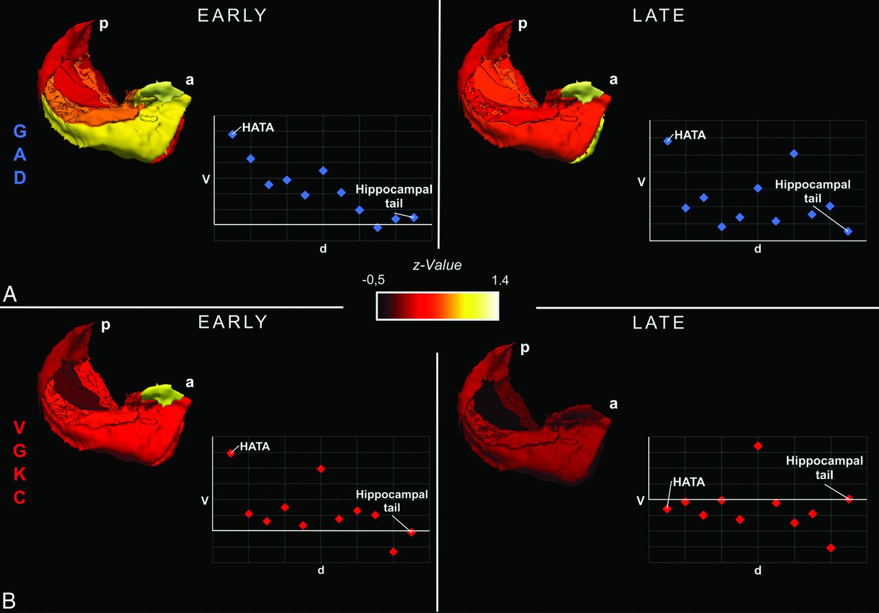
  • Fig 4.
    • Download figure
    • Open in new tab
    • Download powerpoint
    Fig 4.

    3D visualization showing hippocampal subfield volume differences between patients and controls. 3D visualization of group differences between all GAD–LE and controls (A) as well as between VGKC-LE and controls (B) using hippocampal subfield volumes from the affected hemisphere in early and late stages. Schematic scatterplots show Z values of volumes as a function of the approximate spatial distance from the amygdala. P indicates posterior; a, anterior; d, distance; V, volume. Please note the anterior-to-posterior gradient of subfield volume alterations in early GAD-LE and how the gradient tapers off in late GAD-LE.

Tables

  • Figures
    • View popup
    Table 1:

    Demographic and clinical characteristics of subgroups of patients with limbic encephalitis and their corresponding control groups

    GAD-LEVGKC-LEGAD-CONVGKC-CON
    Early group
     No. (men)23 (7)25 (16)23 (7)25 (16)
     Age at MR imaging (mean) (yr)34.2 ± 11.259.5 ± 15.235.4 ± 10.657.2 ± 13.2
     Time between onseta and scan (mean) (mo)9.5 ± 7.47.6 ± 6.1NANA
     Interictal EEG lateralization5/12/1/57/8/2/8NANA
    (n = right/left/bilateral/unclear)
     No. of bilateral mesiotemporal FLAIR-T2-hyperintensities25NANA
     No. of first-line immunotherapies39NANA
     No. of second-line immunotherapies01NANA
    Late group
     No. (men)b33 (10)22 (10)33 (10)22 (10)
     Age at MR imaging (mean) (yr)33.9 ± 12.357.0 ± 16.834.4 ± 11.953.2 ± 13.2
     Time between onseta and scan (mean) (mo)62.5 ± 26.261.5 ± 23.6NANA
     Interictal EEG lateralization5/11/3/124/8/1/9NANA
    (n = right/left/bilateral/unclear)
     No. of bilateral mesiotemporal FLAIR-T2-hyperintensities43NANA
     No. of first-line immunotherapies1418NANA
     No. of second-line immunotherapies64NANA
    • Note:—NA indicates not applicable; CON, matched healthy control group.

    • ↵a Onset of the first limbic encephalitis–related symptom.

    • ↵b Patients scanned repeatedly were included in both (early and late) study groups (repeat scans GAD-LE: n = 10; repeat scans VGKC-LE, n = 13).

PreviousNext
Back to top

In this issue

American Journal of Neuroradiology: 40 (12)
American Journal of Neuroradiology
Vol. 40, Issue 12
1 Dec 2019
  • Table of Contents
  • Index by author
  • Complete Issue (PDF)
Advertisement
Print
Download PDF
Email Article

Thank you for your interest in spreading the word on American Journal of Neuroradiology.

NOTE: We only request your email address so that the person you are recommending the page to knows that you wanted them to see it, and that it is not junk mail. We do not capture any email address.

Enter multiple addresses on separate lines or separate them with commas.
Volumetry of Mesiotemporal Structures Reflects Serostatus in Patients with Limbic Encephalitis
(Your Name) has sent you a message from American Journal of Neuroradiology
(Your Name) thought you would like to see the American Journal of Neuroradiology web site.
CAPTCHA
This question is for testing whether or not you are a human visitor and to prevent automated spam submissions.
Cite this article
L. Ernst, B. David, J. Gaubatz, I. Domínguez-Narciso, G. Lüchters, A.J. Becker, B. Weber, E. Hattingen, C.E. Elger, T. Rüber
Volumetry of Mesiotemporal Structures Reflects Serostatus in Patients with Limbic Encephalitis
American Journal of Neuroradiology Dec 2019, 40 (12) 2081-2089; DOI: 10.3174/ajnr.A6289

Citation Manager Formats

  • BibTeX
  • Bookends
  • EasyBib
  • EndNote (tagged)
  • EndNote 8 (xml)
  • Medlars
  • Mendeley
  • Papers
  • RefWorks Tagged
  • Ref Manager
  • RIS
  • Zotero
0 Responses
Respond to this article
Share
Bookmark this article
Volumetry of Mesiotemporal Structures Reflects Serostatus in Patients with Limbic Encephalitis
L. Ernst, B. David, J. Gaubatz, I. Domínguez-Narciso, G. Lüchters, A.J. Becker, B. Weber, E. Hattingen, C.E. Elger, T. Rüber
American Journal of Neuroradiology Dec 2019, 40 (12) 2081-2089; DOI: 10.3174/ajnr.A6289
del.icio.us logo Twitter logo Facebook logo Mendeley logo
  • Tweet Widget
  • Facebook Like
  • Google Plus One
Purchase

Jump to section

  • Article
    • Abstract
    • ABBREVIATIONS:
    • MATERIALS AND METHODS
    • RESULTS
    • DISCUSSION
    • CONCLUSIONS
    • Acknowledgments
    • Footnotes
    • References
  • Figures & Data
  • Supplemental
  • Info & Metrics
  • Responses
  • References
  • PDF

Related Articles

  • PubMed
  • Google Scholar

Cited By...

  • Mesiotemporal Volumetry, Cortical Thickness, and Neuropsychological Deficits in the Long-term Course of Limbic Encephalitis
  • Crossref (6)
  • Google Scholar

This article has been cited by the following articles in journals that are participating in Crossref Cited-by Linking.

  • Clinical Application of Machine Learning Models for Brain Imaging in Epilepsy: A Review
    Daichi Sone, Iman Beheshti
    Frontiers in Neuroscience 2021 15
  • Autoimmune encephalitis: what the radiologist needs to know
    Francesco Sanvito, Anna Pichiecchio, Matteo Paoletti, Giacomo Rebella, Martina Resaz, Luana Benedetti, Federico Massa, Silvia Morbelli, Eduardo Caverzasi, Carlo Asteggiano, Pietro Businaro, Stefano Masciocchi, Lucio Castellan, Diego Franciotta, Matteo Gastaldi, Luca Roccatagliata
    Neuroradiology 2024 66 5
  • Fixel-based analysis links white matter characteristics, serostatus and clinical features in limbic encephalitis
    Tobias Bauer, Leon Ernst, Bastian David, Albert J. Becker, Jan Wagner, Juri-Alexander Witt, Christoph Helmstaedter, Bernd Weber, Elke Hattingen, Christian E. Elger, Rainer Surges, Theodor Rüber
    NeuroImage: Clinical 2020 27
  • Quantitative brain imaging analysis of neurological syndromes associated with anti-GAD antibodies
    Maëlle Dade, Marine Giry, Giulia Berzero, Marion Benazra, Gilles Huberfeld, Delphine Leclercq, Vincent Navarro, Jean-Yves Delattre, Dimitri Psimaras, Agusti Alentorn
    NeuroImage: Clinical 2021 32
  • Mesiotemporal Volumetry, Cortical Thickness, and Neuropsychological Deficits in the Long-term Course of Limbic Encephalitis
    Antonia Harms, Tobias Bauer, Juri-Alexander Witt, Tobias Baumgartner, Randi von Wrede, Attila Racz, Leon Ernst, Albert J. Becker, Christoph Helmstaedter, Rainer Surges, Theodor Rüber
    Neurology Neuroimmunology & Neuroinflammation 2023 10 4
  • Shape description and volumetry of hippocampus and amygdala in temporal lobe epilepsy – A beneficial combination with a clinical perspective
    Antonia Harms, Tobias Bauer, Laura Fischbach, Bastian David, Leon Ernst, Juri-Alexander Witt, Kersten Diers, Tobias Baumgartner, Bernd Weber, Alexander Radbruch, Albert J. Becker, Christoph Helmstaedter, Martin Reuter, Christian E. Elger, Rainer Surges, Theodor Rüber
    Epilepsy & Behavior 2022 128

More in this TOC Section

  • Diagnostic Neuroradiology of Monoclonal Antibodies
  • Clinical Outcomes After Chiari I Decompression
  • Segmentation of Brain Metastases with BLAST
Show more Adult Brain

Similar Articles

Advertisement

Indexed Content

  • Current Issue
  • Accepted Manuscripts
  • Article Preview
  • Past Issues
  • Editorials
  • Editor's Choice
  • Fellows' Journal Club
  • Letters to the Editor
  • Video Articles

Cases

  • Case Collection
  • Archive - Case of the Week
  • Archive - Case of the Month
  • Archive - Classic Case

Special Collections

  • AJNR Awards
  • ASNR Foundation Special Collection
  • Most Impactful AJNR Articles
  • Photon-Counting CT
  • Spinal CSF Leak Articles (Jan 2020-June 2024)

More from AJNR

  • Trainee Corner
  • Imaging Protocols
  • MRI Safety Corner

Multimedia

  • AJNR Podcasts
  • AJNR Scantastics

Resources

  • Turnaround Time
  • Submit a Manuscript
  • Submit a Video Article
  • Submit an eLetter to the Editor/Response
  • Manuscript Submission Guidelines
  • Statistical Tips
  • Fast Publishing of Accepted Manuscripts
  • Graphical Abstract Preparation
  • Imaging Protocol Submission
  • Evidence-Based Medicine Level Guide
  • Publishing Checklists
  • Author Policies
  • Become a Reviewer/Academy of Reviewers
  • News and Updates

About Us

  • About AJNR
  • Editorial Board
  • Editorial Board Alumni
  • Alerts
  • Permissions
  • Not an AJNR Subscriber? Join Now
  • Advertise with Us
  • Librarian Resources
  • Feedback
  • Terms and Conditions
  • AJNR Editorial Board Alumni

American Society of Neuroradiology

  • Not an ASNR Member? Join Now

© 2025 by the American Society of Neuroradiology All rights, including for text and data mining, AI training, and similar technologies, are reserved.
Print ISSN: 0195-6108 Online ISSN: 1936-959X

Powered by HighWire