
In the article “Prevalence of Superior Semicircular Canal Dehiscence on High-Resolution CT Imaging in Patients without Vestibular or Auditory Abnormalities” by A.W. Berning, K. Arani, and B.F. Branstetter, IV (AJNR Am J Neuroradiol 2019;40:709–12), the images and legends for Figs 3 and 4 were incorrectly matched. They should appear as shown below. The authors regret the error.

A thin-but-intact superior semicircular canal. Coronally reformatted CT of the temporal bone demonstrates very thin bone overlying the superior semicircular canal (arrow). This quantity of bone should be classified as intact in patients with and without suggestive symptoms.
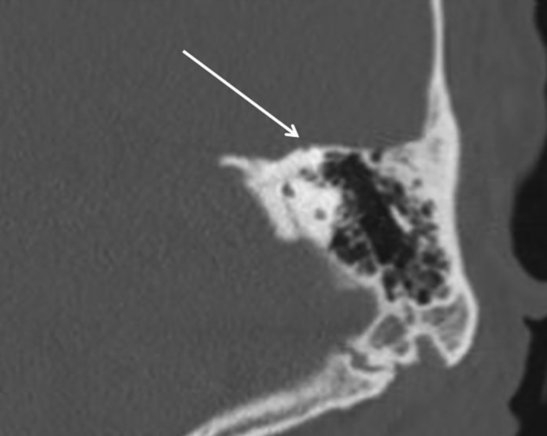
Dehiscent superior semicircular canal. Coronally reformatted CT of the temporal bone demonstrates dehiscence of the roof of the superior semicircular canal (arrow).
- © 2019 by American Journal of Neuroradiology


Jump to section
Related Articles
Cited By...
- No citing articles found.