
Article Figures & Data
Figures
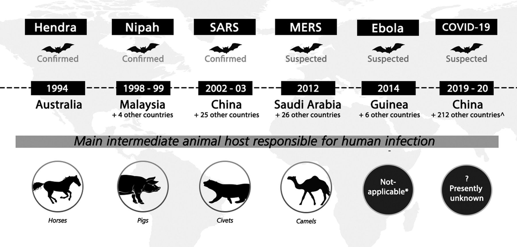
- FIG 1.
Summary of major emerging zoonotic outbreaks related to bats, 1994–2019. Confirmed bat-borne viruses include Hendra, Nipah, and SARS-CoV-1 viruses; bats are also suspected to be viral reservoirs for MERS, Ebola, and SARS-CoV-2 (cause of the current COVID-19 pandemic) viruses. Years when outbreaks occurred and confirmed or suspected intermediate hosts involved in virus spillover are also shown. * indicates that although the 2014 Ebola outbreak was believed to have started with direct bat-to-human transmission, nonhuman primates have been implicated in previous Ebola outbreaks. ∧Data accurate as of May 11, 2020. Reprinted with permission of Duke-NUS Medical School.
- FIG 2.
Patient with Nipah virus infection: initial infection. A, Multiple punctate white matter lesions (arrowheads) are visible on T2-weighted FSE MR image. B, The largest lesion is more prominent on corresponding DWI. Images reprinted from Lim et al.31
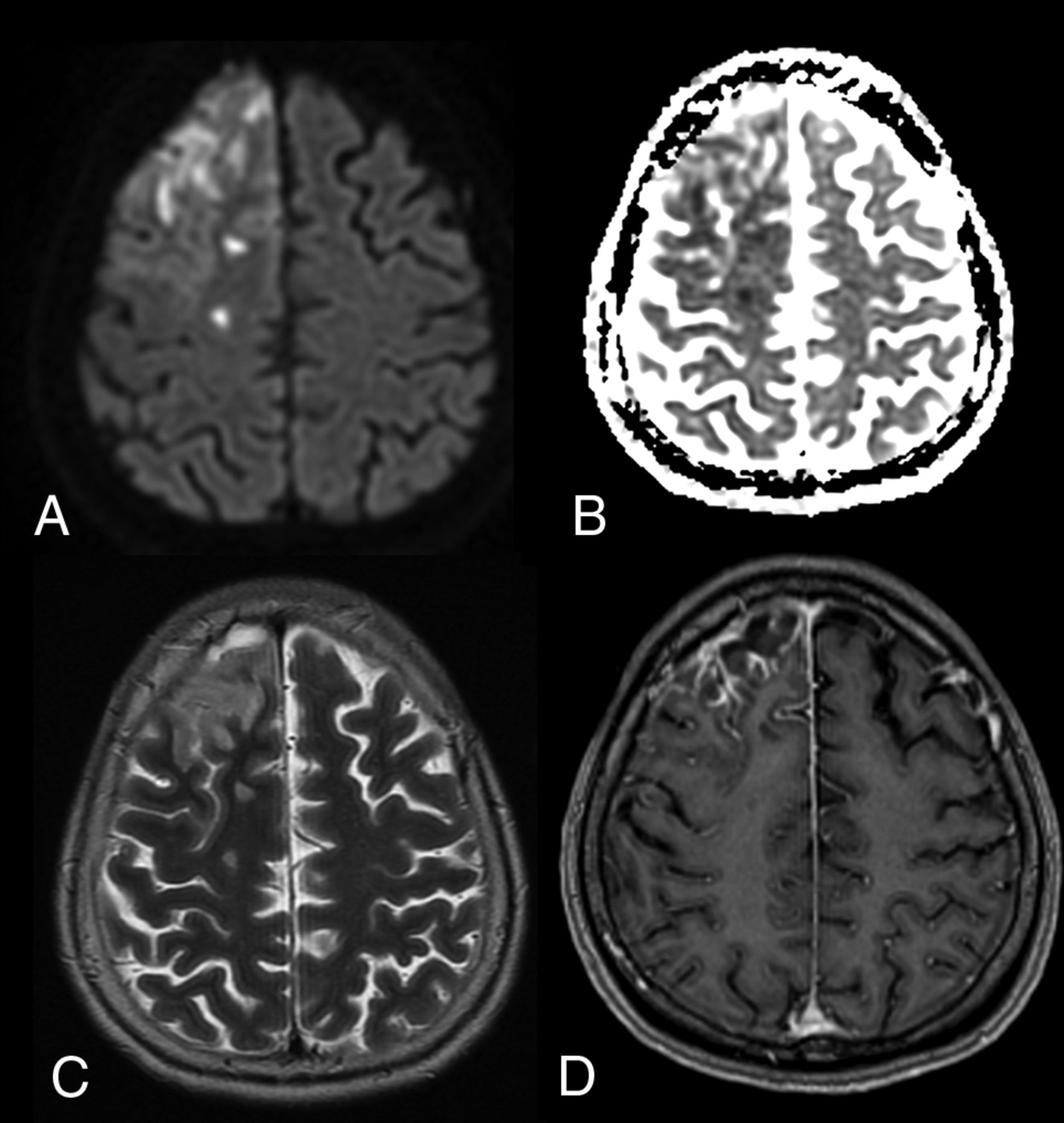
- FIG 3.
Patient with Nipah virus infection 1 month after infection. Selected axial T1-weighted images (upper row) show multifocal punctate high signal intensity on the cortical surfaces (arrows) as well as in the white matter. These did not enhance after contrast injection and disappeared on 6-month and subsequent follow-up MR imaging (not shown). Selected T2-weighted images (lower row) show noncorresponding multiple tiny focal increased signal intensity (arrowheads) in the white matter. These also became smaller or disappeared on follow-up MR imaging (not shown).
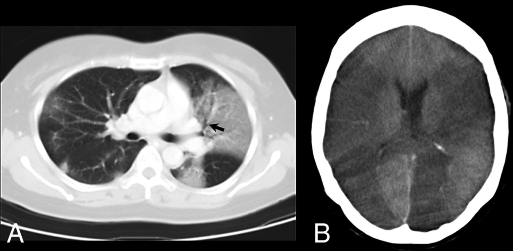
- FIG 4.
Patient with SARS infection. A, Axial 10-mm-section portable chest CT scan shows multifocal and confluent ground-glass opacities bilaterally with air bronchogram (arrow) in the left upper lobe. B, Axial CT scan of the head shows extensive low-attenuation cerebral infarction involving bilateral middle cerebral and left posterior cerebral artery territories.
- FIG 5.
Patient with MERS-CoV infection (on day 28). A, DWI and ADC mapping show diffusion restriction of the multiple white matter lesions. B, Axial FLAIR images show multiple hyperintense lesions in the subcortical areas and deep white matter of the frontal, temporal, and parietal lobes bilaterally as well as in the corpus callosum. Images reprinted from Arabi et al.47
- FIG 6.
Patient with influenza A H1N1 infection. A, Axial T2-weighted image shows symmetric increased signal intensity in the thalami and supratentorial frontal white matter. B, T2-weighted gradient-echo image reveals decreased signal intensity in the central portion of the thalami, indicating hemorrhagic necrosis. C, Axial DWI reveals restricted diffusion, with a concentric pattern, symmetrically involving the thalami. Images reprinted from Ormitti et al.54
- FIG 7.
Patient with group B Streptococcus ST283 meningitis. A, Axial DWI shows multifocal high signal intensity in the cortex, white matter, and subarachnoid space of the right frontal lobe. B, ADC map shows corresponding low ADC. C, T2-weighted images show increased signal. D, After contrast injection, T1-weighted images show leptomeningeal enhancement, typical of meningitis.


Tables
Pathogen Type Animal Reservoir Zoonotic Species or Agent Viral Bats Rabies virus (RABV) Australian bat lyssavirus (ABLV) Hendra virus (HeV) Nipah virus (NiV) SARS-CoV, MERS-CoV, SARS-CoV-2 Bats, primates, duikers Ebola virus (EBOV) Marburg virus (MARV) Primates Herpes B virus Rodents Hantavirus Poultry, swine Influenza A virus (H1N1, H1N2, H3N2, H5N1, H7N9) Bacterial Ruminants Brucella species Leptospira interrogans Coxiella burnetii Rabbits, rodents Francisella tularensis Poultry Chlamydia psittaci Fish Streptococcus iniae Parasitic Felines Toxoplasma gondii Swine Taenia solium Sheep, cattle Echinococcus species Fungal Bats, birds, various mammals Histoplasma capsulatum Cryptococcus neoformans, Cryptococcus gattii Zoonotic Pathogen Brain Neuroimaging Abnormalities Hendra virus (HeV) Widespread cortex21 Nipah virus (NiV) Multifocal tiny white matter and others27,31aWidespread cortex27,34,36 SARS-CoV Large arterial territory infarction (with hemorrhage)45 MERS-CoV Multifocal white matter and others47aLarge arterial territory infarction47aIntracranial hemorrhage48 Ebola virus (EBOV) Multifocal tiny white matter and others52a Influenza A H1N1 virus Bilateral thalamus (with hemorrhage) and others54aMeningeal enhancement55 Streptococcus agalactiae ST283 Multifocal subarachnoid space and others65aMeningeal enhancement65 a Denotes detected on DWI.


Jump to section
Related Articles
Cited By...
- No citing articles found.