
Article Figures & Data
Figures
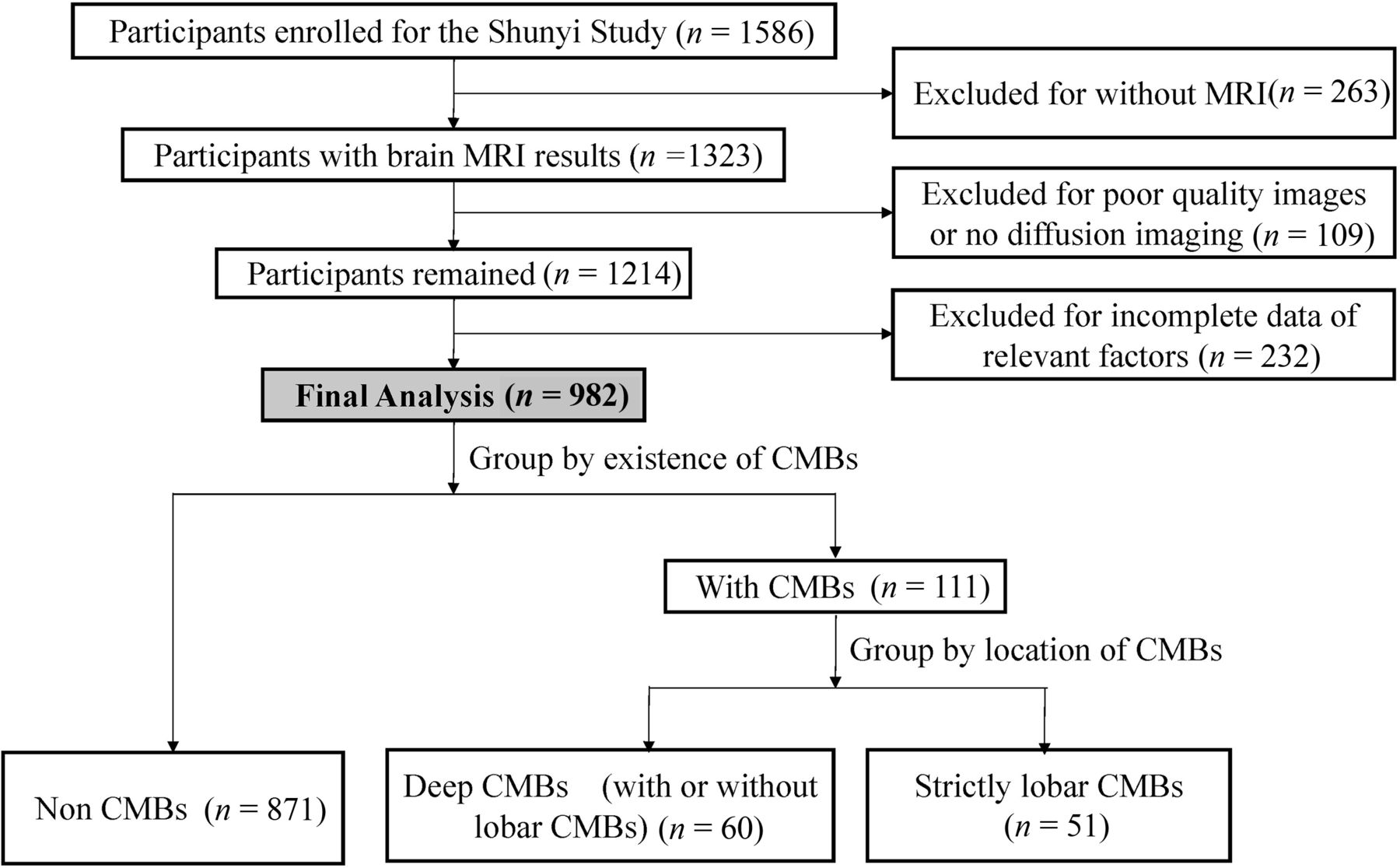
- Fig 1.
Flow diagram of the study population.
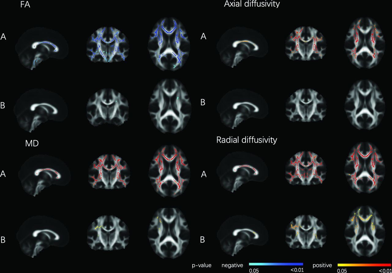
- Fig 2.
DTI parameters related to deep CMBs. Tract-Based Spatial Statistics analysis of FA and the CMB location (deep CMBs with or without lobar CMBs). Deep CMBs were associated with decreased FA and increased MD, axial diffusivity, and radial diffusivity, adjusted for age, sex, hypertension, hyperlipidemia, smoking, and diabetes (A), or age, sex, hypertension, hyperlipidemia, smoking, diabetes, white matter hyperintensity volume, and lacunes (B), thresholded at P < .05, and corrected for multiple comparisons. The red colormap indicates a positive relationship, and the blue colormap indicates a negative relationship. The statistical maps are superimposed onto the spatially normalized (Montreal Neurological Institute stereotactic space) and averaged (n = 931) FA map. Threshold-free cluster enhancement corrected P < .05.
- Fig 3.
DTI parameters related to strictly lobar CMB. Tract-Based Spatial Statistics analysis of FA and CMB location (strictly lobar). Lobar CMBs were associated with decreased FA and increased MD, axial diffusivity, and radial diffusivity, adjusted for age, sex, hypertension, hyperlipidemia, smoking, and diabetes (A), or age, sex, hypertension, hyperlipidemia, smoking, diabetes, white matter hyperintensity volume and lacunes (B), thresholded at P < .05 and corrected for multiple comparisons. The red colormap indicates a positive relationship, and the blue colormap indicates negative relationship. The statistical maps are superimposed onto the spatially normalized (Montreal Neurological Institute stereotactic space) and averaged (n = 922) FA map. Threshold-free cluster enhancement corrected P < .05.
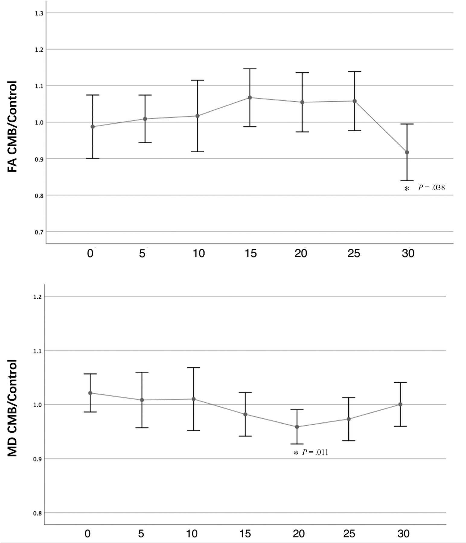
- Fig 4.
FA and MD of the lesioned tract expressed as a ratio relative to the control tract. The x-axis shows the distance in millimeters from the microbleed location. Data show the mean and its 95% CI. The asterisk indicates that the difference was significant between the CMB tract and control tract (P < .05).


Tables
Variables Normal Deep CMBs Lobar CMBs Pb Pc Demographics Age (yr) 55.3 (9.1) 62.6 (7.5) 60.8 (9.0) <.001 .314 Men 296 (34%) 36 (60%) 20 (39%) <.001 .048 Cardiovascular risk factors Hypertension 427 (49%) 48 (80%) 27 (53%) <.001 .002b Diabetes mellites 148 (17%) 14 (23%) 12 (24%) .212 .981 Hyperlipidemia 427 (49%) 29 (48%) 27 (53%) .869 .628 Smoking 231 (26.5%) 25 (41.7%) 15 (29.4%) .038 .180 Neuroimaging characteristics Presence of lacunes 122 (14%) 35 (58%) 15 (29%) <.001 .002d WMH volume (mL) 0.82 (0.25–2.62) 5.82 (2.09–12.81) 3.56 (0.99–7.11) <.001 .086 Total brain volume (mL) 1402.7 (123.2) 1421.0 (132.6) 1424.2 (136.3) .285 .893 CMB numbers – .003d 1 – 29 (48.3%) 36 (70.5%) 2∼3 – 17 (28.3%) 13 (25.5%) >4 – 14 (23.3%) 2 (4.0%) DTI parameters Mean global FA, NAWM 0.37 (0.02) 0.35 (0.03) 0.35 (0.02) <.001 .143 Mean global MD, ×10–3mm2/s, NAWM 0.84 (0.04) 0.89 (0.06) 0.87 (0.05) <.001 .031 Axial diffusivity, ×10−3 mm2/s, NAWM 1.18 (0.04) 1.22 (0.05) 1.20 (0.05) <.001 .023 Radial diffusivity, ×10−3 mm2/s, NAWM 0.67 (0.05) 0.73 (0.07) 0.71 (0.06) <.001 .042 Note:—–indicates no CMBs.
↵a Data represent mean (SD), median (interquartile range), or frequency (percentage).
↵b Significance test among 3 groups, using the ANOVA, χ2 test, or the Kruskal-Wallis test.
↵c Post hoc analysis between the deep CMB and lobar CMB groups.
↵d The difference was significant between deep CMBs and lobar CMBs groups (Bonferroni-corrected, P < .017).
FA MD Axial Diffusivity Radial Diffusivity β P β P β P β P Model 1 −0.450 (−0.616 to −0.285) <.001 0.433 (0.282−0.584) <.001 0.378 (0.221−0.535) <.001 0.446 (0.295−0.597) <.001 Model 2 −0.433 (−0.597 to −0.269) <.001 0.420 (0.271−0.570) <.001 0.368 (0.212−0.524) <.001 0.432 (0.282−0.581) <.001 Model 3 −0.238 (−0.394 to −0.083) .003 0.204 (0.068−0.340) .003 0.165 (0.019−0.310) .026 0.217 (0.081−0.352) .002 Model 4 −0.241 (−0.396 to −0.086) .002 0.206 (0.070−0.342) .003 0.167 (0.021−0.312) .025 0.218 (0.083−0.354) .002 Note:—β indicates regression coefficient.
↵a Values represent differences in z scores of mean FA, MD, axial diffusivity, and radial diffusivity of NAWM for the presence of any microbleeds compared with no microbleeds. Model 1: adjusted for age and sex; model 2: same as model 1, additionally adjusted for hypertension, hyperlipidemia, smoking, diabetes; model 3: adjusted for age, sex, lacunes, and white matter hyperintensity volume (log-transformed); model 4: adjusted for sex, cardiovascular risk factors as in model 2, and CSVD imaging markers as in model 3.
FA MD Axial Diffusivity Radial Diffusivity β P β P β P β P None vs lobar Model 1 −0.396 (−0.622 to −0.169) .001 0.339 (0.135−0.543) .001 0.270 (0.055−0.484) .014 0.363 (0.159−0.566) <.001 Model 2 −0.396 (−0.619 to −0.173) .001 0.340 (0.138−0.541) .001 0.270 (0.058−0.485) .013 0.362 (0.162−0.563) <.001 Model 3 −0.258 (−0.467 to −0.048) .016 0.191 (0.010−0.372) .038 0.131 (−0.065−0.327) .191 0.215 (0.034−0.395) .020 Model 4 −0.264 (−0.472 to −0.056) .013 0.195 (0.014−0.376) .034 0.134 (−0.062−0.330) .180 0.218 (0.039−0.398) .017 None vs deep Model 1 −0.509 (−0.726 to −0.293) <.001 0.530 (0.335−0.725) <.001 0.486 (0.283−0.689) <.001 0.534 (0.339−0.729) <.001 Model 2 −0.481 (−0.697 to −0.266) .001 0.512 (0.317−0.706) <.001 0.472 (0.268−0.675) <.001 0.514 (0.319−0.708) <.001 Model 3 −0.244 (−0.450 to −0.038) .020 0.243 (0.065−0.421) .008 0.218 (0.028−0.408) .025 0.246 (0.068−0.424) .007 Model 4 −0.242 (−0.448 to −0.036) .021 0.245 (0.071−0.424) .007 0.220 (0.029−0.411) .024 0.248 (0.069−0.426) .007 Note:—β indicates regression coefficient.
↵a Values represent differences in z scores of mean FA, MD, axial diffusivity and radial diffusivity of NAWM for the presence of any microbleeds by their location compared with no microbleeds. Model 1: adjusted for age and sex; model 2: same as model 1, additionally adjusted for hypertension, hyperlipidemia, smoking, diabetes; model 3: adjusted for age, sex, lacunes, white matter hyperintensity volume (log-transformed); model 4: adjusted for sex, cardiovascular risk factors as in model 2, and CSVD imaging markers as in model 3.


Jump to section
Related Articles
Cited By...
- Variations in perfusion detectable in advance of microstructure in white matter aging
- Association of Cerebral Microbleeds and Risk of Stroke and Mortality in Posterior Circulation Cerebral Infarction
- Protocol for Multi-modality MEdical imaging sTudy bAsed on KaiLuan Study (META-KLS): rationale, design and database building