Advertisement

Main menu

  • Home
  • Content
    • Current Issue
    • Accepted Manuscripts
    • Article Preview
    • Past Issue Archive
    • Video Articles
    • AJNR Case Collection
    • Case of the Week Archive
    • Case of the Month Archive
    • Classic Case Archive
  • Special Collections
    • AJNR Awards
    • ASNR Foundation Special Collection
    • Most Impactful AJNR Articles
    • Photon-Counting CT
    • Spinal CSF Leak Articles (Jan 2020-June 2024)
  • Multimedia
    • AJNR Podcasts
    • AJNR SCANtastic
    • Trainee Corner
    • MRI Safety Corner
    • Imaging Protocols
  • For Authors
    • Submit a Manuscript
    • Submit a Video Article
    • Submit an eLetter to the Editor/Response
    • Manuscript Submission Guidelines
    • Statistical Tips
    • Fast Publishing of Accepted Manuscripts
    • Graphical Abstract Preparation
    • Imaging Protocol Submission
    • Author Policies
  • About Us
    • About AJNR
    • Editorial Board
    • Editorial Board Alumni
  • More
    • Become a Reviewer/Academy of Reviewers
    • Subscribers
    • Permissions
    • Alerts
    • Feedback
    • Advertisers
    • ASNR Home

User menu

  • Alerts
  • Log in

Search

  • Advanced search
American Journal of Neuroradiology

American Journal of Neuroradiology

ASHNR American Society of Functional Neuroradiology ASHNR American Society of Pediatric Neuroradiology ASSR
  • Alerts
  • Log in

Advanced Search

  • Home
  • Content
    • Current Issue
    • Accepted Manuscripts
    • Article Preview
    • Past Issue Archive
    • Video Articles
    • AJNR Case Collection
    • Case of the Week Archive
    • Case of the Month Archive
    • Classic Case Archive
  • Special Collections
    • AJNR Awards
    • ASNR Foundation Special Collection
    • Most Impactful AJNR Articles
    • Photon-Counting CT
    • Spinal CSF Leak Articles (Jan 2020-June 2024)
  • Multimedia
    • AJNR Podcasts
    • AJNR SCANtastic
    • Trainee Corner
    • MRI Safety Corner
    • Imaging Protocols
  • For Authors
    • Submit a Manuscript
    • Submit a Video Article
    • Submit an eLetter to the Editor/Response
    • Manuscript Submission Guidelines
    • Statistical Tips
    • Fast Publishing of Accepted Manuscripts
    • Graphical Abstract Preparation
    • Imaging Protocol Submission
    • Author Policies
  • About Us
    • About AJNR
    • Editorial Board
    • Editorial Board Alumni
  • More
    • Become a Reviewer/Academy of Reviewers
    • Subscribers
    • Permissions
    • Alerts
    • Feedback
    • Advertisers
    • ASNR Home
  • Follow AJNR on Twitter
  • Visit AJNR on Facebook
  • Follow AJNR on Instagram
  • Join AJNR on LinkedIn
  • RSS Feeds

AJNR is seeking candidates for the AJNR Podcast Editor. Read the position description.

Review ArticleAdult Brain
Open Access

Radiologically Isolated Syndrome: A Review for Neuroradiologists

M. Hosseiny, S.D. Newsome and D.M. Yousem
American Journal of Neuroradiology September 2020, 41 (9) 1542-1549; DOI: https://doi.org/10.3174/ajnr.A6649
M. Hosseiny
aFrom the Department of Radiological Sciences (M.H.), David Geffen School of Medicine at the University of California, Los Angeles, Los Angeles, California
  • Find this author on Google Scholar
  • Find this author on PubMed
  • Search for this author on this site
  • ORCID record for M. Hosseiny
S.D. Newsome
bDepartment of Neurology (S.D.N.), Johns Hopkins University School of Medicine, Baltimore, Maryland
  • Find this author on Google Scholar
  • Find this author on PubMed
  • Search for this author on this site
  • ORCID record for S.D. Newsome
D.M. Yousem
cRussell H. Morgan Department of Radiology and Radiological Sciences (D.M.Y.), Johns Hopkins Medical Institution, Baltimore, Maryland.
  • Find this author on Google Scholar
  • Find this author on PubMed
  • Search for this author on this site
  • ORCID record for D.M. Yousem
  • Article
  • Figures & Data
  • Supplemental
  • Info & Metrics
  • Responses
  • References
  • PDF
Loading

Article Figures & Data

Figures

  • Tables
  • FIG 1.
    • Download figure
    • Open in new tab
    • Download powerpoint
    FIG 1.

    A, Axial T2-weighted image in a 21-year-old patient presenting to the emergency department with the worst headache of her life shows left periventricular posterior fossa lesions (arrow). B, The FLAIR scan demonstrates periventricular (Dawson fingers-like) and juxtacortical lesions (arrows). C, None of the lesions show gadolinium enhancement.
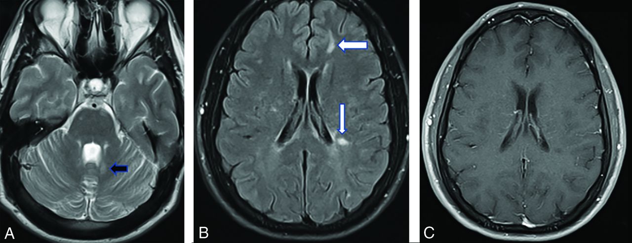
  • FIG 2.
    • Download figure
    • Open in new tab
    • Download powerpoint
    FIG 2.

    A, A 32-year-old woman being evaluated for benign positional vertigo. The FLAIR scans have severe confluent periventricular disease and a few juxtacortical foci (arrows) of high signal. B, On postgadolinium scanning, numerous ring-enhancing demyelinating lesions are present, both periventricular-periatrial on the right and juxtacortical bilaterally.

Tables

  • Figures
    • View popup
    Table 1:

    Proposed diagnostic criteria for RIS by Okuda et al9

    Inclusion CriteriaExclusion Criteria
    CNS white matter lesions on MR imaging that are ovoid, well-circumscribed, >3 mm, and homogeneously hyperintense on T2-weighted images with or without involvement of the corpus callosumCNS lesions in a vascular pattern
    Historical accounts of remitting clinical symptoms consistent with neurologic dysfunction
    CNS lesions fulfill 3 of 4 Barkof criteria
    1) One gadolinium-enhancing lesion or 9 T2-hyperintense lesions if no gadolinium-enhancing lesions
    2) At least 1 infratentorial lesion
    3) At least 1 juxtacortical lesion, and
    4) At least 3 periventricular lesions
    MR imaging anomalies can be explained by the direct physiologic effects of substances (recreational drug abuse, toxic exposure) or a medical condition
    MR imaging phenotypes suggestive of leukoaraiosis (small-vessel ischemic disease) or extensive white matter pathology lacking involvement of the corpus callosum
    MR imaging anomalies do not account for clinically apparent impairment in social, occupational, or generalized areas of functioningWhite matter lesions are better accounted for by another medical condition
    • View popup
    Table 2:

    Modified criteria for the diagnosis of RIS2

    Inclusion CriteriaExclusion Criteria
    ≥1 T2-hyperintense lesions on T2-weighted scans involving at least 2 of the following 4 locations:
    1. Periventricular white matter

    2. Cortical/juxtacortical

    3. Spinal cord

    4. Infratentorial

    Neurologic dysfunction suggestive of MS based on historical symptoms and/or objective signs
    MR imaging abnormalities explained by other disease processes, especially considering age, vascular, toxins, or drug-related abnormalities
    • View popup
    Table 3:

    Selected entities often mistaken for RIS

    EntityDistinguishing Clinical FeaturesDistinguishing Imaging FeaturesImportant Keys to Diagnosis
    Toxins/drug-relatedAltered mental status changes during intoxication, history of substance abuseDeep gray matter frequently involved, symmetric lesionsUrine and serum toxicology tests
    Age-related leukoaraiosisOlder individual, cardiovascular risk factorsSmall (<3 mm), nonenhancing lesions in periventricular and deep white matter, coexistent striatocapsular lacunar disease, absence of callososeptal lesions/Dawson fingers–type lesionsLacunar disease and atypical white matter lesions for RIS in an older individual
    MigrainesHeadache or aura predominatesPredominantly subcortical white matter lesions that are small (<3 mm) and do not enhance, few periventricular lesionsHeadache history and lack of typical imaging features consistent with RIS
    VasculitisEpisodic neurologic symptoms with superimposed strokesGray and white matter lesions coexist, may have enhancing vessel wall and/or leptomeningeal enhancement, MRA with stenosesSystemic symptoms present, elevated erythrocyte sedimentation rate and/or C-reactive protein level, extra-/intracranial vessels abnormal, brain biopsy
    CADASILStrokelike episodes, family history of similar clinical syndromeWhite matter disease favoring anterior temporal tip subcortical regions, external capsule, presence of lacunar infarctsGenetic testing diagnostic
    Collagen vascular diseasesClinical history of arthritis, long-standing chronic disease, episodicGray and white matter lesions ± vasculitis, occasional encephalitisClinical symptoms of a systemic disorder and presence of serologic autoantibodies/inflammatory markers
    ADEMEncephalitis, seizures, children > adults, history of viral/vaccine prodromeGray matter disease predominates, more diffuse enhancement, may have positive findings on DWIHistory of prodromal virus infection or vaccination, encephalopathic
    PosttraumaticHistory of ≥1 traumatic, sports-related eventFavors gray-white matter junction, hemorrhagic products present, classic tears in splenium-brain stem deep gray matterHemorrhage and stereotypical locations of disease at shearing sites
    • Note:—ADEM indicates acute disseminated encephalomyelitis.

PreviousNext
Back to top

In this issue

American Journal of Neuroradiology: 41 (9)
American Journal of Neuroradiology
Vol. 41, Issue 9
1 Sep 2020
  • Table of Contents
  • Index by author
  • Complete Issue (PDF)
Advertisement
Print
Download PDF
Email Article

Thank you for your interest in spreading the word on American Journal of Neuroradiology.

NOTE: We only request your email address so that the person you are recommending the page to knows that you wanted them to see it, and that it is not junk mail. We do not capture any email address.

Enter multiple addresses on separate lines or separate them with commas.
Radiologically Isolated Syndrome: A Review for Neuroradiologists
(Your Name) has sent you a message from American Journal of Neuroradiology
(Your Name) thought you would like to see the American Journal of Neuroradiology web site.
CAPTCHA
This question is for testing whether or not you are a human visitor and to prevent automated spam submissions.
Cite this article
M. Hosseiny, S.D. Newsome, D.M. Yousem
Radiologically Isolated Syndrome: A Review for Neuroradiologists
American Journal of Neuroradiology Sep 2020, 41 (9) 1542-1549; DOI: 10.3174/ajnr.A6649

Citation Manager Formats

  • BibTeX
  • Bookends
  • EasyBib
  • EndNote (tagged)
  • EndNote 8 (xml)
  • Medlars
  • Mendeley
  • Papers
  • RefWorks Tagged
  • Ref Manager
  • RIS
  • Zotero
0 Responses
Respond to this article
Share
Bookmark this article
Radiologically Isolated Syndrome: A Review for Neuroradiologists
M. Hosseiny, S.D. Newsome, D.M. Yousem
American Journal of Neuroradiology Sep 2020, 41 (9) 1542-1549; DOI: 10.3174/ajnr.A6649
del.icio.us logo Twitter logo Facebook logo Mendeley logo
  • Tweet Widget
  • Facebook Like
  • Google Plus One
Purchase

Jump to section

  • Article
    • Abstract
    • ABBREVIATIONS:
    • CONCLUSIONS
    • Footnotes
    • References
  • Figures & Data
  • Supplemental
  • Info & Metrics
  • Responses
  • References
  • PDF

Related Articles

  • PubMed
  • Google Scholar

Cited By...

  • Early detection of white matter hyperintensities using SHIVA-WMH detector
  • Portable, Low-Field Magnetic Resonance Imaging Sensitively Detects and Accurately Quantifies Multiple Sclerosis Lesions
  • The immune signatures of multiple sclerosis: Lessons from twin studies
  • Crossref (11)
  • Google Scholar

This article has been cited by the following articles in journals that are participating in Crossref Cited-by Linking.

  • Cerebrospinal fluid inflammatory biomarkers for disease progression in Alzheimer’s disease and multiple sclerosis: a systematic review
    Joke Temmerman, Sebastiaan Engelborghs, Maria Bjerke, Miguel D’haeseleer
    Frontiers in Immunology 2023 14
  • The role of non-coding RNAs in neuroinflammatory process in multiple sclerosis
    Anna Nowak, Zofia Wicik, Marta Wolska, Andleeb Shahzadi, Piotr Szwed, Joanna Jarosz-Popek, Jeffrey Palatini, Marek Postula, Anna Czlonkowska, Dagmara Mirowska-Guzel, Ceren Eyileten
    Molecular Neurobiology 2022 59 8
  • Early detection of white matter hyperintensities usingSHIVA‐WMHdetector
    Ami Tsuchida, Philippe Boutinaud, Violaine Verrecchia, Christophe Tzourio, Stéphanie Debette, Marc Joliot
    Human Brain Mapping 2024 45 1
  • The immune signatures of multiple sclerosis: Lessons from twin studies
    Pablo Villoslada, Scott S. Zamvil
    Proceedings of the National Academy of Sciences 2020 117 39
  • Predicting multiple sclerosis disease progression and outcomes with machine learning and MRI-based biomarkers: a review
    Hibba Yousef, Brigitta Malagurski Tortei, Filippo Castiglione
    Journal of Neurology 2024 271 10
  • Predictive Factors for Magnetic Resonance Imaging Changes Suggestive of Demyelination in Adult Patients with Uveitis Scanned Prior to Commencing Adalimumab Therapy
    Priyanka Sanghi, Joshua Luis, Sofia Ajamil, Ian YL Yeung, Edward Hindle, Selina Sandhu, Said Hassan, Ben Turner, Angela Rees, Mark Westcott
    Ocular Immunology and Inflammation 2023 31 9
  • Understanding Magnetic Resonance Imaging in Multiple Sclerosis (UMIMS): Development and Piloting of an Online Education Program About Magnetic Resonance Imaging for People With Multiple Sclerosis
    Magalie Freund, Insa Schiffmann, Anne Christin Rahn, Declan Chard, Carsten Lukas, Jutta Scheiderbauer, Anna Sippel, Christoph Heesen
    Frontiers in Neurology 2022 13
  • Conversion from Radiologically Isolated Syndrome to Multiple Sclerosis: Single Center Retrospective Study
    İbrahim Acır, Alperen Güngör, Abbas Musa Yılmaz, Abdurrahman Yusuf Çevik, Esra Derya Dinç Polat, Vildan Yayla
    Medical Science and Discovery 2023 10 8
  • Fallo terapéutico en esclerosis múltiple remitente-recurrente: definición y algoritmo para su manejo, un consenso de expertos
    Jimena García, Lina Gómez, Carlos Alberto Navas Cardozo, María Isabel Zuluaga Rodas, Adriana Casallas Vanegas, Simón Cárdenas-Robledo, Jairo Alonso Quiñones Bautista, Jahir Miranda Acuña, Marco Aurelio Reyes Guerrero, César Augusto Franco Ruiz, Jorge Andrés Jiménez Arango, Paola Andrea Ortiz Salas
    Acta Neurológica Colombiana 2025 41 1
  • Multiple sclerosis
    Hiromasa Tsuda
    Equilibrium Research 2022 81 4

More in this TOC Section

  • Diagnostic Neuroradiology of Monoclonal Antibodies
  • Clinical Outcomes After Chiari I Decompression
  • Segmentation of Brain Metastases with BLAST
Show more Adult Brain

Similar Articles

Advertisement

Indexed Content

  • Current Issue
  • Accepted Manuscripts
  • Article Preview
  • Past Issues
  • Editorials
  • Editor's Choice
  • Fellows' Journal Club
  • Letters to the Editor
  • Video Articles

Cases

  • Case Collection
  • Archive - Case of the Week
  • Archive - Case of the Month
  • Archive - Classic Case

Special Collections

  • AJNR Awards
  • ASNR Foundation Special Collection
  • Most Impactful AJNR Articles
  • Photon-Counting CT
  • Spinal CSF Leak Articles (Jan 2020-June 2024)

More from AJNR

  • Trainee Corner
  • Imaging Protocols
  • MRI Safety Corner

Multimedia

  • AJNR Podcasts
  • AJNR Scantastics

Resources

  • Turnaround Time
  • Submit a Manuscript
  • Submit a Video Article
  • Submit an eLetter to the Editor/Response
  • Manuscript Submission Guidelines
  • Statistical Tips
  • Fast Publishing of Accepted Manuscripts
  • Graphical Abstract Preparation
  • Imaging Protocol Submission
  • Evidence-Based Medicine Level Guide
  • Publishing Checklists
  • Author Policies
  • Become a Reviewer/Academy of Reviewers
  • News and Updates

About Us

  • About AJNR
  • Editorial Board
  • Editorial Board Alumni
  • Alerts
  • Permissions
  • Not an AJNR Subscriber? Join Now
  • Advertise with Us
  • Librarian Resources
  • Feedback
  • Terms and Conditions
  • AJNR Editorial Board Alumni

American Society of Neuroradiology

  • Not an ASNR Member? Join Now

© 2025 by the American Society of Neuroradiology All rights, including for text and data mining, AI training, and similar technologies, are reserved.
Print ISSN: 0195-6108 Online ISSN: 1936-959X

Powered by HighWire