Advertisement

Main menu

  • Home
  • Content
    • Current Issue
    • Publication Preview--Ahead of Print
    • Past Issue Archive
    • Case of the Week Archive
    • Classic Case Archive
    • Case of the Month Archive
    • COVID-19 Content and Resources
  • For Authors
  • About Us
    • About AJNR
    • Editors
    • American Society of Neuroradiology
  • Submit a Manuscript
  • Podcasts
    • Subscribe on iTunes
    • Subscribe on Stitcher
  • More
    • Subscribers
    • Permissions
    • Advertisers
    • Alerts
    • Feedback
  • Other Publications
    • ajnr

User menu

  • Subscribe
  • Alerts
  • Log in

Search

  • Advanced search
American Journal of Neuroradiology

American Journal of Neuroradiology

  • Subscribe
  • Alerts
  • Log in

Advanced Search

  • Home
  • Content
    • Current Issue
    • Publication Preview--Ahead of Print
    • Past Issue Archive
    • Case of the Week Archive
    • Classic Case Archive
    • Case of the Month Archive
    • COVID-19 Content and Resources
  • For Authors
  • About Us
    • About AJNR
    • Editors
    • American Society of Neuroradiology
  • Submit a Manuscript
  • Podcasts
    • Subscribe on iTunes
    • Subscribe on Stitcher
  • More
    • Subscribers
    • Permissions
    • Advertisers
    • Alerts
    • Feedback
  • Follow AJNR on Twitter
  • Visit AJNR on Facebook
  • Follow AJNR on Instagram
  • Join AJNR on LinkedIn
  • RSS Feeds
Research ArticleAdult Brain

Pretreatment ADC Histogram Analysis as a Prognostic Imaging Biomarker for Patients with Recurrent Glioblastoma Treated with Bevacizumab: A Systematic Review and Meta-analysis

R. Kurokawa, A. Baba, M. Kurokawa, A. Capizzano, O. Hassan, T. Johnson, Y. Ota, J. Kim, A. Hagiwara, T. Moritani and A. Srinivasan
American Journal of Neuroradiology February 2022, 43 (2) 202-206; DOI: https://doi.org/10.3174/ajnr.A7406
R. Kurokawa
aFrom the Division of Neuroradiology (R.K., A.B., M.K., A.C., O.H., Y.O., J.K., T.M., A.S.), Department of Radiology, University of Michigan, Ann Arbor, Michigan
  • Find this author on Google Scholar
  • Find this author on PubMed
  • Search for this author on this site
  • ORCID record for R. Kurokawa
A. Baba
aFrom the Division of Neuroradiology (R.K., A.B., M.K., A.C., O.H., Y.O., J.K., T.M., A.S.), Department of Radiology, University of Michigan, Ann Arbor, Michigan
  • Find this author on Google Scholar
  • Find this author on PubMed
  • Search for this author on this site
  • ORCID record for A. Baba
M. Kurokawa
aFrom the Division of Neuroradiology (R.K., A.B., M.K., A.C., O.H., Y.O., J.K., T.M., A.S.), Department of Radiology, University of Michigan, Ann Arbor, Michigan
  • Find this author on Google Scholar
  • Find this author on PubMed
  • Search for this author on this site
  • ORCID record for M. Kurokawa
A. Capizzano
aFrom the Division of Neuroradiology (R.K., A.B., M.K., A.C., O.H., Y.O., J.K., T.M., A.S.), Department of Radiology, University of Michigan, Ann Arbor, Michigan
  • Find this author on Google Scholar
  • Find this author on PubMed
  • Search for this author on this site
  • ORCID record for A. Capizzano
O. Hassan
aFrom the Division of Neuroradiology (R.K., A.B., M.K., A.C., O.H., Y.O., J.K., T.M., A.S.), Department of Radiology, University of Michigan, Ann Arbor, Michigan
  • Find this author on Google Scholar
  • Find this author on PubMed
  • Search for this author on this site
  • ORCID record for O. Hassan
T. Johnson
bDepartment of Biostatistics (T.J.), University of Michigan School of Public Health, Ann Arbor, Michigan
  • Find this author on Google Scholar
  • Find this author on PubMed
  • Search for this author on this site
  • ORCID record for T. Johnson
Y. Ota
aFrom the Division of Neuroradiology (R.K., A.B., M.K., A.C., O.H., Y.O., J.K., T.M., A.S.), Department of Radiology, University of Michigan, Ann Arbor, Michigan
  • Find this author on Google Scholar
  • Find this author on PubMed
  • Search for this author on this site
  • ORCID record for Y. Ota
J. Kim
aFrom the Division of Neuroradiology (R.K., A.B., M.K., A.C., O.H., Y.O., J.K., T.M., A.S.), Department of Radiology, University of Michigan, Ann Arbor, Michigan
  • Find this author on Google Scholar
  • Find this author on PubMed
  • Search for this author on this site
  • ORCID record for J. Kim
A. Hagiwara
cDepartment of Radiology (A.H.), Juntendo University School of Medicine, Tokyo, Japan
  • Find this author on Google Scholar
  • Find this author on PubMed
  • Search for this author on this site
  • ORCID record for A. Hagiwara
T. Moritani
aFrom the Division of Neuroradiology (R.K., A.B., M.K., A.C., O.H., Y.O., J.K., T.M., A.S.), Department of Radiology, University of Michigan, Ann Arbor, Michigan
  • Find this author on Google Scholar
  • Find this author on PubMed
  • Search for this author on this site
  • ORCID record for T. Moritani
A. Srinivasan
aFrom the Division of Neuroradiology (R.K., A.B., M.K., A.C., O.H., Y.O., J.K., T.M., A.S.), Department of Radiology, University of Michigan, Ann Arbor, Michigan
  • Find this author on Google Scholar
  • Find this author on PubMed
  • Search for this author on this site
  • ORCID record for A. Srinivasan
  • Article
  • Figures & Data
  • Supplemental
  • Info & Metrics
  • References
  • PDF
Loading

Abstract

BACKGROUND: The mean ADC value of the lower Gaussian curve (ADCL) derived from the bi-Gaussian curve-fitting histogram analysis has been reported as a predictive/prognostic imaging biomarker in patients with recurrent glioblastoma treated with bevacizumab; however, its systematic summary has been lacking.

PURPOSE: We applied a systematic review and meta-analysis to investigate the predictive/prognostic performance of ADCL in patients with recurrent glioblastoma treated with bevacizumab.

DATA SOURCES: We performed a literature search using PubMed, Scopus, and EMBASE.

STUDY SELECTION: A total of 1344 abstracts were screened, of which 83 articles were considered potentially relevant. Data were finally extracted from 6 studies including 578 patients.

DATA ANALYSIS: Forest plots were generated to illustrate the hazard ratios of overall survival and progression-free survival. The heterogeneity across the studies was assessed using the Cochrane Q test and I2 values.

DATA SYNTHESIS: The pooled hazard ratios for overall survival and progression-free survival in patients with an ADCL lower than the cutoff values were 1.89 (95% CI, 1.53–2.31) and 1.98 (95% CI, 1.54–2.55) with low heterogeneity among the studies. Subgroup analysis of the bevacizumab-free cohort showed a pooled hazard ratio for overall survival of 1.20 (95% CI, 1.08–1.34) with low heterogeneity.

LIMITATIONS: The conclusions are limited by the difference in the definition of recurrence among the included studies.

CONCLUSIONS: This systematic review with meta-analysis supports the prognostic value of ADCL in patients with recurrent glioblastoma treated with bevacizumab, with a low ADCL demonstrating decreased overall survival and progression-free survival. On the other hand, the predictive role of ADCL for bevacizumab treatment was not confirmed.

ABBREVIATIONS:

ADCL
mean ADC value of the lower Gaussian curve
HR
hazard ratio
OS
overall survival
PFS
progression-free survival
QUADAS-2
Quality Assessment of Diagnostic Accuracy Studies 2
VEGF
vascular endothelial growth factor

Glioblastoma remains the most common and lethal primary malignant tumor of the CNS, with a median overall survival of 8–14 months despite aggressive surgery, chemotherapy, and radiation.1,2 Histologically, glioblastoma is characterized by tumor cell anaplasia, necrosis, and prominent angiogenesis mediated by the vascular endothelial growth factor (VEGF), constituting a rationale for targeted therapy.3 Bevacizumab is a humanized anti-VEGF monoclonal immunoglobulin 1 antibody, and its use for recurrent glioblastoma was approved by the US Food and Drug Administration in 2009. Although several clinical trials failed to demonstrate its contribution to extending patient survival in newly diagnosed glioblastoma or progressive glioblastoma,4⇓-6 prolonged progression-free survival (PFS) and overall survival (OS) have been reported in the recurrence setting with either bevacizumab alone or in combination with other chemotherapy.7

MR imaging is an essential imaging technique for diagnosis, treatment planning, and evaluation of therapeutic effects in patients with glioblastoma. A meta-analysis by Choi et al8 demonstrated the benefit of perfusion MR imaging, including dynamic susceptibility contrast MR imaging and dynamic contrast-enhanced MR imaging, as a predictive and prognostic imaging biomarker in patients with recurrent glioblastoma treated with bevacizumab. There have also been many studies investigating the performance of DWI as a predictive/prognostic imaging biomarker in recurrent glioblastoma.9⇓⇓⇓⇓⇓⇓⇓⇓-18 In particular, several studies have reported the usefulness of the mean ADC value of the lower Gaussian curve (ADCL) derived from the bi-Gaussian curve-fitting histogram analysis;9⇓⇓⇓⇓-14 however, a systematic summary of this topic has been lacking.

The purpose of this study was to systematically review the literature and investigate the predictive/prognostic role of pretreatment DWI, especially ADCL, in patients with recurrent glioblastoma receiving bevacizumab treatment.

MATERIALS AND METHODS

Protocol

This study was performed in accordance with the Preferred Reporting Items for Systematic Reviews and Meta-Analyses 2020 statement.19

Study Selection

We searched the PubMed, Scopus, and EMBASE data bases using the following search terms on August 24, 2021, without any language or date limits:

  • ((glioblastoma)OR(GBM)) AND ((avastin) OR (bevacizumab)) AND ((ADC) OR (apparent diffusion coefficient)) for PubMed

  • (glioblastoma OR gbm) AND (avastin OR bevacizumab) AND (adc OR (apparent AND diffusion AND coefficient)) for EMBASE

  • ALL ((glioblastoma OR gbm) AND (avastin OR bevacizumab) AND (adc OR (apparent AND diffusion AND coefficient) AND (LIMIT-TO (SUBJAREA, “MEDI”)) for Scopus

Publications that met all of the following criteria were considered eligible:

  • Participants: patients were clinically, radiologically, and/or histologically diagnosed with recurrent glioblastoma treated with bevacizumab and underwent pretreatment MRI

  • Index test: ADCL

  • Outcome: hazard ratio (HR) for PFS or OS between patients with high and low ADCL

  • Study design: retrospective or prospective observational studies and clinical trials.

Exclusion criteria were as follows:

  • The full text was unavailable.

  • It was not a peer-reviewed journal publication.

  • The relationship between ADC and survival was not analyzed, or the HRs were not calculated or could not be estimated.

  • The index was not ADCL.

  • Possible duplication of patients: the study with a smaller number of patients was excluded.

  • Review, systematic review, or meta-analysis.

Non-English references were translated into English using Google Translate (translate.google.com) and examined.

Data Analyses

Two board-certified radiologists with 9 and 6 years of experience in neuroradiology reviewed the full text of the eligible studies by consensus. Any disagreements were resolved by another board-certified radiologist with 13 years of experience in neuroradiology. We collected authors names, publication year, the region where patients were included, period of patient inclusion, study methods, trial name, number of patients, mean or median age, sex, the definition of recurrence of glioblastoma, treatment regimen, MR imaging vendor/model/magnetic field strength, b-values, ROI placement method, ADC type, the cutoff value of ADC, how the cutoff value was determined, and outcomes (HRs for OS and PFS). When the original study did not report the 95% confidence intervals of HRs, we estimated them using all the available data from the reported statistics.

Quality and Risk-of-Bias Assessment

We used the Quality Assessment of Diagnostic Accuracy Studies 2 (QUADAS-2).20 QUADAS-2 is based on the 4 domains (patient selection, index test, reference standard, and flow and timing). These domains are assessed regarding the risk of bias, and the first 3 domains are also assessed in terms of applicability.

Statistics

A forest plot was generated to illustrate the HRs for OS and PFS in patients with an ADCL lower than the cutoff values along with upper and lower 95% CIs. The pooled HR with 95% CIs was calculated using the fixed-effects model. The heterogeneity across the studies was assessed using the Cochrane Q test and I2 values. I2 values were interpreted as follows: 0%–29%, low; 30%–49%, moderate; and 50%–90%, considerable heterogeneity. P values < .05 were considered statistically significant. Statistical analyses were performed using Review Manual (RevMan, Version 5.4; https://training.cochrane.org/online-learning/core-software-cochrane-reviews/revman).

RESULTS

Study Selection

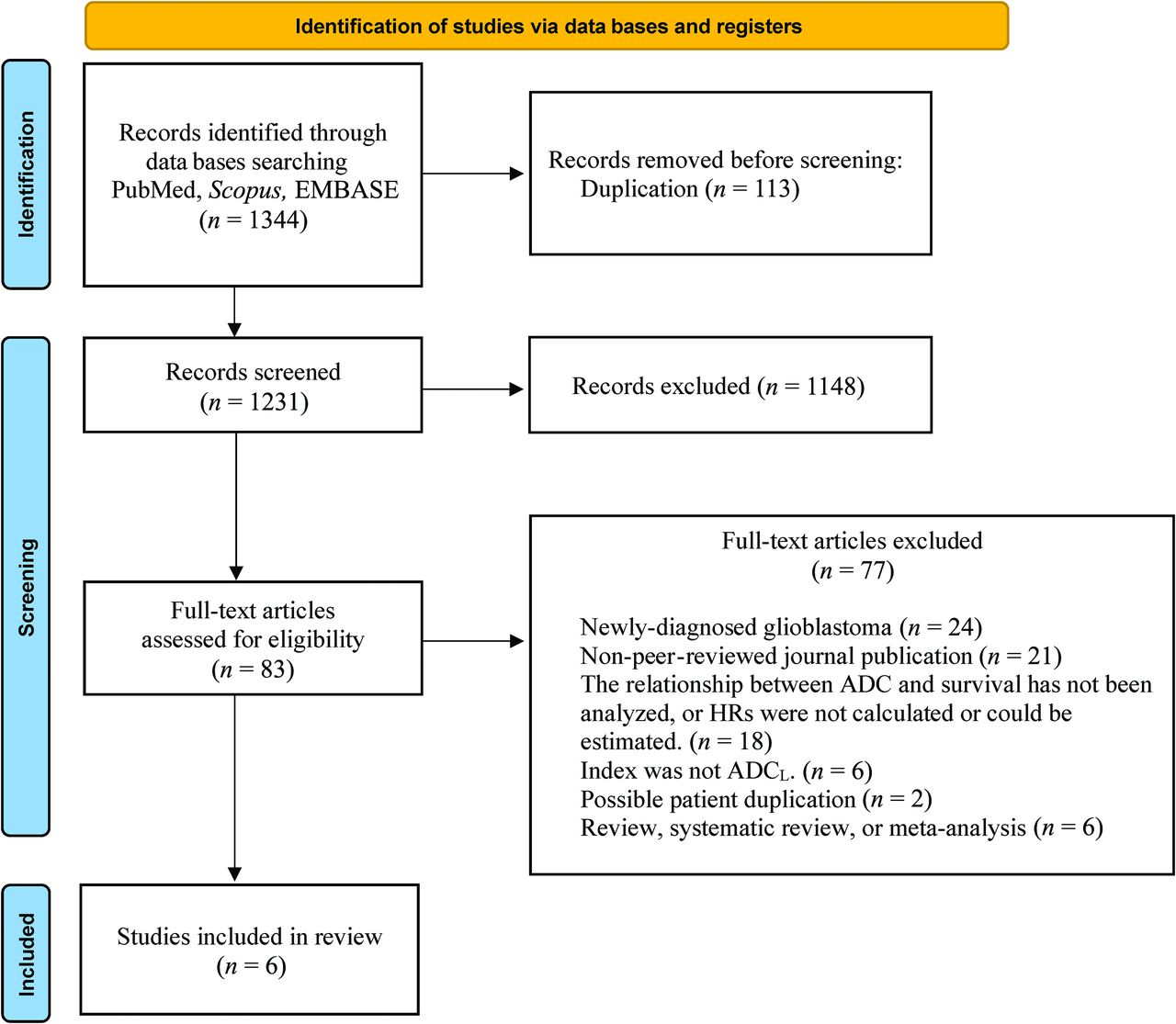
A total of 1344 abstracts were screened, of which 113 duplications were excluded. A total of 1148 articles were excluded by title and abstract screening. After excluding 77 articles according to the inclusion/exclusion criteria, finally, 6 studies including a total of 578 patients met the selection criteria for the systematic review.9⇓⇓⇓-14 The process of study selection is summarized in Fig 1. Publications in this systematic review ranged from 2012 to 2020.

FIG 1.
  • Download figure
  • Open in new tab
  • Download powerpoint
FIG 1.

Flow diagram of study selection.

Study Characteristics

The individual study characteristics are summarized in the Online Supplemental Data. A total of 578 patients with recurrent glioblastoma treated with bevacizumab and 236 patients treated without bevacizumab (hereafter, the bevacizumab-free cohort) were included. The mean or median patient age was approximately 50 years in each study. The VOI was acquired in the enhancing tumor areas, and ADC histogram analysis was performed with the bi-Gaussian fitting model, though 1 study used the 3 peaks model.10 The ADCL was generated on the basis of the fitting curve, and OS and PFS were compared using cutoff values of 1.050–1.240 × 10−3mm2/s and 1.050–1.241 × 10−3mm2/s, respectively. The cutoff value was determined by averaging,9,13,17 the hierarchical Bayesian method,14 where the OS difference among patient cohorts was the largest,12 or based on empiric thresholds identified in previous studies,11 though the method was not described in detail in 1 study.10 These studies excluded the areas of macroscopic cystic, hemorrhagic, and necrotic changes from the VOIs for ADC histogram analyses. Three studies11,12,14 compared the OS of the bevacizumab-free cohort depending on the ADCL, though only 2 studies compared PFS.11,14

Quality and Risk-of-Bias Assessment

The results of QUADAS-2 are summarized in the Online Supplemental Data. Most studies had a low risk of bias in terms of patient selection, index test, reference standard, and flow and timing. Studies that did not describe whether the participants were consecutive, randomized, or neither were considered to have an uncertain risk of bias regarding the patient selection. A study in which the method of the definition of ADCL and the cutoff value determination were not described in detail was considered to have an uncertain risk of bias regarding the index test and reference standard.

Meta-analysis

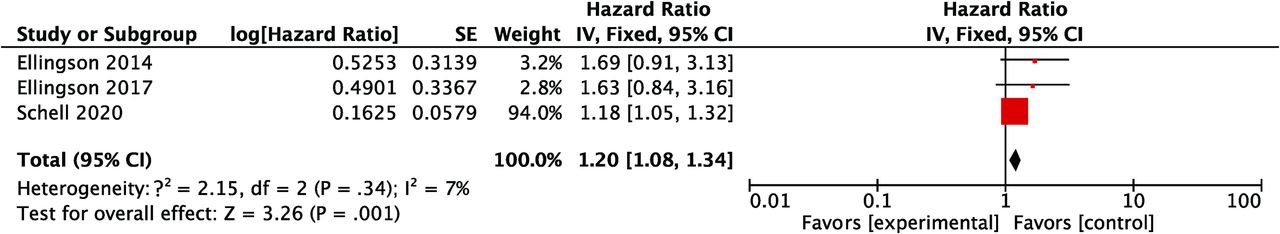
HRs for OS in patients with an ADCL lower than the cutoff values were available in all of the 6 studies, and the pooled HR of the lower ADCL was 1.89 (95% CI, 1.53–2.32), indicating worse survival (Fig 2). The lower 95% CI of HRs for OS was >1.00 in all studies. The heterogeneity of HRs for OS was considered low with the Q value in the Cochran Q test of 5.38 (P = .37) and an I2 of 7%. The comparison of HRs for PFS was available in 5 studies, and the pooled HR with an ADCL lower than the cutoff values was 1.98 (95% CI, 1.54–2.55) (Fig 3). Although the lower 95% CI of HR for PFS was <1.00 in 1 study,10 the heterogeneity of HRs for PFS was low, with the Q value in the Cochran Q test of 5.63 (P = .23) and an I2 of 29%. For studies with a bevacizumab-free cohort, no differences in OS or PFS were found, depending on the ADCL in 2 of 3 studies,11,12 whereas the significant differences in OS and PFS were retained in the bevacizumab-free cohort in 1 study.14 The pooled HR of the lower ADCL for OS in this subgroup was 1.20 (95% CI, 1.08–1.34) with a low heterogeneity (the Q value in Cochran Q test was 2.15 [P = .34] and the I2 was 7%; Fig 4).

FIG 2.
  • Download figure
  • Open in new tab
  • Download powerpoint
FIG 2.

A forest plot summarizing the HR of OS in patients treated with bevacizumab with the ADCL lower than the cutoff values compared with those with the ADCL higher than the cutoff values. SE indicates standard error; IV, instrumental variable.

FIG 3.
  • Download figure
  • Open in new tab
  • Download powerpoint
FIG 3.

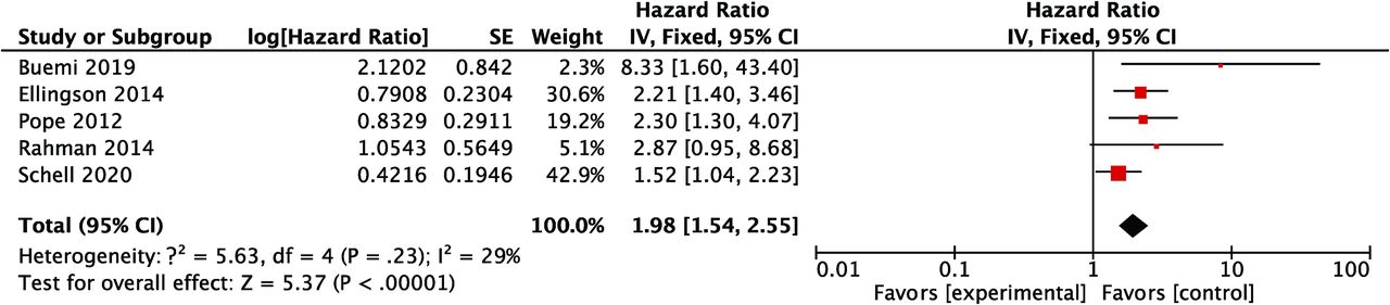
A forest plot summarizing the HR of PFS in patients treated with bevacizumab with the ADCL lower than the cutoff values compared with those with the ADCL higher than the cutoff values. SE indicates standard error; IV, instrumental variable.

FIG 4.
  • Download figure
  • Open in new tab
  • Download powerpoint
FIG 4.

A forest plot summarizing the HR of OS in the bevacizumab-free cohort with the ADCL lower than the cutoff values compared with those with the ADCL higher than the cutoff values. SE indicates standard error; IV, instrumental variable.

DISCUSSION

DWI is a unique technique that allows noninvasive observation of the microstructure of tumors and surrounding brain tissues and is widely used in daily clinical practice. In this study, the high prognostic performance of the ADCL for survival was confirmed in patients with recurrent glioblastoma treated with bevacizumab. The meta-analysis demonstrated that a lower ADCL on pretreatment MR imaging was related to unfavorable survival with pooled HRs of 1.89 (95% CI, 1.53–2.32) for OS and 1.98 (95% CI, 1.54–2.55) for PFS, with low heterogeneity among the studies. However, the subgroup analysis with the patients treated without bevacizumab (the bevacizumab-free cohort) also showed a high pooled HR for OS of 1.20 (95% CI, 1.08–1.34), indicating that although the ADCL has a prognostic value, uncertainty remains as to whether the ADCL has a predictive value for bevacizumab treatment.

Noninvasive characterization of glioblastoma using MR imaging has been extensively studied to predict the treatment effect and subsequent patient survival in patients with recurrent glioblastoma treated with bevacizumab. Focused sequences included perfusion MR imaging,8,21⇓⇓-24 31P MR spectroscopy,25,26 and ADC.9⇓⇓⇓⇓⇓⇓⇓⇓-18 Choi et al8 demonstrated that the pooled HRs between responders and nonresponders to bevacizumab, as defined by the relative CBV on dynamic susceptibility contrast MR imaging, were 0.47 (95% CI, 0.29–0.76) for OS and 0.46 (95% CI, 0.28–0.76) for PFS, indicating that tumor perfusion was decreased in responders, resulting in longer survival. The results of their meta-analysis showed the utility of perfusion MR imaging in patients with recurrent glioblastoma being treated with bevacizumab, though the timing of perfusion MR imaging differed among the studies (ie, changes of relative CBV values and posttreatment relative CBV were assessed simultaneously). The results in the present study may be more uniformly applicable to individual cases, given that all the timing of ADCL values was during pretreatment.

The biologic mechanisms of the worse survival in patients with tumors showing a low ADCL have not been pathologically proved. One hypothesis is that a more hypoxic or hypercellular nature of the tumor is represented by a low ADCL, making bevacizumab treatment less effective and the tumor more aggressive, as Ellingson et al11 pointed out. Indeed, the correlation between high tumor cellularity and low ADC values is known. However, ADC values fluctuate depending on the degree of intratumoral vascular edema, cystic change, and necrosis.27 The evaluated studies in this meta-analysis excluded the areas of macroscopic cystic, hemorrhagic, and necrotic changes from the VOIs for ADC histogram analysis, but intratumoral microscopic changes and vascular edema might have affected the results.

In their study with vestibular schwannomas, Plotkin et al28 reported that the higher the pretreatment ADC value, the greater was the bevacizumab-induced tumor shrinkage, suggesting that the higher ADC was associated with a higher degree of intratumoral vascular edema, which is more likely to respond to anti-VEGF therapy. Similarly, in recurrent glioblastoma, intratumoral vascular edema and microscopic cystic/necrotic changes, as well as tumor cellularity, might have affected the ADC values. To elucidate this, further studies with radiopathologic correlation will be necessary.

Not only for patients with recurrent glioblastoma but also for those with newly diagnosed glioblastoma, the prognostic performance of pretreatment ADCL has been reported.29⇓-31 Notably, these studies showed opposite results from each other as well as from this study; ie, the study by Wirsching et al29 demonstrated that a longer OS was associated with a higher ADCL, whereas the other 2 studies30,31 reported a trend in which longer OS was associated with a lower ADCL or a lower mean ADC, respectively. It remains to be seen whether the differences in the results among these studies and the present study reflect differences in the nature of newly diagnosed and recurrent glioblastoma and/or the effect of heterogeneity exaggerated by the limited number of studies. Further studies of newly diagnosed glioblastoma treated with bevacizumab and its relationship with pretreatment ADC value are warranted to clarify this issue.

There are some limitations to this study. First, like all meta-analyses, the conclusions of this study are limited by the heterogeneity of the included studies, such as patient age, the regimen, and different factors used in the multivariate analyses, though the results of Cochran Q tests and I2 values for OS and PFS indicated low heterogeneity, implying that the pooled results were robust. Second, the definition of the recurrence of glioblastoma varied among the included studies. Third, the definition of glioblastoma could be different from that of the 2021 World Health Organization Classification.32 Further investigation is needed to determine the role of ADCL in glioblastoma in the new definition. Third, unclear risk of bias remained in 1–2 studies in terms of patient selection, index test, and reference standard. Finally, the methodology of the meta-analysis was limited by the inability to obtain 1 potentially relevant reference for a full-text review.

CONCLUSIONS

The systematic review and meta-analysis of this study support the prognostic value of ADCL in patients with recurrent glioblastoma treated with bevacizumab, with a low ADCL demonstrating decreased OS and PFS. On the other hand, the role of ADCL as a predictive imaging biomarker was not confirmed.

Footnotes

  • Disclosure forms provided by the authors are available with the full text and PDF of this article at www.ajnr.org.

References

  1. 1.↵
    1. Ostrom QT,
    2. Patil N,
    3. Cioffi G, et al
    . CBTRUS Statistical Report: Primary Brain and Other Central Nervous System Tumors Diagnosed in the United States in 2013-2017. Neuro Oncol 2020;22:iv1–96 doi:10.1093/neuonc/noaa200 pmid:33123732
    CrossRefPubMed
  2. 2.↵
    1. Van Meir EG,
    2. Hadjipanayis CG,
    3. Norden AD, et al
    . Exciting new advances in neuro-oncology: the avenue to a cure for malignant glioma. CA Cancer J Clin 2010;60:166–93 doi:10.3322/caac.20069 pmid:20445000
    CrossRefPubMed
  3. 3.↵
    1. Lu-Emerson C,
    2. Duda DG,
    3. Emblem KE, et al
    . Lessons from anti-vascular endothelial growth factor and anti-vascular endothelial growth factor receptor trials in patients with glioblastoma. J Clin Oncol 2015;33:1197–213 doi:10.1200/JCO.2014.55.9575 pmid:25713439
    Abstract/FREE Full Text
  4. 4.↵
    1. Chinot OL,
    2. Wick W,
    3. Mason W, et al
    . Bevacizumab plus radiotherapy-temozolomide for newly diagnosed glioblastoma. N Engl J Med 2014;370:709–22 doi:10.1056/NEJMoa1308345 pmid:24552318
    CrossRefPubMed
  5. 5.↵
    1. Wick W,
    2. Gorlia T,
    3. Bendszus M, et al
    . Lomustine and bevacizumab in progressive glioblastoma. N Engl J Med 2017;377:1954–63 doi:10.1056/NEJMoa1707358 pmid:29141164
    CrossRefPubMed
  6. 6.↵
    1. Gilbert MR,
    2. Dignam JJ,
    3. Armstrong TS, et al
    . A randomized trial of bevacizumab for newly diagnosed glioblastoma. N Engl J Med 2014;370:699–708 doi:10.1056/NEJMoa1308573 pmid:24552317
    CrossRefPubMed
  7. 7.↵
    1. Diaz RJ,
    2. Ali S,
    3. Qadir MG, et al
    . The role of bevacizumab in the treatment of glioblastoma. J Neurooncol 2017;133:455–67 doi:10.1007/s11060-017-2477-x pmid:28527008
    CrossRefPubMed
  8. 8.↵
    1. Choi SH,
    2. Jung SC,
    3. Kim KW, et al
    . Perfusion MRI as the predictive/prognostic and pharmacodynamic biomarkers in recurrent malignant glioma treated with bevacizumab: a systematic review and a time-to-event meta-analysis. J Neurooncol 2016;128:185–94 doi:10.1007/s11060-016-2102-4 pmid:27108275
    CrossRefPubMed
  9. 9.↵
    1. Pope WB,
    2. Qiao XJ,
    3. Kim HJ, et al
    . Apparent diffusion coefficient histogram analysis stratifies progression-free and overall survival in patients with recurrent GBM treated with bevacizumab: a multi-center study. J Neurooncol 2012;108:491–98 doi:10.1007/s11060-012-0847-y pmid:22426926
    CrossRefPubMed
  10. 10.↵
    1. Rahman R,
    2. Hamdan A,
    3. Zweifler R, et al
    . Histogram analysis of apparent diffusion coefficient within enhancing and nonenhancing tumor volumes in recurrent glioblastoma patients treated with bevacizumab. J Neurooncol 2014;119:149–58 doi:10.1007/s11060-014-1464-8 pmid:24805151
    CrossRefPubMed
  11. 11.↵
    1. Ellingson BM,
    2. Sahebjam S,
    3. Kim HJ, et al
    . Pretreatment ADC histogram analysis is a predictive imaging biomarker for bevacizumab treatment but not chemotherapy in recurrent glioblastoma. AJNR Am J Neuroradiol 2014;35:673–79 doi:10.3174/ajnr.A3748 pmid:24136647
    Abstract/FREE Full Text
  12. 12.↵
    1. Ellingson BM,
    2. Gerstner ER,
    3. Smits M, et al
    . Diffusion MRI phenotypes predict overall survival benefit from anti-VEGF monotherapy in recurrent glioblastoma: converging evidence from phase II trials. Clin Cancer Res 2017;23:5745–56 doi:10.1158/1078-0432.CCR-16-2844 pmid:28655794
    Abstract/FREE Full Text
  13. 13.↵
    1. Buemi F,
    2. Guzzardi G,
    3. Del Sette B, et al
    . Apparent diffusion coefficient and tumor volume measurements help stratify progression-free survival of bevacizumab-treated patients with recurrent glioblastoma multiforme. Neuroradiol J 2019;32:241–49 doi:10.1177/1971400919847184 pmid:31066622
    CrossRefPubMed
  14. 14.↵
    1. Schell M,
    2. Pflüger I,
    3. Brugnara G, et al
    . Validation of diffusion MRI phenotypes for predicting response to bevacizumab in recurrent glioblastoma: post-hoc analysis of the EORTC-26101 trial. Neuro Oncol 2020;22:1667–76 doi:10.1093/neuonc/noaa120 pmid:32393964
    CrossRefPubMed
  15. 15.↵
    1. Pope WB,
    2. Kim HJ,
    3. Huo J, et al
    . Recurrent glioblastoma multiforme: ADC histogram analysis predicts response to bevacizumab treatment. Radiology 2009;252:182–89 doi:10.1148/radiol.2521081534 pmid:19561256
    CrossRefPubMed
  16. 16.↵
    1. Patel KS,
    2. Everson RG,
    3. Yao J, et al
    . Diffusion magnetic resonance imaging phenotypes predict overall survival benefit from bevacizumab or surgery in recurrent glioblastoma with large tumor burden. Neurosurg 2020;87:931–38 doi:10.1093/neuros/nyaa135 pmid:32365185
    CrossRefPubMed
  17. 17.↵
    1. Park JE,
    2. Kim HS,
    3. Park SY, et al
    . Identification of early response to anti-angiogenic therapy in recurrent glioblastoma: amide proton transfer-weighted and perfusion-weighted MRI compared with diffusion-weighted MRI. Radiology 2020;295:397–406 doi:10.1148/radiol.2020191376 pmid:32154775
    CrossRefPubMed
  18. 18.↵
    1. Zhang M,
    2. Gulotta B,
    3. Thomas A, et al
    . Large-volume low apparent diffusion coefficient lesions predict poor survival in bevacizumab-treated glioblastoma patients. Neuro Oncol 2016;18:735–43 doi:10.1093/neuonc/nov268 pmid:26538618
    CrossRefPubMed
  19. 19.↵
    1. Page MJ,
    2. McKenzie JE,
    3. Bossuyt PM, et al
    . The PRISMA 2020 statement: an updated guideline for reporting systematic reviews. BMJ 2021;372:n71 doi:10.1136/bmj.n71 pmid:33782057
    FREE Full Text
  20. 20.↵
    1. Whiting PF,
    2. Rutjes AW,
    3. Westwood ME, et al
    ; QUADAS-2 Group. QUADAS-2: a revised tool for the quality assessment of diagnostic accuracy studies. Ann Intern Med 2011;155:529–36 doi:10.7326/0003-4819-155-8-201110180-00009 pmid:22007046
    CrossRefPubMed
  21. 21.↵
    1. Aquino D,
    2. Di Stefano AL,
    3. Scotti A, et al
    . Parametric response maps of perfusion MRI may identify recurrent glioblastomas responsive to bevacizumab and irinotecan. PLoS One 2014;9:e90535 doi:10.1371/journal.pone.0090535 pmid:24675671
    CrossRefPubMed
  22. 22.↵
    1. Harris RJ,
    2. Cloughesy TF,
    3. Hardy AJ, et al
    . MRI perfusion measurements calculated using advanced deconvolution techniques predict survival in recurrent glioblastoma treated with bevacizumab. J Neurooncol 2015;122:497–505 doi:10.1007/s11060-015-1755-8 pmid:25773062
    CrossRefPubMed
  23. 23.↵
    1. Kickingereder P,
    2. Wiestler B,
    3. Burth S, et al
    . Relative cerebral blood volume is a potential predictive imaging biomarker of bevacizumab efficacy in recurrent glioblastoma. Neuro Oncol 2015;17:1139–47 doi:10.1093/neuonc/nov028 pmid:25754089
    CrossRefPubMed
  24. 24.↵
    1. Leu K,
    2. Enzmann DR,
    3. Woodworth DC, et al
    . Hypervascular tumor volume estimated by comparison to a large-scale cerebral blood volume radiographic atlas predicts survival in recurrent glioblastoma treated with bevacizumab. Cancer Imaging 2014;14:31 doi:10.1186/s40644-014-0031-z pmid:25608485
    CrossRefPubMed
  25. 25.↵
    1. Wenger KJ,
    2. Hattingen E,
    3. Franz K, et al
    . Intracellular pH measured by 31 P-MR-spectroscopy might predict site of progression in recurrent glioblastoma under antiangiogenic therapy. J Magn Reson Imaging 2017;46:1200–08 doi:10.1002/jmri.25619 pmid:28165649
    CrossRefPubMed
  26. 26.↵
    1. Hattingen E,
    2. Bähr O,
    3. Rieger J, et al
    . Phospholipid metabolites in recurrent glioblastoma: in vivo markers detect different tumor phenotypes before and under antiangiogenic therapy. PLoS One 2013;8:e56439 doi:10.1371/journal.pone.0056439 pmid:23520454
    CrossRefPubMed
  27. 27.↵
    1. Huang WY,
    2. Wen JB,
    3. Wu G, et al
    . Diffusion-weighted imaging for predicting and monitoring primary central nervous system lymphoma treatment response. AJNR Am J Neuroradiol 2016;37:2010–18 doi:10.3174/ajnr.A4867 pmid:27390318
    Abstract/FREE Full Text
  28. 28.↵
    1. Plotkin SR,
    2. Stemmer-Rachamimov AO,
    3. Barker FG, 2nd., et al
    . Hearing improvement after bevacizumab in patients with neurofibromatosis type 2. N Engl J Med 2009;361:358–67 doi:10.1056/NEJMoa0902579 pmid:19587327
    CrossRefPubMed
  29. 29.↵
    1. Wirsching HG,
    2. Roelcke U,
    3. Weller J, et al
    . MRI and 18FET-PET predict survival benefit from bevacizumab plus radiotherapy in patients with isocitrate dehydrogenase wild-type glioblastoma: results from the randomized ARTE trial. Clin Cancer Res 2021;27:179–88 doi:10.1158/1078-0432.CCR-20-2096 pmid:32967939
    Abstract/FREE Full Text
  30. 30.↵
    1. Omuro A,
    2. Beal K,
    3. Gutin P, et al
    . Phase II study of bevacizumab, temozolomide, and hypofractionated stereotactic radiotherapy for newly diagnosed glioblastoma. Clin Cancer Res 2014;20:5023–31 doi:10.1158/1078-0432.CCR-14-0822 pmid:25107913
    Abstract/FREE Full Text
  31. 31.↵
    1. Pope WB,
    2. Lai A,
    3. Mehta R, et al
    . Apparent diffusion coefficient histogram analysis stratifies progression-free survival in newly diagnosed bevacizumab-treated glioblastoma. AJNR Am J Neuroradiol 2011;32:882–89 doi:10.3174/ajnr.A2385 pmid:21330401
    Abstract/FREE Full Text
  32. 32.↵
    1. Louis DN,
    2. Perry A,
    3. Wesseling P, et al
    . The 2021 WHO Classification of Tumors of the Central Nervous System: a summary. Neuro Oncol 2021;23:1231–51 doi:10.1093/neuonc/noab106 pmid:34185076
    CrossRefPubMed
  • Received September 8, 2021.
  • Accepted after revision November 15, 2021.
  • © 2022 by American Journal of Neuroradiology
PreviousNext
Back to top

In this issue

American Journal of Neuroradiology: 43 (2)
American Journal of Neuroradiology
Vol. 43, Issue 2
1 Feb 2022
  • Table of Contents
  • Index by author
  • Complete Issue (PDF)
Advertisement
Print
Download PDF
Email Article

Thank you for your interest in spreading the word on American Journal of Neuroradiology.

NOTE: We only request your email address so that the person you are recommending the page to knows that you wanted them to see it, and that it is not junk mail. We do not capture any email address.

Enter multiple addresses on separate lines or separate them with commas.
Pretreatment ADC Histogram Analysis as a Prognostic Imaging Biomarker for Patients with Recurrent Glioblastoma Treated with Bevacizumab: A Systematic Review and Meta-analysis
(Your Name) has sent you a message from American Journal of Neuroradiology
(Your Name) thought you would like to see the American Journal of Neuroradiology web site.
CAPTCHA
This question is for testing whether or not you are a human visitor and to prevent automated spam submissions.
Citation Tools
Pretreatment ADC Histogram Analysis as a Prognostic Imaging Biomarker for Patients with Recurrent Glioblastoma Treated with Bevacizumab: A Systematic Review and Meta-analysis
R. Kurokawa, A. Baba, M. Kurokawa, A. Capizzano, O. Hassan, T. Johnson, Y. Ota, J. Kim, A. Hagiwara, T. Moritani, A. Srinivasan
American Journal of Neuroradiology Feb 2022, 43 (2) 202-206; DOI: 10.3174/ajnr.A7406

Citation Manager Formats

  • BibTeX
  • Bookends
  • EasyBib
  • EndNote (tagged)
  • EndNote 8 (xml)
  • Medlars
  • Mendeley
  • Papers
  • RefWorks Tagged
  • Ref Manager
  • RIS
  • Zotero
Share
Pretreatment ADC Histogram Analysis as a Prognostic Imaging Biomarker for Patients with Recurrent Glioblastoma Treated with Bevacizumab: A Systematic Review and Meta-analysis
R. Kurokawa, A. Baba, M. Kurokawa, A. Capizzano, O. Hassan, T. Johnson, Y. Ota, J. Kim, A. Hagiwara, T. Moritani, A. Srinivasan
American Journal of Neuroradiology Feb 2022, 43 (2) 202-206; DOI: 10.3174/ajnr.A7406
del.icio.us logo Digg logo Reddit logo Twitter logo CiteULike logo Facebook logo Google logo Mendeley logo
  • Tweet Widget
  • Facebook Like
  • Google Plus One
Purchase

Jump to section

  • Article
    • Abstract
    • ABBREVIATIONS:
    • MATERIALS AND METHODS
    • RESULTS
    • DISCUSSION
    • CONCLUSIONS
    • Footnotes
    • References
  • Figures & Data
  • Supplemental
  • Info & Metrics
  • References
  • PDF

Related Articles

  • PubMed
  • Google Scholar

Cited By...

  • No citing articles found.
  • Crossref
  • Google Scholar

This article has not yet been cited by articles in journals that are participating in Crossref Cited-by Linking.

More in this TOC Section

Adult Brain

  • Incidental Findings from 16,400 Brain MRI Examinations of Research Volunteers
  • Leptomeningeal Interfoliar Enhancement on Vessel Wall MR Imaging as a Unique Radiologic Finding of Susac Syndrome
  • Edge-Enhancing Gradient-Echo MP2RAGE for Clinical Epilepsy Imaging at 7T
Show more Adult Brain

Functional

  • Phenotyping Superagers Using Resting-State fMRI
  • Validity Assessment of an Automated Brain Morphometry Tool for Patients with De Novo Memory Symptoms
  • Identification of the Language Network from Resting-State fMRI in Patients with Brain Tumors: How Accurate Are Experts?
Show more Functional

Similar Articles

Advertisement

News and Updates

  • Lucien Levy Best Research Article Award
  • Thanks to our 2022 Distinguished Reviewers
  • Press Releases

Resources

  • Evidence-Based Medicine Level Guide
  • How to Participate in a Tweet Chat
  • AJNR Podcast Archive
  • Ideas for Publicizing Your Research
  • Librarian Resources
  • Terms and Conditions

Opportunities

  • Share Your Art in Perspectives
  • Get Peer Review Credit from Publons
  • Moderate a Tweet Chat

American Society of Neuroradiology

  • Neurographics
  • ASNR Annual Meeting
  • Fellowship Portal
  • Position Statements

© 2023 by the American Society of Neuroradiology | Print ISSN: 0195-6108 Online ISSN: 1936-959X

Powered by HighWire