Advertisement

Main menu

  • Home
  • Content
    • Current Issue
    • Accepted Manuscripts
    • Article Preview
    • Past Issue Archive
    • Video Articles
    • AJNR Case Collection
    • Case of the Week Archive
    • Case of the Month Archive
    • Classic Case Archive
  • Special Collections
    • AJNR Awards
    • ASNR Foundation Special Collection
    • Most Impactful AJNR Articles
    • Photon-Counting CT
    • Spinal CSF Leak Articles (Jan 2020-June 2024)
  • Multimedia
    • AJNR Podcasts
    • AJNR SCANtastic
    • Trainee Corner
    • MRI Safety Corner
    • Imaging Protocols
  • For Authors
    • Submit a Manuscript
    • Submit a Video Article
    • Submit an eLetter to the Editor/Response
    • Manuscript Submission Guidelines
    • Statistical Tips
    • Fast Publishing of Accepted Manuscripts
    • Graphical Abstract Preparation
    • Imaging Protocol Submission
    • Author Policies
  • About Us
    • About AJNR
    • Editorial Board
    • Editorial Board Alumni
  • More
    • Become a Reviewer/Academy of Reviewers
    • Subscribers
    • Permissions
    • Alerts
    • Feedback
    • Advertisers
    • ASNR Home

User menu

  • Alerts
  • Log in

Search

  • Advanced search
American Journal of Neuroradiology

American Journal of Neuroradiology

ASHNR American Society of Functional Neuroradiology ASHNR American Society of Pediatric Neuroradiology ASSR
  • Alerts
  • Log in

Advanced Search

  • Home
  • Content
    • Current Issue
    • Accepted Manuscripts
    • Article Preview
    • Past Issue Archive
    • Video Articles
    • AJNR Case Collection
    • Case of the Week Archive
    • Case of the Month Archive
    • Classic Case Archive
  • Special Collections
    • AJNR Awards
    • ASNR Foundation Special Collection
    • Most Impactful AJNR Articles
    • Photon-Counting CT
    • Spinal CSF Leak Articles (Jan 2020-June 2024)
  • Multimedia
    • AJNR Podcasts
    • AJNR SCANtastic
    • Trainee Corner
    • MRI Safety Corner
    • Imaging Protocols
  • For Authors
    • Submit a Manuscript
    • Submit a Video Article
    • Submit an eLetter to the Editor/Response
    • Manuscript Submission Guidelines
    • Statistical Tips
    • Fast Publishing of Accepted Manuscripts
    • Graphical Abstract Preparation
    • Imaging Protocol Submission
    • Author Policies
  • About Us
    • About AJNR
    • Editorial Board
    • Editorial Board Alumni
  • More
    • Become a Reviewer/Academy of Reviewers
    • Subscribers
    • Permissions
    • Alerts
    • Feedback
    • Advertisers
    • ASNR Home
  • Follow AJNR on Twitter
  • Visit AJNR on Facebook
  • Follow AJNR on Instagram
  • Join AJNR on LinkedIn
  • RSS Feeds

AJNR is seeking candidates for the AJNR Podcast Editor. Read the position description.

Research ArticleBRAIN

Atypical Rathke's Cleft Cyst Associated with Ossification

Yoko Nakasu, Satoshi Nakasu, Masayuki Nakajima, Ryuta Itoh and Masayuki Matsuda
American Journal of Neuroradiology July 1999, 20 (7) 1287-1289;
Yoko Nakasu
  • Find this author on Google Scholar
  • Find this author on PubMed
  • Search for this author on this site
Satoshi Nakasu
  • Find this author on Google Scholar
  • Find this author on PubMed
  • Search for this author on this site
Masayuki Nakajima
  • Find this author on Google Scholar
  • Find this author on PubMed
  • Search for this author on this site
Ryuta Itoh
  • Find this author on Google Scholar
  • Find this author on PubMed
  • Search for this author on this site
Masayuki Matsuda
  • Find this author on Google Scholar
  • Find this author on PubMed
  • Search for this author on this site
  • Article
  • Figures & Data
  • Info & Metrics
  • Responses
  • References
  • PDF
Loading

Abstract

Summary: We report a case of symptomatic Rathke's cleft cyst with ossification. CT scans showed curvilinear calcification on the wall of the cyst. MR images revealed a cystic sellar lesion with a nodular solid mass extending to the floor of the third ventricle. This case shows that calcification of the suprasellar cyst does not always suggest craniopharyngioma. Rathke's cysts should be histologically differentiated from craniopharyngiomas because their treatments are different.

Symptomatic Rathke's cleft cysts are rare epithelial cysts that have been recognized increasingly on MR images (1–3). Calcification is rare in Rathke's cleft cysts, and its presence is used as a presurgical diagnostic clue in the diagnosis of craniopharyngioma. We present a case of Rathke's cleft cyst associated with mature ossification. Because extensive surgical removal or adjuvant therapy is not necessary for Rathke's cleft cysts, careful diagnosis should be made on calcified sellar lesions.

Case Report

A 23-year-old woman had suffered from secondary amenorrhea for a year. A gynecologist prescribed for her a regimen of human-chorionic gonadotropin–human-menopausal gonadotropin that restored regular menstruation. Endocrinologic examination showed normal base values except for low luteinizing hormone levels. MR imaging of the brain revealed a cyst in the sella with suprasellar extension.

Neurologic examination revealed no visual disturbance. She had no features of abnormal pituitary function. Her blood hormone levels were as follows: growth hormone 1.8 µg/l (normal <5.0 µg/l); prolactin 7.3 µg/l (normal <15.0 µg/l); thyrotropin 1.0 mU/l (normal 0.1–4.0 mU/l); luteinizing hormone 4.00 IU/l (normal 15–67 IU/l); follicle-stimulating hormone 8.50 IU/l (normal 20–40 IU/l); adrenocorticotropic hormone 16.3 pg/ml (normal 4.4–48.0 pg/ml); and cortisol 10.8 nmol/L (normal 3.2–13.9 nmol/L). A skull radiograph showed slight enlargement of the sella, but no calcification in or around the sella. A precontrast CT scan revealed a cystic lesion with a linear crescent-like calcification along the wall of the cyst in the suprasellar cistern (Fig 1A). A postcontrast CT scan showed thin-ring enhancement (Fig 1B). MR images showed a cyst of homogeneous high-intensity signal in the sella and suprasellar regions and a solid mass of slightly increased intensity in the anteroventral portion in the cyst (Fig 1C, D).

fig 1.
  • Download figure
  • Open in new tab
  • Download powerpoint
fig 1.

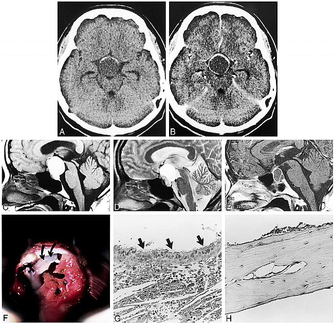
Calcification in Rathke's cleft cyst in a 23-year-old woman.

A, Presurgical axial CT scan of head shows crescent-like calcification in wall of suprasellar cyst.

B, Postcontrast CT scan reveals thin-ring enhancement of wall of cyst.

C, Presurgical sagittal T1-weighted MR image (400/15/2 [TR/TE/excitations]) shows hyperintense cyst with small nodule in inferior wall of cavity. Character of cyst wall is not apparent on this image.

D, Sagittal T2-weighted MR image (5000/90/1) depicts a small, nodular lesion in the anterior ventral portion of cyst. Note that cyst wall is not well defined on MR images.

E, T1-weighted MR image (400/14/2), after contrast administration prior to second surgery, shows an intra- and suprasellar cyst with enhancing thin wall and septum.

F, lntraoperative photograph shows a bony plate (curved arrows) in cystic cavity underneath incised dura of sellar floor (transsphenoidal approach).

G, Photomicrographs of biopsy specimen. Cyst is lined by a layer of columnar cells with cilia (arrows) (hematoxylin-eosin–stained section, × 200).

H, Photomicrographs of a plate of mature bone removed from cyst cavity in hematoxylin-eosin–stained section (× 200).

Preoperative diagnosis was an intrasellar craniopharyngioma or a Rathke's cleft cyst. In 1994, the patient underwent pituitary surgery; a transsphenoidal approach was applied. Thick, yellow fluid and small, solid fragments were removed from the cyst. The cavity was thoroughly irrigated. The diaphragma sellae was covered with calcification and did not appear to have descended into the sella during surgery. Only a small part of the cyst wall was removed from the floor of the sella. Histologic examination revealed nonspecific degenerative tissue with xanthogranulomatous change. We had suggested the preoperative diagnosis of craniopharyngioma because of the CT findings of calcification and from the intraoperative observation of the sella, but we reserved complete transcranial resection or irradiation therapy because of inconclusive histopathologic findings.

The patient was discharged and regular follow-up MR imaging was scheduled. She received another course of treatment that was identical to that of her preoperative gonadotropin regimen. The cyst recurred 2 years after the first surgery, and MR imaging showed a gradual increase in size (Fig 1E). On admission, she had no abnormal neurologic findings. Endocrinologic examination showed similar results to the previous hospitalizaion. Skull radiography revealed a thin linear calcification in the suprasellar region.

Transsphenoidal surgery was performed 4 years after the patient's initial operation. The cyst contained thick, yellow fluid with glistening particles. A plate of bone was firmly attached to the diaphragma in the anterior part of the sella. Approximately half of the calcification along the cyst wall was removed from the cavity (Fig 1F). A part of the capsule was removed from the sellar floor. Neither anterior nor posterior lobes of the pituitary gland were identified in the sella. The diaphragma sellae did not descend into the sella. Dissection on the posterior wall of the cyst caused CSF leakage, which was controlled with an autologous fat implant. The patient's postoperative course was uneventful, and she was discharged for further gynecologic treatment.

Histologic study of the cyst wall revealed a layer of columnar epithelial cells with cilia, leading to the diagnosis of Rathke's cleft cyst (Fig 1G). The columnar cells covered the connective tissue with nonspecific inflammatory cells. These tissues have no calcification or bone formation. Histologic examination also showed a plate of mature bone without neoplastic cells (Fig 1H).

Discussion

Rathke's cleft cyst may arise between the anterior and intermediate lobes of the pituitary gland if the lumen of Rathke's pouch, which develops as an out-pouching of stomodeum, is not obliterated by the twelfth week of gestation (3). Other authors have suggested an origin arising from neuroepithelium, endoderm, or reverse metaplasia (3, 4). Histopathologically, the cyst is composed of a layer of cuboidal or columnar epithelium on a basement membrane without neoplastic transformation (5). The CT feature of Rathke's cleft cyst consists of a low-density, well-circumscribed, nonenhancing sellar mass with suprasellar extension. Several unusual findings have been reported: precontrast iso- or hyperdense and mixed lesions, cyst wall enhancement, and cyst wall calcification (1, 6). On MR images, Rathke's cleft cysts show various signal intensities (2, 3, 6). Because of their frequent presentation and characteristically benign nature, Rathke's cleft cysts should be included in the differential diagnosis of any sellar or suprasellar cyst.

The majority of sellar cysts are craniopharyngiomas, Rathke's cleft cysts, cystic pituitary adenomas, and arachnoid cysts. Preoperative differential diagnosis is important, because the appropriate treatment for these lesions differs according to type. Calcification is considered an important clue to differentiate craniopharyngiomas from other cystic lesions.

Craniopharyngiomas have a high frequency of calcification on CT scans (7). Ossification and teeth formations have been reported in several cases of adamantinous-type craniopharyngioma (8, 9). On the other hand, calcification was reported in only 0.2% to 8% of pituitary adenomas in radiologic studies (10), and ossification in adenomas was reported in two cases (11, 12). Though Rathke's cleft cysts are long-standing benign cysts, they rarely present with histologic findings of calcification or bone formation. It should be recognized, however, that a small percentage of Rathke's cleft cysts may have mural calcification. Eight cases with calcification have been reported in the literature (13–18), and two of those eight, along with our case, presented with histologic findings of bone formation (16, 18).

Calcification in suprasellar tumors has been classified into three types according to the shape and histologic distribution: nodular or floccular, more common cystic or curvilinear, and mixed (19). The calcification in the Rathke's cleft cysts of all reported cases, including ours, is exclusively curvilinear in shape, except for one speck-like calcification in a 74-year-old man (15). Alternatively, the majority of craniopharyngiomas have floccular-type calcification (19). Therefore, the shape of calcification may help differentiate between Rathke's cleft cyst and craniopharyngioma.

Mural calcification may represent nonspecific deposits originating from meningeal tissue or epithelial lining of the cysts. On the other hand, bone formation may be caused by osteoid metaplasia in mesenchymal cells by inflammatory, chemical, or mechanical stimulation (12). Chronic degenerative change and mechanical stimulation in the cyst might cause metaplastic ossification of the wall, as in the present case.

If a Rathke's cleft cyst is symptomatic, it should be treated carefully with simple evacuation, irrigation, and biopsy via a transsphenoidal route to avoid postoperative pituitary insufficiency. Craniopharyngiomas, however, require a different treatment strategy, including the choice of meticulous dissection from the hypothalamus and other surrounding structures or irradiation or both. Our case demonstrates that although a coexisting cyst and calcification may lead to speculation of a craniopharyngioma by either radiologic or intraoperative findings, the diagnosis and decision for extensive surgery must be made on the basis of histopathologic analysis.

Footnotes

  • ↵1 Address reprint requests to Y. Nakasu, M.D., Department of Neurosurgery, Shiga University of Medical Science, Shiga, 520–2192 Japan.

References

  1. ↵
    Kucharczyk W, Peck WW, Kelly WM, Norman D, Newton TH. Rathke cleft cysts: CT, MR imaging and pathologic features. Radiology 1987;165:491-495
    PubMed
  2. ↵
    Nakasu Y, Isozumi T, Nakasu S, Handa J. Rathke's cleft cyst: Computed tomographic scan and magnetic resonance imaging. Acta Neurochir (Wien) 1990;103:99-104
    CrossRefPubMed
  3. ↵
    Ross DA, Norman D, Wilson CB. Radiological characteristics and results of surgical management of Rathke's cysts in 43 patients. Neurosurgery 1992;30:173-179
    PubMed
  4. Voelker JL, Campbell RL, Muller J. Clinical, radiographic, and pathological features of symptomatic Rathke's cleft cysts. J Neurosurg 1991;74:535-544
    PubMed
  5. ↵
    Keyaki A, Hirano A, Llena JF. Asymptomatic and symptomatic Rathke's cleft cysts. Histological study of 45 cases. Neurol Med Chir (Tokyo) 1989;29:88-93
    PubMed
  6. El-Mahdy W, Powell M. Transsphenoidal management of 28 symptomatic Rathke's cleft cysts, with special reference to visual and hormonal recovery. Neurosurgery 1998;42:7-17
    PubMed
  7. ↵
    Eldevik OP, Blaivas M, Gabrielsen TO, Hald JK, Chandler WF. Craniopharyngioma: radiologic and histologic findings and recurrence. AJNR Am J Neuroradiol 1996;17:1427-1439
    Abstract
  8. ↵
    Alvarez-Garijo JA, Froufe A, Taboada D, Vila M. Successful surgical treatment of an odontogenic ossified craniopharyngioma. J Neurosurg 1981;55:832-835
    PubMed
  9. Palaoglu S, Akbay A, Mocan G, et al. Ossified adamantinous type craniopharyngiomas. A series of 13 patients. Acta Neurochir (Wien) 1994;127:166-169
    PubMed
  10. ↵
    Horiuchi T, Tanaka Y, Kobayashi S, Yokoh A, Unoki T. Total capsular calcification in a prolactinoma. Case report. Neurol Med Chir (Tokyo) 1996;36:729-732
    PubMed
  11. ↵
    Mukada K, Ohta M, Uozumi T, Arita K, Kurisu K, Inai K. Ossified prolactinoma: case report. Neurosurgery 1987;20:473-475
    PubMed
  12. ↵
    Palaoglu S, Bavbek M, Peker S, Onol B, Sungur A, Erbengi A. Ossified somatotropinoma. Surg Neurol 1994;41:143-146
    PubMed
  13. ↵
    Worster-Drought C, Dickson WEC, Archer BWC. Dyspituitarism of the Lorain type, associated with a pituitary cyst arising from Rathke's cleft and secondary lesions in the hypothalamic region and ventricles. Brain 1927;50:704-718
    FREE Full Text
  14. Bayoumi ML. Rathke's cleft and its cysts. Edinburgh Med J 1948;55:745-749
    PubMed
  15. ↵
    Naiken VS, Tellem M, Meranze DR. Pituitary cyst of Rathke's cleft origin with hypopituitarism. J Neurosurg 1961;18:703-708
  16. ↵
    Adelman LS, Post KD. Calcification in Rathke's cleft cysts (Letter). J Neurosurg 1977;47:641
  17. Tajika Y, Kubo O, Kamiya M, et al. Clinico-pathological features of 5 cases of pituitary cyst including Rathke's cleft cyst. Neurol Surg (Tokyo) 1982;10:1055-1064
  18. Matsumori K, Okada T, Nakayama K, Miyasaka Y, Beppu T. A case of calcified prolactinoma combined with Rathke's cleft cyst. Neurol Surg (Tokyo) 1984;12:833-838
  19. ↵
    Burrows EH. Pathologic intracranial calcification: Neoplasms. In: Newton TH, Potts DG. Radiology of the skull and brain, Vol.1. Book 2, The skull. St Louis, MO: Mosby; 1971: 848–873
  • Received October 29, 1998.
  • Accepted after revision March 10, 1999.
  • Copyright © American Society of Neuroradiology
View Abstract
PreviousNext
Back to top

In this issue

American Journal of Neuroradiology
Vol. 20, Issue 7
1 Jul 1999
  • Table of Contents
  • Index by author
Advertisement
Print
Download PDF
Email Article

Thank you for your interest in spreading the word on American Journal of Neuroradiology.

NOTE: We only request your email address so that the person you are recommending the page to knows that you wanted them to see it, and that it is not junk mail. We do not capture any email address.

Enter multiple addresses on separate lines or separate them with commas.
Atypical Rathke's Cleft Cyst Associated with Ossification
(Your Name) has sent you a message from American Journal of Neuroradiology
(Your Name) thought you would like to see the American Journal of Neuroradiology web site.
CAPTCHA
This question is for testing whether or not you are a human visitor and to prevent automated spam submissions.
Cite this article
Yoko Nakasu, Satoshi Nakasu, Masayuki Nakajima, Ryuta Itoh, Masayuki Matsuda
Atypical Rathke's Cleft Cyst Associated with Ossification
American Journal of Neuroradiology Jul 1999, 20 (7) 1287-1289;

Citation Manager Formats

  • BibTeX
  • Bookends
  • EasyBib
  • EndNote (tagged)
  • EndNote 8 (xml)
  • Medlars
  • Mendeley
  • Papers
  • RefWorks Tagged
  • Ref Manager
  • RIS
  • Zotero
0 Responses
Respond to this article
Share
Bookmark this article
Atypical Rathke's Cleft Cyst Associated with Ossification
Yoko Nakasu, Satoshi Nakasu, Masayuki Nakajima, Ryuta Itoh, Masayuki Matsuda
American Journal of Neuroradiology Jul 1999, 20 (7) 1287-1289;
del.icio.us logo Twitter logo Facebook logo Mendeley logo
  • Tweet Widget
  • Facebook Like
  • Google Plus One

Jump to section

  • Article
    • Abstract
    • Case Report
    • Discussion
    • Footnotes
    • References
  • Figures & Data
  • Info & Metrics
  • Responses
  • References
  • PDF

Related Articles

  • No related articles found.
  • PubMed
  • Google Scholar

Cited By...

  • No citing articles found.
  • Crossref
  • Google Scholar

This article has not yet been cited by articles in journals that are participating in Crossref Cited-by Linking.

More in this TOC Section

  • Usefulness of Quantitative Susceptibility Mapping for the Diagnosis of Parkinson Disease
  • White Matter Alterations in the Brains of Patients with Active, Remitted, and Cured Cushing Syndrome: A DTI Study
  • Qualitative and Quantitative Analysis of MR Imaging Findings in Patients with Middle Cerebral Artery Stroke Implanted with Mesenchymal Stem Cells
Show more BRAIN

Similar Articles

Advertisement

Indexed Content

  • Current Issue
  • Accepted Manuscripts
  • Article Preview
  • Past Issues
  • Editorials
  • Editor's Choice
  • Fellows' Journal Club
  • Letters to the Editor
  • Video Articles

Cases

  • Case Collection
  • Archive - Case of the Week
  • Archive - Case of the Month
  • Archive - Classic Case

Special Collections

  • AJNR Awards
  • ASNR Foundation Special Collection
  • Most Impactful AJNR Articles
  • Photon-Counting CT
  • Spinal CSF Leak Articles (Jan 2020-June 2024)

More from AJNR

  • Trainee Corner
  • Imaging Protocols
  • MRI Safety Corner

Multimedia

  • AJNR Podcasts
  • AJNR Scantastics

Resources

  • Turnaround Time
  • Submit a Manuscript
  • Submit a Video Article
  • Submit an eLetter to the Editor/Response
  • Manuscript Submission Guidelines
  • Statistical Tips
  • Fast Publishing of Accepted Manuscripts
  • Graphical Abstract Preparation
  • Imaging Protocol Submission
  • Evidence-Based Medicine Level Guide
  • Publishing Checklists
  • Author Policies
  • Become a Reviewer/Academy of Reviewers
  • News and Updates

About Us

  • About AJNR
  • Editorial Board
  • Editorial Board Alumni
  • Alerts
  • Permissions
  • Not an AJNR Subscriber? Join Now
  • Advertise with Us
  • Librarian Resources
  • Feedback
  • Terms and Conditions
  • AJNR Editorial Board Alumni

American Society of Neuroradiology

  • Not an ASNR Member? Join Now

© 2025 by the American Society of Neuroradiology All rights, including for text and data mining, AI training, and similar technologies, are reserved.
Print ISSN: 0195-6108 Online ISSN: 1936-959X

Powered by HighWire