Advertisement

Main menu

  • Home
  • Content
    • Current Issue
    • Accepted Manuscripts
    • Article Preview
    • Past Issue Archive
    • Video Articles
    • AJNR Case Collection
    • Case of the Week Archive
    • Case of the Month Archive
    • Classic Case Archive
  • Special Collections
    • AJNR Awards
    • ASNR Foundation Special Collection
    • Most Impactful AJNR Articles
    • Photon-Counting CT
    • Spinal CSF Leak Articles (Jan 2020-June 2024)
  • Multimedia
    • AJNR Podcasts
    • AJNR SCANtastic
    • Trainee Corner
    • MRI Safety Corner
    • Imaging Protocols
  • For Authors
    • Submit a Manuscript
    • Submit a Video Article
    • Submit an eLetter to the Editor/Response
    • Manuscript Submission Guidelines
    • Statistical Tips
    • Fast Publishing of Accepted Manuscripts
    • Graphical Abstract Preparation
    • Imaging Protocol Submission
    • Author Policies
  • About Us
    • About AJNR
    • Editorial Board
    • Editorial Board Alumni
  • More
    • Become a Reviewer/Academy of Reviewers
    • Subscribers
    • Permissions
    • Alerts
    • Feedback
    • Advertisers
    • ASNR Home

User menu

  • Alerts
  • Log in

Search

  • Advanced search
American Journal of Neuroradiology

American Journal of Neuroradiology

ASHNR American Society of Functional Neuroradiology ASHNR American Society of Pediatric Neuroradiology ASSR
  • Alerts
  • Log in

Advanced Search

  • Home
  • Content
    • Current Issue
    • Accepted Manuscripts
    • Article Preview
    • Past Issue Archive
    • Video Articles
    • AJNR Case Collection
    • Case of the Week Archive
    • Case of the Month Archive
    • Classic Case Archive
  • Special Collections
    • AJNR Awards
    • ASNR Foundation Special Collection
    • Most Impactful AJNR Articles
    • Photon-Counting CT
    • Spinal CSF Leak Articles (Jan 2020-June 2024)
  • Multimedia
    • AJNR Podcasts
    • AJNR SCANtastic
    • Trainee Corner
    • MRI Safety Corner
    • Imaging Protocols
  • For Authors
    • Submit a Manuscript
    • Submit a Video Article
    • Submit an eLetter to the Editor/Response
    • Manuscript Submission Guidelines
    • Statistical Tips
    • Fast Publishing of Accepted Manuscripts
    • Graphical Abstract Preparation
    • Imaging Protocol Submission
    • Author Policies
  • About Us
    • About AJNR
    • Editorial Board
    • Editorial Board Alumni
  • More
    • Become a Reviewer/Academy of Reviewers
    • Subscribers
    • Permissions
    • Alerts
    • Feedback
    • Advertisers
    • ASNR Home
  • Follow AJNR on Twitter
  • Visit AJNR on Facebook
  • Follow AJNR on Instagram
  • Join AJNR on LinkedIn
  • RSS Feeds

AJNR is seeking candidates for the AJNR Podcast Editor. Read the position description.

LetterLetter

Schimmelpenning Syndrome: A Kind of Craniofacial Epidermal Nevus Associated with Cerebral and Ocular MR Imaging Abnormalities

C. Amato, M. Elia and C. Schepis
American Journal of Neuroradiology May 2010, 31 (5) E47-E48; DOI: https://doi.org/10.3174/ajnr.A2062
C. Amato
  • Find this author on Google Scholar
  • Find this author on PubMed
  • Search for this author on this site
M. Elia
  • Find this author on Google Scholar
  • Find this author on PubMed
  • Search for this author on this site
C. Schepis
  • Find this author on Google Scholar
  • Find this author on PubMed
  • Search for this author on this site
  • Article
  • Figures & Data
  • Info & Metrics
  • Responses
  • References
  • PDF
Loading

This letter focuses on Schimmelpenning syndrome (SS), a neurocutaneous disorder related to epidermal nevus syndromes and characterized by craniofacial nevus, neurologic anomalies, and ocular pathology.

We discuss clinical and MR imaging features of a 10-year-old boy, pointing out the etiopathologic substratum of this condition and the vascular origin of brain abnormalities; in our case, there is an unusual association of hemimacrocephaly and ipsilateral cerebral hemiatrophy.

Epidermal nevus syndrome is a rare phacomatosis characterized by epidermal nevi in association with neurologic disorders and ocular or skeletal anomalies; other organ systems may be involved with hamartomas. The term “epidermal nevus syndrome” does not indicate 1 disease but rather a heterogeneous group of neurocutaneous disorders with a distinct genetic profile and a similar cutaneous phenotype.1 These conditions may differ from one another either by type and site of nevi or by distinctive clinical correlations.

SS represents the best definition for a craniofacial sebaceus nevus associated with difficult seizures, mental retardation, and ocular lesions, such as coloboma or epibulbar lipodermoid; skull deformity is usually present. Cerebral manifestations include developmental anomalies, vascular lesions, tumors, and tumorlike conditions, which are all generally ipsilateral to the nevi.2,3

The classic form of SS is characterized by craniofacial hemihypertrophy, hemimegalencephaly, and pachygyria-polymicrogyria. The reported case of a large epidermal nevus of the head and neck differs from the classic form and consists of cerebral hemiatrophy with normal cortical organization, vascular anomalies, and giant Virchow-Robin spaces (VRSs).

The pathogenesis and clinical expression of SS and correlate syndromes are based on genomic mosaicism, in which ≥2 genotypical different clones of the same specific cell line coexist in a single individual.

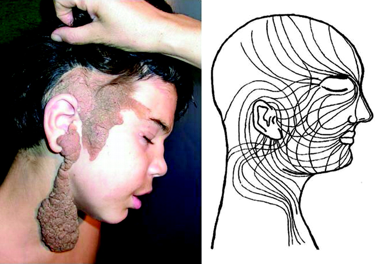
Epidermal and pigmentary cells originate early in the neural crest and, after proliferation, migrate to the periphery of the embryo. In neurocutaneous disorders, a somatic mutation at this time originates an altered cellular clone that will be phenotypically represented along the so-called lines of Blaschko (Fig 1). These lines indicate the pathway of embryonic ectoderm and are clinically apparent when recognizable from surrounding normal cells, thus representing the cutaneous expression of mosaicism. When genetic mutations occur early and their products do not cause the death of the embryo because of involvement of only some cell lineages, by the lethal gene theory,4 skin and the embryogenetically contiguous central nervous system will both be damaged.

Fig 1.
  • Download figure
  • Open in new tab
  • Download powerpoint
Fig 1.

A, Patient at 10 years old with a large, tumefactive, and hyperkeratotic epidermal nevus involving the skull, face, and neck. B, Note lines of Blaschko on the head and neck. Modified from Moss et al.4

The neurologic abnormalities are strictly related to the timing of these events. If mutation occurs during neuronal migration or cortical organization, there will be brain malformations; whereas if it happens later, atrophy and vascular anomalies will be more evident.

In our patient, unilateral hypoplasia of the arterial system, Sylvian stenosis, and ischemic changes in the temporal lobe (Figs 2 and 3) should refer to a malformative vasculopathy that occurred late in utero or at least after brain gyration and seriously reduced hemispheric perfusion. In fact, patients with SS are at risk of vascular anomalies, as reported in approximately one-third of published cases; among these anomalies are aortic coarctation and aneurysm, and renal artery and carotid stenosis.5

Fig 2.
  • Download figure
  • Open in new tab
  • Download powerpoint
Fig 2.

MR angiography of the patient at 2 years old shows a hypoplastic right siphon and middle cerebral artery, with a stenotic Sylvian bifurcation.

Fig 3.
  • Download figure
  • Open in new tab
  • Download powerpoint
Fig 3.

A, Coronal T2-weighted image of the patient at 20 days old shows a right cerebral hemiatrophy with cortical thinning and mild parenchymal change in the temporal lobe (arrow). On the same side, macrocrania and dilated liquoral spaces are present. B, Coronal T2-weighted image of the patient at 2 years old shows hypomyelination and an old infarct (arrow) in the right temporal lobe. Next to the ventricle, confluent and enlarged VRSs are recognizable. C, MR image of the patient at 10 years old. Coronal T2-weighted image shows, on the right side, stable cerebral hemiatrophy, a giant VRS next to the ventricle, and old ischemic changes within the hypomyelinated temporal lobe. D, High-resolution axial T2-weighted image shows a scleroatrophic right eyeball with a hypoplastic optic nerve. Lipodermoid tissue in the lacrimal fossa is present (arrow).

In conclusion, our patient exhibited typical features of SS with craniofacial epidermal nevus, ipsilateral cerebral abnormalities, difficult epilepsy, mental retardation, and ocular pathology; such characteristics give us the possibility of distinguishing this condition from the other related disorders. The relevance of this case is in the unusual association of hemimacrocrania and ipsilateral cerebral atrophy; moreover, brain abnormalities are the result of an in utero vasculopathy and do not include cortical dysplasia as in most of the previous reports.

References

  1. 1.↵
    1. Happle R
    . Epidermal nevus syndrome. Semin Dermatol 1995;14:111–21
    CrossRefPubMed
  2. 2.↵
    1. Sugarman JL
    . Epidermal nevus syndrome. Semin Cutan Med Surg 2007;26:221–30
    CrossRefPubMed
  3. 3.↵
    1. Zhang W,
    2. Simos PG,
    3. Ishibashi I,
    4. et al
    . Neuroimaging features of epidermal nevus syndrome. AJNR Am J Neuroradiol 2003;24:1468–70
    Abstract/FREE Full Text
  4. 4.↵
    1. Moss C,
    2. Larkins S,
    3. Stacey M,
    4. et al
    . Epidermal mosaicism and Blaschko's lines. J Med Genet 1993;30:752–55
    Abstract/FREE Full Text
  5. 5.↵
    1. Greene AK,
    2. Rogers GF,
    3. Mulliken JB
    . Schimmelpenning syndrome: an association with vascular anomalies. Cleft Palate Craniofac J 2007;44:208–15
    CrossRefPubMed
  • Copyright © American Society of Neuroradiology
PreviousNext
Back to top

In this issue

American Journal of Neuroradiology: 31 (5)
American Journal of Neuroradiology
Vol. 31, Issue 5
1 May 2010
  • Table of Contents
  • Index by author
Advertisement
Print
Download PDF
Email Article

Thank you for your interest in spreading the word on American Journal of Neuroradiology.

NOTE: We only request your email address so that the person you are recommending the page to knows that you wanted them to see it, and that it is not junk mail. We do not capture any email address.

Enter multiple addresses on separate lines or separate them with commas.
Schimmelpenning Syndrome: A Kind of Craniofacial Epidermal Nevus Associated with Cerebral and Ocular MR Imaging Abnormalities
(Your Name) has sent you a message from American Journal of Neuroradiology
(Your Name) thought you would like to see the American Journal of Neuroradiology web site.
CAPTCHA
This question is for testing whether or not you are a human visitor and to prevent automated spam submissions.
Cite this article
C. Amato, M. Elia, C. Schepis
Schimmelpenning Syndrome: A Kind of Craniofacial Epidermal Nevus Associated with Cerebral and Ocular MR Imaging Abnormalities
American Journal of Neuroradiology May 2010, 31 (5) E47-E48; DOI: 10.3174/ajnr.A2062

Citation Manager Formats

  • BibTeX
  • Bookends
  • EasyBib
  • EndNote (tagged)
  • EndNote 8 (xml)
  • Medlars
  • Mendeley
  • Papers
  • RefWorks Tagged
  • Ref Manager
  • RIS
  • Zotero
0 Responses
Respond to this article
Share
Bookmark this article
Schimmelpenning Syndrome: A Kind of Craniofacial Epidermal Nevus Associated with Cerebral and Ocular MR Imaging Abnormalities
C. Amato, M. Elia, C. Schepis
American Journal of Neuroradiology May 2010, 31 (5) E47-E48; DOI: 10.3174/ajnr.A2062
del.icio.us logo Twitter logo Facebook logo Mendeley logo
  • Tweet Widget
  • Facebook Like
  • Google Plus One
Purchase

Jump to section

  • Article
    • References
  • Figures & Data
  • Info & Metrics
  • Responses
  • References
  • PDF

Related Articles

  • No related articles found.
  • PubMed
  • Google Scholar

Cited By...

  • Schimmelpenning-Feuerstein-Mims syndrome with benign enlargement of subarachnoid space in infancy
  • Oral HRAS Mutation in Orofacial Nevus Sebaceous Syndrome (Schimmelpenning-Feuerstein-Mims-Syndrome): A Case Report With a Literature Survey
  • Crossref (7)
  • Google Scholar

This article has been cited by the following articles in journals that are participating in Crossref Cited-by Linking.

  • Multilineage somatic activating mutations in HRAS and NRAS cause mosaic cutaneous and skeletal lesions, elevated FGF23 and hypophosphatemia
    Young H. Lim, Diana Ovejero, Jeffrey S. Sugarman, Cynthia M.C. DeKlotz, Ann Maruri, Lawrence F. Eichenfield, Patrick K. Kelley, Harald Jüppner, Michael Gottschalk, Cynthia J. Tifft, Rachel I. Gafni, Alison M. Boyce, Edward W. Cowen, Nisan Bhattacharyya, Lori C. Guthrie, William A. Gahl, Gretchen Golas, Erin C. Loring, John D. Overton, Shrikant M. Mane, Richard P. Lifton, Moise L. Levy, Michael T. Collins, Keith A. Choate
    Human Molecular Genetics 2014 23 2
  • Ocular manifestations of genetic skin disorders
    Melinda Jen, Sudha Nallasamy
    Clinics in Dermatology 2016 34 2
  • Do you know this syndrome? Schimmelpenning-Feuerstein-Mims syndrome
    Clarissa Patias Lena, Rogério Nabor Kondo, Théo Nicolacópulos
    Anais Brasileiros de Dermatologia 2019 94 2
  • Clinicopathological and molecular insights into odontogenic tumors associated with syndromes: A comprehensive review
    Lauren Frenzel Schuch, Felipe Martins Silveira, Vanesa Pereira-Prado, Estefania Sicco, Deepak Pandiar, Mariana Villarroel-Dorrego, Ronell Bologna-Molina
    World Journal of Experimental Medicine 2024 14 4
  • Schimmelpenning-Feuerstein-Mims syndrome with benign enlargement of subarachnoid space in infancy
    Shadab B Maldar, Christopher Jude Pinto, Uday Kumar, Swathi Pitchiammal Vairaprakasam
    BMJ Case Reports 2023 16 11
  • Cutaneous findings in children with intellectual disabilities
    Carmelo Schepis
    The Journal of Dermatology 2013 40 1
  • Unveiling the complexity of Schimmelpenning–Feuerstein–Mims syndrome: A comprehensive case study
    Guillermo Raul Vera-Duarte, Ruth Eskenazi-Betech, Isabel De la Fuente-Batta, David Carreño-Bolaños, Oscar F. Chacón-Camacho, Juan C. Zenteno, Enrique O. Graue-Hernandez
    American Journal of Ophthalmology Case Reports 2025 38

More in this TOC Section

  • Brain AVM’s Nidus: What if We Hadn’t Understood Anything?
  • Letter to the Editor regarding “Automated Volumetric Software in Dementia: Help or Hindrance to the Neuroradiologist?”
  • Reply:
Show more Letters

Similar Articles

Advertisement

Indexed Content

  • Current Issue
  • Accepted Manuscripts
  • Article Preview
  • Past Issues
  • Editorials
  • Editor's Choice
  • Fellows' Journal Club
  • Letters to the Editor
  • Video Articles

Cases

  • Case Collection
  • Archive - Case of the Week
  • Archive - Case of the Month
  • Archive - Classic Case

Special Collections

  • AJNR Awards
  • ASNR Foundation Special Collection
  • Most Impactful AJNR Articles
  • Photon-Counting CT
  • Spinal CSF Leak Articles (Jan 2020-June 2024)

More from AJNR

  • Trainee Corner
  • Imaging Protocols
  • MRI Safety Corner

Multimedia

  • AJNR Podcasts
  • AJNR Scantastics

Resources

  • Turnaround Time
  • Submit a Manuscript
  • Submit a Video Article
  • Submit an eLetter to the Editor/Response
  • Manuscript Submission Guidelines
  • Statistical Tips
  • Fast Publishing of Accepted Manuscripts
  • Graphical Abstract Preparation
  • Imaging Protocol Submission
  • Evidence-Based Medicine Level Guide
  • Publishing Checklists
  • Author Policies
  • Become a Reviewer/Academy of Reviewers
  • News and Updates

About Us

  • About AJNR
  • Editorial Board
  • Editorial Board Alumni
  • Alerts
  • Permissions
  • Not an AJNR Subscriber? Join Now
  • Advertise with Us
  • Librarian Resources
  • Feedback
  • Terms and Conditions
  • AJNR Editorial Board Alumni

American Society of Neuroradiology

  • Not an ASNR Member? Join Now

© 2025 by the American Society of Neuroradiology All rights, including for text and data mining, AI training, and similar technologies, are reserved.
Print ISSN: 0195-6108 Online ISSN: 1936-959X

Powered by HighWire