
Article Figures & Data
Figures
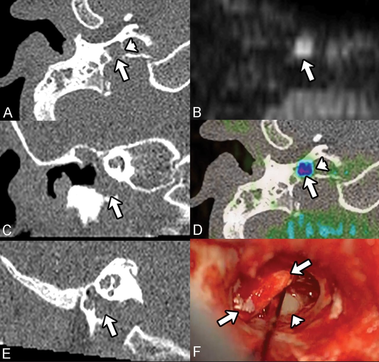
- FIGURE.
Example of rMEC leading to a carotid canal erosion. Recurrent middle ear cholesteatoma (axial b=1000 non-EPI MR imaging, B, arrow) probably located (rated Likert 4) in the hypotympanum, on uncoregistered dataset (A, B, C, E, arrows). The hypotympanum location is clear (rated Likert 5), considering that the fused dataset (b=1000/CT in axial plane, D, arrow) and the vertical carotid canal lysis (A and D, arrowheads) can be seen. The perioperative surgical findings (F) confirmed the hypotympanum rMEC location (arrows) and the carotid canal lysis (arrowhead). In that case, the presurgical image fusion allowed the surgeon to adapt his approach, thus lowering the surgical risks.

Tables
T1WI T2WI SE DWI 3D-T1 VIBE TR (ms) 610 4460 2000 9.19 TE (ms) 14 95 105 3.34 FOV (mm) 150 × 150 166 × 129 220 × 220 172 × 200 NEX 2 2 8 2 Slice thickness (mm) 2 2 3 0.7 Acquisition time (min:sec) 2:11 2 3:12 3:23 Matrix size 358 × 512 344 × 384 144 × 192 213 × 288 B-values b=1000 Note:—SE indicates spin-echo.
Anatomic Location No.a PREV UD Exact FD Exact UD Reliab FD Reliab Epitympanic recess 18 83% 94% 100% 83% 97% Posterior epitympanic recess 18 78% 100% 100% 92% 100% Aditus of mastoid antrum 18 72% 72% 100% 72% 94% Tegmen antri 18 67% 67% 100% 64% 97% Anterior epitympanic recess 18 67% 67% 100% 83% 83% Tegmen tympani 18 61% 83% 100% 81% 94% Mastoidectomy cavity 18 56% 78% 100% 89% 94% Facial nerve: tympanic segment 18 44% 67% 100% 64% 89% Promontory of tympanic cavity 18 33% 94% 100% 86% 97% Oval window/stapes 18 33% 83% 100% 83% 97% Mesotympanum 18 28% 72% 100% 69% 100% Vestibule 18 22% 78% 100% 97% 89% Facial nerve: second genu 18 22% 83% 100% 81% 97% Sinus tympani 18 17% 83% 94% 86% 97% Eustachian tube 18 11% 89% 100% 86% 100% Dura mater 18 11% 89% 100% 94% 94% Round window 18 6% 83% 100% 78% 100% Carotid canal 18 6% 94% 100% 94% 97% Facial nerve: geniculum 18 6% 89% 100% 83% 97% Cochlea 18 0% 100% 100% 100% 100% Temporal lobe 18 0% 100% 100% 97% 100% Facial nerve: mastoid segment 18 0% 100% 100% 94% 100% Note:—PREV indicates surgical prevalence; Exact, correctly identified and correctly rejected findings (ie, true-positive + true-negative considering surgical findings as the criterion standard); Reliab, reliability score.
↵a Number of lesions.
Mean Range (%) No. of patients 20 Mean age (yr) 41.2 (11.1–70.9) Female/male (%) 35/65 Surgery No. of previous interventions 2.9 (1–8) Right/left (%) 55/45 Time to recurrence (mo) 70.6 (9–430) MRI to surgery (mo) 2.1 (0–10) MRI True-positive in DWI 18 90% False-negative in DWI 2 10% Fusion quality Good 9 50% Perfect 9 50% ICC ICC-CI Preci Interpret P Valuea Obs1 (vol 1–vol 3) 0.93 0.82–0.97 8% Almost perfect <.001 Obs2 (vol 2–vol 4) 0.86 0.67–0.95 14% Almost perfect <.001 Unfused (vol 1–vol 2) 0.91 0.78–0.97 10% Almost perfect <.001 Fused (vol 3–vol 4) 0.95 0.88–0.99 6% Almost perfect <.001 Note:—Preci indicates precision of ICC estimate; Interpret, interpretation of ICC; Obs, observer; vol, volume.
↵a Statistical significance using an ANOVA model for ICC.


Jump to section
Related Articles
Cited By...
- Diffusion Analysis of Intracranial Epidermoid, Head and Neck Epidermal Inclusion Cyst, and Temporal Bone Cholesteatoma
- Fusion of middle ear optical coherence tomography and computed tomography in three ears
- Comparison of the Utility of High-Resolution CT-DWI and T2WI-DWI Fusion Images for the Localization of Cholesteatoma