
Article Figures & Data
Figures
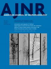
- FIG 1.
Structural abnormalities in fetuses with brain injury on T2- and T1-weighted images. A, Coronal T2 HASTE in a 35.3-week fetus (subject 5, scan 1) shows localized T2 prolongation (arrow), volume loss, and ventriculomegaly. B, Coronal T2 HASTE in a 21.9-week fetus (subject 3, scan 2) shows T2 hypointensity in the periventricular region (arrow). C, Coronal T2 HASTE shows periventricular cystic change (arrow) in a 29.4-week fetus (subject 2, scan 2). Coronal T2 HASTE (D) and coronal T1 VIBE (E) in a 33-week fetus (subject 6, scan 3) show generalized T2 prolongation and cerebral edema (asterisk), periventricular T2 hypointensity (arrow in D), and corresponding T1 hyperintensity (arrow in E).
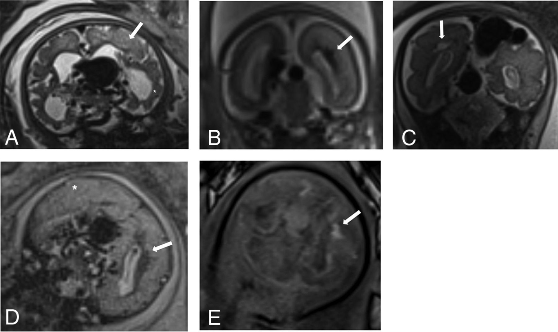
- FIG 2.
Abnormalities on T2*-weighted echo-planar sequences in fetuses with brain injury. A, Axial image in a 28.6-week fetus (subject 2, scan 1) shows blooming in the periventricular regions (arrows). B, Axial image in a 21.9-week fetus (subject 3, scan 2) shows blooming in the periventricular regions following the expected distribution of the proliferative compartments (germinal matrix [arrows]). C, Axial image in a 35.3-week fetus (subject 5, scan 1) shows generalized signal drop throughout the parenchyma.
- FIG 3.
Diffusion abnormalities in fetuses with brain injury. ADC (A) and diffusion trace (B) in a 35.3-week fetus (subject 5, scan 1) show localized restricted diffusion in the left frontal lobe (arrows). ADC (C) and diffusion trace (D) in a 33-week fetus (subject 6, scan 3) show generalized restricted diffusion throughout the parenchyma (manual ROI measurements revealed ADC in C < 700 mm2/s in the deep gray nuclei and white matter).
- FIG 4.
Progression of brain injury in 3 patients who underwent serial fetal MRIs. A and B, Subject 3, scan 1 and 2, at 20.4 weeks and then at 21.9 weeks when there is evidence of increased periventricular T2 hypointensity. C and D, Subject 2, scan 1 and 2, at 28.6 weeks and then at 29.4 weeks when there is evidence of a cystic change in the periventricular white matter and worsening of the T2 signal abnormality. E and F, Subject 6, scan 2 and 3, at 31.7 weeks and then at 33 weeks when there is generalized brain swelling and effacement of the extra-axial CSF in a pattern consistent with diffuse injury.


Tables
MRI Feature % (No.) Fetal brain parenchymal injury (n = 8) 26 (8/31) Structural abnormality (n = 8) 100 (8/8) Low volume 88 (7/8) Ventriculomegaly 50 (4/8) Signal abnormality (T1WI or T2WI) 88 (7/8) DWI abnormality (n = 5) 71 (5/7)a DWI data available for only 7 of 8 subjects T2* Abnormality (n = 5) 100 (5/5)a T2* data available for only 5 of 8 subjects Progression (n = 7) 100 (7/7)a ↵a Repeat scan data are available for only 7 of 8 subjects.
Subject Dx GA (wk) Structural Abnormality Low Volume Ventriculomegaly Signal Abnormality DWI Abnormality T2* Abnormality Documented Progression 1 VOGM 36.9 Yes Yes Yes Yes 2 NG-AVF 28.6 Yes No No Yes Yes Yes Fetal 29.4 Yes No No Yes No Yes 3 VOGM 20.4 Yes No No Yes Fetal 21.9 Yes Yes No Yes No Yes 4 VOGM 32.3 Yes Yes No No No Postnatala 5 VOGM 35.3 Yes Yes Yes Yes Yes Yes Postnatala 6 VOGM 29.3 Yes Yes No Yes No Yes Fetal 31.7 Yes Yes Yes Yes No Yes 33 Yes No No Yes Yes Yes 7 VOGM 26.7 No No No No Yes Fetal 30.3 Yes Yes Yes Yes No 8 NG-AVF 32.0 Yes Yes No Yes Yes Yes Fetal 35.0 Yes Yes No Yes No Yes Note:—Dx indicates diagnosis; GA, gestational age; wk, weeks.
↵a Immediate postnatal exam.


Jump to section
Related Articles
Cited By...
- Cerebrovascular Anomalies in the Fetus
- Reversal of vein of Galen aneurysmal malformation by stimulation of flow-mediated vessel fusion
- Comparing Vascular Morphology and Hemodynamics in Patients with Vein of Galen Malformations Using Intracranial 4D Flow MRI
- Fetal Brain Growth in the Early Second Trimester
- Overcoming roadblocks in clinical innovation via high fidelity simulation: use of a phantom simulator to achieve FDA and IRB approval of a clinical trial of fetal embolization of vein of Galen malformations