Advertisement

Main menu

  • Home
  • Content
    • Current Issue
    • Accepted Manuscripts
    • Article Preview
    • Past Issue Archive
    • Video Articles
    • AJNR Case Collection
    • Case of the Week Archive
    • Case of the Month Archive
    • Classic Case Archive
  • Special Collections
    • AJNR Awards
    • ASNR Foundation Special Collection
    • Most Impactful AJNR Articles
    • Photon-Counting CT
    • Spinal CSF Leak Articles (Jan 2020-June 2024)
  • Multimedia
    • AJNR Podcasts
    • AJNR SCANtastic
    • Trainee Corner
    • MRI Safety Corner
    • Imaging Protocols
  • For Authors
    • Submit a Manuscript
    • Submit a Video Article
    • Submit an eLetter to the Editor/Response
    • Manuscript Submission Guidelines
    • Statistical Tips
    • Fast Publishing of Accepted Manuscripts
    • Graphical Abstract Preparation
    • Imaging Protocol Submission
    • Author Policies
  • About Us
    • About AJNR
    • Editorial Board
    • Editorial Board Alumni
  • More
    • Become a Reviewer/Academy of Reviewers
    • Subscribers
    • Permissions
    • Alerts
    • Feedback
    • Advertisers
    • ASNR Home

User menu

  • Alerts
  • Log in

Search

  • Advanced search
American Journal of Neuroradiology

American Journal of Neuroradiology

ASHNR American Society of Functional Neuroradiology ASHNR American Society of Pediatric Neuroradiology ASSR
  • Alerts
  • Log in

Advanced Search

  • Home
  • Content
    • Current Issue
    • Accepted Manuscripts
    • Article Preview
    • Past Issue Archive
    • Video Articles
    • AJNR Case Collection
    • Case of the Week Archive
    • Case of the Month Archive
    • Classic Case Archive
  • Special Collections
    • AJNR Awards
    • ASNR Foundation Special Collection
    • Most Impactful AJNR Articles
    • Photon-Counting CT
    • Spinal CSF Leak Articles (Jan 2020-June 2024)
  • Multimedia
    • AJNR Podcasts
    • AJNR SCANtastic
    • Trainee Corner
    • MRI Safety Corner
    • Imaging Protocols
  • For Authors
    • Submit a Manuscript
    • Submit a Video Article
    • Submit an eLetter to the Editor/Response
    • Manuscript Submission Guidelines
    • Statistical Tips
    • Fast Publishing of Accepted Manuscripts
    • Graphical Abstract Preparation
    • Imaging Protocol Submission
    • Author Policies
  • About Us
    • About AJNR
    • Editorial Board
    • Editorial Board Alumni
  • More
    • Become a Reviewer/Academy of Reviewers
    • Subscribers
    • Permissions
    • Alerts
    • Feedback
    • Advertisers
    • ASNR Home
  • Follow AJNR on Twitter
  • Visit AJNR on Facebook
  • Follow AJNR on Instagram
  • Join AJNR on LinkedIn
  • RSS Feeds

AJNR is seeking candidates for the AJNR Podcast Editor. Read the position description.

Research ArticleADULT BRAIN
Open Access

Cerebellar Hypoperfusion in Migraine Attack: Incidence and Significance

F. Kellner-Weldon, M. El-Koussy, S. Jung, M. Jossen, P.P. Klinger-Gratz and R. Wiest
American Journal of Neuroradiology January 2018, DOI: https://doi.org/10.3174/ajnr.A5508
F. Kellner-Weldon
aFrom the Support Center for Advanced Neuroimaging (F.K.-W., M.E.-K., M.J., P.P.K.-G., R.W.), Institute for Diagnostic and Interventional Neuroradiology
  • Find this author on Google Scholar
  • Find this author on PubMed
  • Search for this author on this site
  • ORCID record for F. Kellner-Weldon
M. El-Koussy
aFrom the Support Center for Advanced Neuroimaging (F.K.-W., M.E.-K., M.J., P.P.K.-G., R.W.), Institute for Diagnostic and Interventional Neuroradiology
  • Find this author on Google Scholar
  • Find this author on PubMed
  • Search for this author on this site
  • ORCID record for M. El-Koussy
S. Jung
bDepartment of Neurology (S.J.), University Hospital Inselspital, Bern, Switzerland
  • Find this author on Google Scholar
  • Find this author on PubMed
  • Search for this author on this site
  • ORCID record for S. Jung
M. Jossen
aFrom the Support Center for Advanced Neuroimaging (F.K.-W., M.E.-K., M.J., P.P.K.-G., R.W.), Institute for Diagnostic and Interventional Neuroradiology
  • Find this author on Google Scholar
  • Find this author on PubMed
  • Search for this author on this site
  • ORCID record for M. Jossen
P.P. Klinger-Gratz
aFrom the Support Center for Advanced Neuroimaging (F.K.-W., M.E.-K., M.J., P.P.K.-G., R.W.), Institute for Diagnostic and Interventional Neuroradiology
cDepartment of Radiology (P.P.K.-G.), University of Basel, Basel, Switzerland.
  • Find this author on Google Scholar
  • Find this author on PubMed
  • Search for this author on this site
  • ORCID record for P.P. Klinger-Gratz
R. Wiest
aFrom the Support Center for Advanced Neuroimaging (F.K.-W., M.E.-K., M.J., P.P.K.-G., R.W.), Institute for Diagnostic and Interventional Neuroradiology
  • Find this author on Google Scholar
  • Find this author on PubMed
  • Search for this author on this site
  • ORCID record for R. Wiest
  • Article
  • Figures & Data
  • Info & Metrics
  • Responses
  • References
  • PDF
Loading

Abstract

BACKGROUND AND PURPOSE: Patients diagnosed with migraine with aura have an increased lifetime risk of ischemic stroke. It is not yet clear whether prolonged cortical hypoperfusion during an aura increases the immediate risk of cerebellar infarction because it may induce crossed cerebellar diaschisis and subsequent tissue damage. To address this question, we retrospectively analyzed potential relationships between cortical oligemia and cerebellar hypoperfusion in patients with migraine with aura and their potential relation to small infarct-like cerebellar lesions.

MATERIALS AND METHODS: One hundred six migraineurs who underwent MR imaging, including DSC perfusion, were included in the study. In patients with apparent perfusion asymmetry, we used ROI analysis encompassing 18 infra- and supratentorial ROIs to account for differences in regional cerebral blood flow and volume. The presence of cerebellar hypoperfusion was calculated using an asymmetry index, with values of >10% being considered significant.

RESULTS: We observed perfusion asymmetries in 23/106 patients, 22 in patients with migraine with aura (20.8%). Cerebellar hypoperfusion was observed in 12/23 patients (52.2%), and crossed cerebellar diaschisis, in 9/23 patients (39.1%) with abnormal perfusion. In none of the 106 patients were DWI restrictions observed during migraine with aura.

CONCLUSIONS: Cerebellar hypoperfusion and crossed cerebellar diaschisis are common in patients with migraine with aura and cortical perfusion abnormalities. Crossed cerebellar diaschisis in migraine with aura may be considered a benign phenomenon because we observed no association with DWI restriction or manifest cerebellar infarctions, even in patients with prolonged symptom-related perfusion abnormalities persisting for up to 24 hours.

ABBREVIATIONS:

AI
asymmetry index
CCD
crossed cerebellar diaschisis
HCH
hypoperfused cerebellar hemisphere
MwA
migraine with aura
rCBF
regional CBF
rCBV
regional CBV

Migraine is the most frequent primary headache and affects up to 12% of the general population,1 causing disabling symptoms during attacks. Migraine without aura is defined as a recurrent headache with attacks lasting between 4 and 72 hours, typically unilateral, of pulsatile quality, with moderate severity that is aggravated by routine physical activity and associated with nausea and/or photo- and phonophobia.2 In contrast, migraine with aura (MwA) presents with transient focal neurologic symptoms that may mimic acute ischemic stroke, particularly if not immediately followed by a typical headache.1 Patients who have MwA have an increased lifetime risk of latent cerebellar infarctions that is twice as high as that in the unaffected population.3 In patients with MwA, most WM lesions (80%) are observed in the posterior cerebral artery territory, most pronounced along vascular borderzones. Spreading oligemia associated with neuronal depression—a condition induced by a slowly propagating wave of neuronal and glial depolarization within the cerebral cortex—has been postulated as the most likely cause.4 Hemodynamic responses because of spreading depression may, like ictal and postictal neuronal activation and deactivation, induce regional brain perfusion changes that are sensitive to MR perfusion imaging. Variant patterns of perfusion changes have been observed during onset, spread, and termination of migraine attacks.5⇓⇓–8

Cortical hypoperfusion may also be associated with crossed cerebellar diaschisis (CCD), a depression of CBF and metabolism in the cerebellar hemisphere contralateral to a supratentorial neuronal deactivation. CCD is observed in several disorders that result in neuronal damage or depletion, such as acute ischemic motor stroke,9,10 hemorrhage,11,12 and epilepsy with disturbance of the motor network.13 So far, CCD in migraine has only been described anecdotally14,15 as posing a risk of subsequent brain infarctions for patients with CCD. The aim of our study was to investigate the association between cortical oligemia and cerebellar hypoperfusion in patients with MwA and their potential relation to small infarct-like cerebellar lesions.

Materials and Methods

Study Population

We retrospectively identified patients with a proved history of migraine, admitted to our tertiary care stroke center between October 2010 and October 2013 who were referred for MR imaging within 24 hours of onset of symptoms of suspected stroke. Patients were diagnosed with migraine if they fulfilled the criteria specified in the International Classification of Headache Disorders.2 The diagnosis was confirmed by a board-certified neurologist either at discharge or, in cases of a first episode, after confirmation during follow-up visits. The demographic characteristics and clinical history were obtained from the case records. The study was approved by the local ethics committee. Inclusion criteria were the following: 1) confirmed migraine according to the International Headache Society1; 2) MR imaging within 24 hours of the onset of symptoms according to the standardized stroke protocol, which included perfusion imaging. Exclusion criteria were any cause of secondary headache (eg, cerebral vascular ischemia, sinus venous thrombosis, tumor, hemorrhage, dissection, or encephalitis). Due to the retrospective nature of this study, we did not exclude patients who were taking medication.

Image Acquisition

The imaging studies were performed with either a 1.5T or 3T scanner (Magnetom Avanto, Magnetom Aera, or Magnetom Verio; Siemens, Erlangen, Germany) using a standard 12-channel head coil. Gadobutrol (Gadovist; Bayer Schering Pharma, Berlin, Germany) at 0.1 mL/kg body weight was injected before the perfusion sequence. Perfusion parameters were as follows: 1.5T: TR, 1410 ms; TE, 30 ms; averages, 1; FOV read, 230 mm; FOV phase, 100%; voxel size, 1.8 × 1.8 × 5.0 mm; flip angle, 90°; 80 repetitions; acquisition time, 1:59 minutes; 3T: TR, 1400 ms; TE, 29 ms; averages, 1; FOV read, 230 mm; FOV phase, 100%; voxel size, 1.8 × 1.8 × 5.0 mm; flip angle, 90°; 80 repetitions; acquisition time, 1:59 minutes. Nineteen parallel images were acquired with a slice thickness of 5.0 mm. Time-of-flight axial 3D MR angiography was performed with the following parameters for 1.5T: TR, 23 ms; TE, 7 ms; flip angle, 25°; FOV read, 230 mm; FOV phase, 94%; voxel size, 1.3 × 1.2 × 1.0 mm; number of slices, 78; number of averages, 1; for 3T: TR, 22 ms; TE, 3.6 ms; flip angle, 18°; FOV read, 181 mm; FOV phase, 90%; voxel size, 1.2 × 1.2 × 0.5 mm; number of slices, 78; number of averages, 1.

Image Analysis

Perfusion Analysis.

Postprocessing for perfusion image analysis was performed with an FDA-approved postprocessing software package Olea Sphere 2.0 (Olea Medical, La Ciotat, France). The arterial input function was selected automatically using a cluster analysis algorithm,16 and the deconvoluted perfusion parameters were calculated using the oscillation index-type extension of the block-circulant singular value decomposition technique.17 Two radiologists with >15 years of experience qualitatively analyzed the perfusion data of all patients with the intention of depicting perfusion disturbances in any supratentorial area of the brain parenchyma on TTP, CBF, or CBV maps once consensus had been reached. The window width was manually adjusted to maximize visual contrast. The imaging data of patients with perfusion asymmetries were further analyzed in a semiquantitative fashion.

Semiquantitative Imaging Analysis.

To account for focal changes of perfusion, ROI positioning followed the ASPECTS score18,19 with 1 additional slice covering the sensory and motor cortex and frontal superior gyrus. ROIs were semiautomatically positioned on the postprocessed parameter maps, and the numeric values were extracted for TTP, MTT, regional CBF (rCBF), regional CBV (rCBV), and time-to-maximum of the tissue residue function time-to-maximum.16 For each hemisphere, 18 ROIs were placed as in Fig 1.

Fig 1.
  • Download figure
  • Open in new tab
  • Download powerpoint
Fig 1.

ROI analysis for perfusion parameters: CER indicates cerebellum; Put, putamen; I, insula; T, thalamus; A, arterial territory of the anterior cerebral artery; M, arterial territory of the middle cerebral artery; P, arterial territory of the posterior cerebral artery; PREC, precentral gyrus; POSTC, postcentral gyrus.

Because DSC MR perfusion imaging must be considered a semiquantitative method, we calculated an asymmetry index (AI) for the paired ROIs of the parameters rCBF and rCBV.20,21 The apparently more severely hypoperfused hemisphere was identified by experts. The AI was calculated using the following formula: AI = 200 × (ROInormal − ROIhypoperfused)/(ROInormal +ROIcontralateral).22 We heuristically set the cutoff at >10% for asymmetric perfusion, in line with previous work on perfusion imaging in patients with epilepsy.23,24

CCD was defined as involving 2 conditions: 1) an AI value of >10% within 1 hypoperfused cerebellar hemisphere (HCH), and 2) an asymmetry in the contralateral hemisphere with an AI value of >10% in a majority of any of the supratentorial ROIs.

A sample description is provided in Fig 2.

Fig 2.
  • Download figure
  • Open in new tab
  • Download powerpoint
Fig 2.

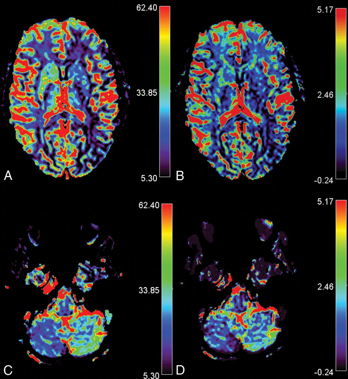
CCD in a 14-year-old girl presenting with MwA and CCD. Slices A and C show rCBF images of the infra- and supratentorial brain, and slices B and D, rCBV images respectively. The patient presented with HCH in the right cerebellum with overt hemispheric perfusion asymmetry. Fourteen of 17 supratentorial ROIs in the left supratentorial hemisphere exceeded the AI threshold with lateralization of hypoperfusion contralateral to the cerebellum. Bars indicate relative values for CBV and CBF.

Vertebral Artery Configuration

The vertebral arteries were analyzed on TOF angiograms to look for patency or hypoplasia to exclude perfusion abnormalities in the PICA territory related to an occluded or hypoplastic vessel. Vertebral artery hypoplasia was defined as a diameter of the vertebral artery of <1 mm.

Statistics

We used SPSS (Version 21; IBM, Armonk, New York) for statistical analysis. Descriptive data are presented as mean and SD or median. We used the Student t test for normally distributed supratentorial data and the Mann-Whitney U test for not normally distributed infratentorial data to compare numbers of ROIs with AI > 10% and mean values of AI (%) between groups. A P value < .05 was considered significant.

Results

We identified 191 patients with migraine from our data base. Of these, 106 patients underwent a dedicated stroke protocol within 24 hours of onset of symptoms and 98 were finally diagnosed with MwA. In 23/106 patients (21.69%) patients, we identified focal cortical hypoperfusion. None of the patients presented with a DWI restriction. Fifteen of the 23 were female. Mean age was 28.8 years (range, 13–77 years). The laterality of the clinical symptoms was left-sided in 8 patients and right-sided in 11. Alternating clinical patterns were observed in 2, and nonspecific symptoms, in 2 patients. MwA was the predominant final diagnosis in the patients with perfusion asymmetry, 22/23 patients.

For 13 of these 23 patients, the time between onset of symptoms and imaging was precisely documented (mean, 212 minutes; range, 95–429 minutes; median, 180 minutes). For the remaining 10 patients, the records documented MR imaging being performed within 12 hours of symptom onset. Of the 83 patients who had no perfusion alterations, the time between the onset of symptoms and MR imaging was precisely documented for 45 patients (mean, 384 minutes; range 55–1400 minutes; median, 315 minutes). For the remaining 38 patients, the onset of symptoms occurred up to 24 hours before the MR imaging. See Table 1 for clinical information on all patients and Table 2 for information on the subgroup of patients with and without CCD. Patients with cortical oligemia presented more often with aura symptoms than patients with normal perfusion. In patients with cortical oligemia, language and sensory symptoms were reported more frequently than visual and motor symptoms. Patients with HCH and the subgroup with CCD had a slightly lower frequency of sensory symptoms than the non-HCH group, but other aura symptoms were equally distributed between the groups (Tables 1 and 2). Aura symptoms were not a predictor of HCH or CCD (Table 2).

View this table:
  • View inline
  • View popup
Table 1:

Clinical characteristics

View this table:
  • View inline
  • View popup
Table 2:

Predominant clinical symptoms in patients with abnormal cortical perfusion

Semiquantitative Evaluation

Overall, we included 18 paired ROIs in the analysis. The size of each ROI was kept uniform for all patients but varied among regions to account for regional anatomy (mean, 291 ± 100 mm2; range, 132–471 mm2). All 23 patients presented with at least 1 supratentorial ROI with an AI value of >10% rCBF and rCBV.

Twelve patients had HCH. Nine of these patients presented with reduced rCBF contralateral to the ill-perfused cerebellar hemisphere (CCD-positive); in 3 patients, we detected cortical rCBF reduction ipsilateral to the cerebellum. All patients with HCH presented with clinical symptoms attributable to a region of cortical hypoperfusion in the contralateral hemisphere (Table 3).

View this table:
  • View inline
  • View popup
Table 3:

The number of ROIs with an AI of > 10% in supratentorial oligemia (maximum, 17 per patient), in the group with hemispheric cerebellar hypoperfusion (154/204) and in the group with symmetric cerebellar perfusion (145/187)

When HCH was present, the number of ROIs with an AI > 10% found contralateral to the hypoperfused cerebellar hemisphere (103 for rCBF and 101 for rCBV) was nearly twice the number of ROIs which exceeded the threshold of 10% in the ipsilateral hemisphere (51 for rCBF and 54 for rCBV).

We observed no significant differences between HCH and non-HCH groups for mean AI values of rCBF and rCBV in the supratentorial hemisphere (Table 4).

View this table:
  • View inline
  • View popup
Table 4:

Mean AI for hypoperfused cerebellar hemisphere and non-HCH (rCBF and rCBV)a

Vertebral Artery Configuration

In 22/23 patients, the vertebral artery appeared normal. One patient with CCD-positive MwA had a hypoplastic vertebral artery, which tapered into the PICA on the side of the hypoperfused cerebellum.

Discussion

In this retrospective single-center study, we aimed to investigate the association between oligemia and HCH in patients with migraine and its potential relation to small infarct-like cerebellar lesions. We observed cortical oligemia in 23/106 (21.69%) patients who underwent MR imaging to rule out ischemic stroke. Fifty-two percent of patients with oligemia presented with abnormal cerebellar perfusion, and 39.1% fulfilled the criteria for CCD, rendering it a frequent consequence of cortical spreading depression unrelated to vertebral artery hypoplasia.25

Cerebellar hypoperfusion in patients with stroke is associated with a worse clinical outcome than if it was not present, related to deafferentation and transneural metabolic depression.26 In seizure-related CCD, the outcome is variable, ranging from complete neurologic recovery to permanent disability depending on the duration of prolonged excitatory synaptic activity affecting cortico-ponto-cerebellar pathways.27⇓⇓⇓–31 While diffusion restriction is a common consequence of perfusion changes and related metabolic depletion in patients with stroke and epilepsy, this should be considered an exceptional phenomenon in migraine. In our cohort, none of the 106 patients had restricted diffusion or preexisting cerebellar infarct-like lesions. Although CCD was frequent, none of our patients with migraine experienced permanent deficits. We conclude that unlike in stroke- and seizure-related CCD, CCD in MwA may be considered a benign phenomenon.

Cerebellar hypoperfusion has been addressed as a potential cause of cerebellar infarct-like lesions in MwA in previous studies.3 The findings of our retrospective study do not support the hypothesis of an increased risk of episode-triggered and oligemia-related hypoxic cerebellar infarctions. In our series, none of the patients with oligemia reached the ischemic threshold. In patients with MwA, the severity of cortical oligemia, expressed as increasing AI, was not directly associated with HCH or CCD, in contrast to observations in patients with ischemic stroke.30 The size of the supratentorial oligemic area, expressed by the number of supratentorial ROIs beyond the threshold AI of >10%, also did not predict the likelihood of HCH or CCD. From a methodologic point of view, we excluded the transition parameters (TTP and MTT) from our perfusion analysis. Our findings were in line with those of Förster et al,32 who demonstrated that perfusion abnormalities in patients with MwA are predominantly substantiated by CBF and CBV changes. TTP analysis of the cerebellar hemispheres is hampered by variant vertebral artery configurations that directly influence the TTP and MTT maps, while no such detrimental effects have been reported for CBF analysis.24

Furthermore, it is not yet known whether the development of infarct-like lesions is facilitated by multifactorial interactions of severe oligemia with other impeding factors that are not detected by perfusion MR imaging (eg, increased vulnerability to blood flow reduction). An increased susceptibility to acute ischemic injury has been demonstrated in patients with migraine, due to an unstable penumbra, especially in those with MwA.33 Greater susceptibility to spreading depolarization in migraine-susceptible brains may be another potential mechanism for increased cerebral vulnerability to ischemia and stroke. Similar findings have been reported in an animal study.34 Thus, typical episodes with regularly experienced aura symptoms may reflect benign oligemia not related to tissue damage, while prolonged and severe hypoxic episodes may ultimately induce parenchymal damage, which was not observed in our cohort.

This study has some limitations. Due to its retrospective design, there was no healthy control group. Because expert analysis was performed to select apparent perfusion deficits in patients with migraine, a rater-dependent bias cannot be fully excluded. However, in healthy volunteers, interhemispheric asymmetry is minimal (1.01%–3.14%) and symmetric rCBF distribution can be assumed between homologous regions, independent of age.35 Thus, our ROI-based semiquantitative analysis with the rather strict threshold of >10%, as commonly used in stroke or epilepsy studies, may be considered appropriate to balance measurements of random asymmetry and spreading oligemia.22⇓–24 Finally, all patients were symptom-free during follow-up. However, we cannot fully exclude the possibility that asymptomatic parenchymal damage may have remained undetected. Further studies would be required to answer this question.

Conclusions

HCH and CCD are frequently observed in patients presenting with acute MwA. Cerebellar hypoperfusion in patients with migraine appears, in contrast to stroke- and seizure-related CCD, to be a benign phenomenon. Our study could not provide evidence for a link between cerebellar hypoperfusion and cortical hypoperfusion in the pathogenesis of cerebellar infarctions but encourages future investigations that pay special attention to factors other than hypoperfusion alone to better understand the cause of infarct-like lesions in the cerebellum of patients who have MwA.

Footnotes

  • Disclosures: Roland Wiest—UNRELATED: Grants/Grants Pending: Swiss National Foundation, Swiss Heart Foundation.

  • This study was partially supported by Bayer (Swiss) AG, Zurich, Switzerland.

Indicates open access to non-subscribers at www.ajnr.org

References

  1. 1.↵
    1. Lipton RB,
    2. Stewart WF,
    3. Diamond S, et al
    . Prevalence and burden of migraine in the United States: data from the American Migraine Study II. Headache 2001;41:646–57 doi:10.1046/j.1526-4610.2001.041007646.x pmid:11554952
    CrossRefPubMed
  2. 2.↵
    Headache Classification Subcommittee of the International Headache Society. The International Classification of Headache Disorders: 2nd ed. Cephalalgia 2004;24(Suppl 1):9–160 doi:10.1111/j.1468-2982.2003.00824.x pmid:14979299
    CrossRefPubMed
  3. 3.↵
    1. Kruit MC,
    2. van Buchem MA,
    3. Hofman PA, et al
    . Migraine as a risk factor for subclinical brain lesions. JAMA 2004;291:427–34 doi:10.1001/jama.291.4.427 pmid:14747499
    CrossRefPubMed
  4. 4.↵
    1. Kruit MC,
    2. Launer LJ,
    3. Ferrari MD, et al
    . Infarcts in the posterior circulation territory in migraine: the population-based MRI CAMERA study. Brain 2005;128(Pt 9):2068–77 doi:10.1093/brain/awh542 pmid:16006538
    CrossRefPubMed
  5. 5.↵
    1. Olesen J,
    2. Larsen B,
    3. Lauritzen M
    . Focal hyperemia followed by spreading oligemia and impaired activation of rCBF in classic migraine. Ann Neurol 1981;9:344–52 doi:10.1002/ana.410090406 pmid:6784664
    CrossRefPubMed
  6. 6.↵
    1. Kapinos G,
    2. Fischbein NJ,
    3. Zaharchuk G, et al
    . Migraine-like headache with visual deficit and perfusion abnormality on MRI. Neurology 2010;25;74:1743–45 doi:10.1212/WNL.0b013e3181e042e4 pmid:20498443
    CrossRefPubMed
  7. 7.↵
    1. Fernandes PM,
    2. Whiteley WN,
    3. Hart SR, et al
    . Strokes: mimics and chameleons. Pract Neurol 2013:21–28 doi:10.1136/practneurol-2012-000465 pmid:23315456
    CrossRefPubMed
  8. 8.↵
    1. Magauran BG Jr.,
    2. Nitka M
    . Stroke mimics. Emerg Med Clin North Am 2012;30:795–804 doi:10.1016/j.emc.2012.06.006 pmid:22974649
    CrossRefPubMed
  9. 9.↵
    1. Yamauchi H,
    2. Fukuyama H,
    3. Nagahama Y, et al
    . Uncoupling of oxygen and glucose metabolism in persistent crossed cerebellar diaschisis. Stroke 1999;30:1424–28 doi:10.1161/01.STR.30.7.1424 pmid:10390317
    Abstract/FREE Full Text
  10. 10.↵
    1. Yamauchi H,
    2. Fukuyama H,
    3. Kimura J
    , Hemodynamic and metabolic changes in crossed cerebellar hypoperfusion. Stroke 1992;23:855–60 doi:10.1161/01.STR.23.6.855 pmid:1595105
    Abstract/FREE Full Text
  11. 11.↵
    1. Noguchi T,
    2. Nishihara M,
    3. Egashira Y, et al
    . Arterial spin-labeling MR imaging of cerebral hemorrhages. Neuroradiology 2015;57:1135–44 doi:10.1007/s00234-015-1574-9 pmid:26280515
    CrossRefPubMed
  12. 12.↵
    1. Fu J,
    2. Chen WJ,
    3. Wu GY, et al
    . Whole-brain 320-detector row dynamic volume CT perfusion detected crossed cerebellar diaschisis after spontaneous intracerebral hemorrhage. Neuroradiology 2015;57:179–87 doi:10.1007/s00234-014-1456-6 pmid:25381579
    CrossRefPubMed
  13. 13.↵
    1. Nakae S,
    2. Inamasu J,
    3. Ohmi T, et al
    . Relative vasodilatory change in seizure-induced crossed cerebellar diaschisis. Intern Med 2016;55:713–14 doi:10.2169/internalmedicine.55.5958 pmid:26984098
    CrossRefPubMed
  14. 14.↵
    1. Dodick DW,
    2. Roarke MC
    . Crossed cerebellar diaschisis during migraine with prolonged aura: a possible mechanism for cerebellar infarctions. Cephalalgia 2008;28:83–86 doi:10.1111/j.1468-2982.2007.01458.x pmid:17970765
    CrossRefPubMed
  15. 15.↵
    1. Crawford JS,
    2. Konkol RJ
    . Familial hemiplegic migraine with crossed cerebellar diaschisis and unilateral meningeal enhancement. Headache 1997;37:590–93 doi:10.1046/j.1526-4610.1997.3709590.x pmid:9385760
    CrossRefPubMed
  16. 16.↵
    1. Mouridsen K,
    2. Christensen S,
    3. Gyldensted L, et al
    . Automatic selection of arterial input function using cluster analysis. Magn Reson Med 2006;55:524–31 doi:10.1002/mrm.20759 pmid:16453314
    CrossRefPubMed
  17. 17.↵
    1. Wu O,
    2. Østergaard L,
    3. Weisskoff RM, et al
    . Tracer arrival timing-insensitive technique for estimating flow in MR perfusion-weighted imaging using singular value decomposition with a block-circulant deconvolution matrix. Magn Reson Med 2003;50:164–74 doi:10.1002/mrm.10522 pmid:12815691
    CrossRefPubMed
  18. 18.↵
    1. Aviv RI,
    2. Mandelcorn J,
    3. Chakraborty S, et al
    . Alberta Stroke Program Early CT Scoring of CT perfusion in early stroke visualization and assessment. AJNR Am J Neuroradiol 2007;28:1975–80 doi:10.3174/ajnr.A0689 pmid:17921237
    Abstract/FREE Full Text
  19. 19.↵
    1. Puetz V,
    2. Khomenko A,
    3. Hill MD, et al
    . Basilar Artery International Cooperation Study (BASICS) Group: extent of hypoattenuation on CT angiography source images in basilar artery occlusion—prognostic value in the Basilar Artery International Cooperation Study. Stroke 2011;42:3454–59 doi:10.1161/STROKEAHA.111.622175 pmid:21960577
    Abstract/FREE Full Text
  20. 20.↵
    1. Strother MK,
    2. Buckingham C,
    3. Faraco CC, et al
    . Crossed cerebellar diaschisis after stroke identified noninvasively with cerebral blood flow-weighted arterial spin labeling MRI. Eur J Radiol 2016;85:136–42 doi:10.1016/j.ejrad.2015.11.003 pmid:26724658
    CrossRefPubMed
  21. 21.↵
    1. Madai VI,
    2. Altaner A,
    3. Stengl KL, et al
    . Crossed cerebellar diaschisis after stroke: can perfusion-weighted MRI show functional inactivation? J Cereb Blood Flow Metab 2011;31:1493–500 doi:10.1038/jcbfm.2011.15 pmid:21386854
    CrossRefPubMed
  22. 22.↵
    1. Takano A,
    2. Shiga T,
    3. Kobayashi J, et al
    . Thalamic asymmetry on interictal SPECT in patients with frontal lobe epilepsy. Nucl Med Commun 2001;22:319–24 doi:10.1097/00006231-200103000-00009 pmid:11314765
    CrossRefPubMed
  23. 23.↵
    1. Wiest R,
    2. von Bredow F,
    3. Schindler K, et al
    . Detection of regional blood perfusion changes in epileptic seizures with dynamic brain perfusion CT: a pilot study. Epilepsy Res 2006;72:102–10 doi:10.1016/j.eplepsyres.2006.07.017 pmid:17118628
    CrossRefPubMed
  24. 24.↵
    1. Hauf M,
    2. Slotboom J,
    3. Nirkko A, et al
    . Cortical regional hyperperfusion in nonconvulsive status epilepticus measured by dynamic brain perfusion CT. AJNR Am J Neuroradiol 2009;30:693–98 doi:10.3174/ajnr.A1456 pmid:19213823
    Abstract/FREE Full Text
  25. 25.↵
    1. Thierfelder KM,
    2. Baumann AB,
    3. Sommer WH, et al
    . Vertebral artery hypoplasia: frequency and effect on cerebellar blood flow characteristics. Stroke 2014;45:1363–68 doi:10.1161/STROKEAHA.113.004188 pmid:24699051
    Abstract/FREE Full Text
  26. 26.↵
    1. Takasawa M,
    2. Watanabe M,
    3. Yamamoto S, et al
    . Prognostic value of subacute crossed cerebellar diaschisis: single-photon emission CT study in patients with middle cerebral artery territory infarct. AJNR Am J Neuroradiol 2002;23:189–93 pmid:11847040
    Abstract/FREE Full Text
  27. 27.↵
    1. Graffeo CS,
    2. Snyder KA,
    3. Nasr DM, et al
    . Prognostic and mechanistic factors characterizing seizure-associated crossed cerebellar diaschisis. Neurocrit Care 2016;24:258–63 doi:10.1007/s12028-015-0155-4 pmid:26063348
    CrossRefPubMed
  28. 28.↵
    1. Kato T,
    2. Okumura A,
    3. Hayakawa F, et al
    . Transient reduced diffusion in the cortex in a child with prolonged febrile seizures. Brain Dev 2012;34:773–75 doi:10.1016/j.braindev.2011.12.006 pmid:22236656
    CrossRefPubMed
  29. 29.↵
    1. Massaro AM
    . Teaching neuroimages: crossed cerebellar diaschisis in hemispheric status epilepticus. Neurology 2012;79:e182 doi:10.1212/WNL.0b013e318274de51 pmid:23150537
    Abstract/FREE Full Text
  30. 30.↵
    1. Zaidi SA,
    2. Haq MA,
    3. Bindman D, et al
    . Crossed cerebellar diaschisis: a radiological finding in status epilepticus not to miss. BMJ Case Rep 2013;2013. pii: bcr2013200478 doi:10.1136/bcr-2013-200478 pmid:24234428
    CrossRefPubMed
  31. 31.↵
    1. Ohe Y,
    2. Hayashi T,
    3. Deguchi I, et al
    . A case of nonconvulsive status epilepticus with a reversible contralateral cerebellar lesion: temporal changes in magnetic resonance imaging and single-photon emission computed tomography finding. J Stroke Cerebrovasc Dis 2013;22:e639–42 doi:10.1016/j.jstrokecerebrovasdis.2013.03.015 pmid:23910513
    CrossRefPubMed
  32. 32.↵
    1. Förster A,
    2. Wenz H,
    3. Kerl HU, et al
    . Perfusion patterns in migraine with aura. Cephalalgia 2014;34:870–76 doi:10.1177/0333102414523339 pmid:24554619
    CrossRefPubMed
  33. 33.↵
    1. Mawet J,
    2. Eikermann-Haerter K,
    3. Park KY, et al
    . Sensitivity to acute cerebral ischemic injury in migraineurs: a retrospective case-control study. Neurology 2015;85:1945–49 doi:10.1212/WNL.0000000000002166 pmid:26537055
    Abstract/FREE Full Text
  34. 34.↵
    1. Eikermann-Haerter K,
    2. Lee JH,
    3. Yalcin N, et al
    . Migraine prophylaxis, ischemic depolarizations, and stroke outcomes in mice. Stroke 2015;46:229–36 doi:10.1161/STROKEAHA.114.006982 pmid:25424478
    Abstract/FREE Full Text
  35. 35.↵
    1. Catafau AM,
    2. Lomeña FJ,
    3. Pavia J, et al
    . Regional cerebral blood flow pattern in normal young and aged volunteers: a 99mTc-HMPAO SPET study. Eur J Nucl Med 1996;23:1329–37 doi:10.1007/BF01367588 pmid:8781137
    CrossRefPubMed
  • Received June 28, 2016.
  • Accepted after revision November 5, 2017.
  • © 2018 by American Journal of Neuroradiology
View Abstract
Next
Back to top
Advertisement
Print
Download PDF
Email Article

Thank you for your interest in spreading the word on American Journal of Neuroradiology.

NOTE: We only request your email address so that the person you are recommending the page to knows that you wanted them to see it, and that it is not junk mail. We do not capture any email address.

Enter multiple addresses on separate lines or separate them with commas.
Cerebellar Hypoperfusion in Migraine Attack: Incidence and Significance
(Your Name) has sent you a message from American Journal of Neuroradiology
(Your Name) thought you would like to see the American Journal of Neuroradiology web site.
CAPTCHA
This question is for testing whether or not you are a human visitor and to prevent automated spam submissions.
Cite this article
F. Kellner-Weldon, M. El-Koussy, S. Jung, M. Jossen, P.P. Klinger-Gratz, R. Wiest
Cerebellar Hypoperfusion in Migraine Attack: Incidence and Significance
American Journal of Neuroradiology Jan 2018, DOI: 10.3174/ajnr.A5508

Citation Manager Formats

  • BibTeX
  • Bookends
  • EasyBib
  • EndNote (tagged)
  • EndNote 8 (xml)
  • Medlars
  • Mendeley
  • Papers
  • RefWorks Tagged
  • Ref Manager
  • RIS
  • Zotero
0 Responses
Respond to this article
Share
Bookmark this article
Cerebellar Hypoperfusion in Migraine Attack: Incidence and Significance
F. Kellner-Weldon, M. El-Koussy, S. Jung, M. Jossen, P.P. Klinger-Gratz, R. Wiest
American Journal of Neuroradiology Jan 2018, DOI: 10.3174/ajnr.A5508
del.icio.us logo Twitter logo Facebook logo Mendeley logo
  • Tweet Widget
  • Facebook Like
  • Google Plus One
Purchase

Jump to section

  • Article
    • Abstract
    • ABBREVIATIONS:
    • Materials and Methods
    • Results
    • Discussion
    • Conclusions
    • Footnotes
    • References
  • Figures & Data
  • Info & Metrics
  • Responses
  • References
  • PDF

Related Articles

  • No related articles found.
  • PubMed
  • Google Scholar

Cited By...

  • Reversible Perfusion Changes during Acute Attacks in Glucose Transporter Type 1 Deficiency Syndrome: A Pediatric Case Series
  • Small molecules from Bacopa monneiri as potent inhibitors against Neurodegenerative disorders
  • Crossed Cerebellar Diaschisis in Alzheimers Disease Detected by Arterial Spin-labelling Perfusion MRI
  • Crossref (28)
  • Google Scholar

This article has been cited by the following articles in journals that are participating in Crossref Cited-by Linking.

  • 2021 Guideline for the Prevention of Stroke in Patients With Stroke and Transient Ischemic Attack: A Guideline From the American Heart Association/American Stroke Association
    Dawn O. Kleindorfer, Amytis Towfighi, Seemant Chaturvedi, Kevin M. Cockroft, Jose Gutierrez, Debbie Lombardi-Hill, Hooman Kamel, Walter N. Kernan, Steven J. Kittner, Enrique C. Leira, Olive Lennon, James F. Meschia, Thanh N. Nguyen, Peter M. Pollak, Pasquale Santangeli, Anjail Z. Sharrief, Sidney C. Smith, Tanya N. Turan, Linda S. Williams
    Stroke 2021 52 7
  • Neuroimaging clues of migraine aura
    Nouchine Hadjikhani, Maurice Vincent
    The Journal of Headache and Pain 2019 20 1
  • A novel computed tomography perfusion-based quantitative tool for evaluation of perfusional abnormalities in migrainous aura stroke mimic
    Antonio Granato, Laura D’Acunto, Miloš Ajčević, Giovanni Furlanis, Maja Ukmar, Roberta Antea Pozzi Mucelli, Paolo Manganotti
    Neurological Sciences 2020 41 11
  • Cortical mechanisms in migraine
    Wei Dai, Ren-Hao Liu, Enchao Qiu, Yinglu Liu, Zhiye Chen, Xiaoyan Chen, Ran Ao, Min Zhuo, Shengyuan Yu
    Molecular Pain 2021 17
  • Postural instability during attacks of migraine without aura
    E. Anagnostou, S. Gerakoulis, P. Voskou, E. Kararizou
    European Journal of Neurology 2019 26 2
  • Evaluating migraine with typical aura with neuroimaging
    Nazia Karsan, Elisa Silva, Peter J. Goadsby
    Frontiers in Human Neuroscience 2023 17
  • Recent developments in imaging of epilepsy
    Roland Wiest, Roland Beisteiner
    Current Opinion in Neurology 2019 32 4
  • Specificities of arterial spin labeling (ASL) abnormalities in acute seizure
    Mathieu Schertz, Joseph Benzakoun, Nadya Pyatigorskaya, Samia Belkacem, Melika Sahli-Amor, Vincent Navarro, Clément Cholet, Delphine Leclercq, Didier Dormont, Bruno Law-ye
    Journal of Neuroradiology 2020 47 1
  • Cerebro-Cerebellar Networks in Migraine Symptoms and Headache
    Rodrigo Noseda
    Frontiers in Pain Research 2022 3
  • Findings in susceptibility weighted imaging in pediatric patients with migraine with aura
    Frauke Kellner-Weldon, Vera Franziska Lehmann, Philipe Sebastian Breiding, Lorenz Grunder, Raphaela Muri, Manuela Pastore-Wapp, Sandra Bigi, Roland Wiest, Marwan El-Koussy, Nedelina Slavova
    European Journal of Paediatric Neurology 2020 28

More in this TOC Section

  • Diagnostic Neuroradiology of Monoclonal Antibodies
  • Cerebral ADC Changes in Fabry Disease
  • ML for Glioma Molecular Subtype Prediction
Show more Adult Brain

Similar Articles

Advertisement

Indexed Content

  • Current Issue
  • Accepted Manuscripts
  • Article Preview
  • Past Issues
  • Editorials
  • Editor's Choice
  • Fellows' Journal Club
  • Letters to the Editor
  • Video Articles

Cases

  • Case Collection
  • Archive - Case of the Week
  • Archive - Case of the Month
  • Archive - Classic Case

Special Collections

  • AJNR Awards
  • ASNR Foundation Special Collection
  • Most Impactful AJNR Articles
  • Photon-Counting CT
  • Spinal CSF Leak Articles (Jan 2020-June 2024)

More from AJNR

  • Trainee Corner
  • Imaging Protocols
  • MRI Safety Corner

Multimedia

  • AJNR Podcasts
  • AJNR Scantastics

Resources

  • Turnaround Time
  • Submit a Manuscript
  • Submit a Video Article
  • Submit an eLetter to the Editor/Response
  • Manuscript Submission Guidelines
  • Statistical Tips
  • Fast Publishing of Accepted Manuscripts
  • Graphical Abstract Preparation
  • Imaging Protocol Submission
  • Evidence-Based Medicine Level Guide
  • Publishing Checklists
  • Author Policies
  • Become a Reviewer/Academy of Reviewers
  • News and Updates

About Us

  • About AJNR
  • Editorial Board
  • Editorial Board Alumni
  • Alerts
  • Permissions
  • Not an AJNR Subscriber? Join Now
  • Advertise with Us
  • Librarian Resources
  • Feedback
  • Terms and Conditions
  • AJNR Editorial Board Alumni

American Society of Neuroradiology

  • Not an ASNR Member? Join Now

© 2025 by the American Society of Neuroradiology All rights, including for text and data mining, AI training, and similar technologies, are reserved.
Print ISSN: 0195-6108 Online ISSN: 1936-959X

Powered by HighWire