Advertisement

Main menu

  • Home
  • Content
    • Current Issue
    • Publication Preview--Ahead of Print
    • Past Issue Archive
    • Case of the Week Archive
    • Classic Case Archive
    • Case of the Month Archive
  • For Authors
  • About Us
    • About AJNR
    • Editors
    • American Society of Neuroradiology
  • Submit a Manuscript
  • Podcasts
    • Subscribe on iTunes
    • Subscribe on Stitcher
  • More
    • Subscribers
    • Permissions
    • Advertisers
    • Alerts
    • Feedback
  • Other Publications
    • ajnr

User menu

  • Subscribe
  • Alerts
  • Log in

Search

  • Advanced search
American Journal of Neuroradiology

American Journal of Neuroradiology

  • Subscribe
  • Alerts
  • Log in

Advanced Search

  • Home
  • Content
    • Current Issue
    • Publication Preview--Ahead of Print
    • Past Issue Archive
    • Case of the Week Archive
    • Classic Case Archive
    • Case of the Month Archive
  • For Authors
  • About Us
    • About AJNR
    • Editors
    • American Society of Neuroradiology
  • Submit a Manuscript
  • Podcasts
    • Subscribe on iTunes
    • Subscribe on Stitcher
  • More
    • Subscribers
    • Permissions
    • Advertisers
    • Alerts
    • Feedback
  • Follow AJNR on Twitter
  • Visit AJNR on Facebook
  • Follow AJNR on Instagram
  • Join AJNR on LinkedIn
  • RSS Feeds
Research ArticleADULT BRAIN
Open Access

Cerebellar Hypoperfusion in Migraine Attack: Incidence and Significance

F. Kellner-Weldon, M. El-Koussy, S. Jung, M. Jossen, P.P. Klinger-Gratz and R. Wiest
American Journal of Neuroradiology January 2018, DOI: https://doi.org/10.3174/ajnr.A5508
F. Kellner-Weldon
aFrom the Support Center for Advanced Neuroimaging (F.K.-W., M.E.-K., M.J., P.P.K.-G., R.W.), Institute for Diagnostic and Interventional Neuroradiology
  • Find this author on Google Scholar
  • Find this author on PubMed
  • Search for this author on this site
  • ORCID record for F. Kellner-Weldon
M. El-Koussy
aFrom the Support Center for Advanced Neuroimaging (F.K.-W., M.E.-K., M.J., P.P.K.-G., R.W.), Institute for Diagnostic and Interventional Neuroradiology
  • Find this author on Google Scholar
  • Find this author on PubMed
  • Search for this author on this site
  • ORCID record for M. El-Koussy
S. Jung
bDepartment of Neurology (S.J.), University Hospital Inselspital, Bern, Switzerland
  • Find this author on Google Scholar
  • Find this author on PubMed
  • Search for this author on this site
  • ORCID record for S. Jung
M. Jossen
aFrom the Support Center for Advanced Neuroimaging (F.K.-W., M.E.-K., M.J., P.P.K.-G., R.W.), Institute for Diagnostic and Interventional Neuroradiology
  • Find this author on Google Scholar
  • Find this author on PubMed
  • Search for this author on this site
  • ORCID record for M. Jossen
P.P. Klinger-Gratz
aFrom the Support Center for Advanced Neuroimaging (F.K.-W., M.E.-K., M.J., P.P.K.-G., R.W.), Institute for Diagnostic and Interventional Neuroradiology
cDepartment of Radiology (P.P.K.-G.), University of Basel, Basel, Switzerland.
  • Find this author on Google Scholar
  • Find this author on PubMed
  • Search for this author on this site
  • ORCID record for P.P. Klinger-Gratz
R. Wiest
aFrom the Support Center for Advanced Neuroimaging (F.K.-W., M.E.-K., M.J., P.P.K.-G., R.W.), Institute for Diagnostic and Interventional Neuroradiology
  • Find this author on Google Scholar
  • Find this author on PubMed
  • Search for this author on this site
  • ORCID record for R. Wiest
  • Article
  • Figures & Data
  • Info & Metrics
  • References
  • PDF
Loading

Article Figures & Data

Figures

  • Tables
  • Fig 1.
    • Download figure
    • Open in new tab
    • Download powerpoint
    Fig 1.

    ROI analysis for perfusion parameters: CER indicates cerebellum; Put, putamen; I, insula; T, thalamus; A, arterial territory of the anterior cerebral artery; M, arterial territory of the middle cerebral artery; P, arterial territory of the posterior cerebral artery; PREC, precentral gyrus; POSTC, postcentral gyrus.
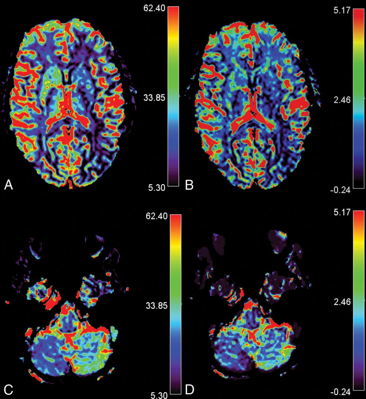
  • Fig 2.
    • Download figure
    • Open in new tab
    • Download powerpoint
    Fig 2.

    CCD in a 14-year-old girl presenting with MwA and CCD. Slices A and C show rCBF images of the infra- and supratentorial brain, and slices B and D, rCBV images respectively. The patient presented with HCH in the right cerebellum with overt hemispheric perfusion asymmetry. Fourteen of 17 supratentorial ROIs in the left supratentorial hemisphere exceeded the AI threshold with lateralization of hypoperfusion contralateral to the cerebellum. Bars indicate relative values for CBV and CBF.

Tables

  • Figures
    • View popup
    Table 1:

    Clinical characteristics

    Predominant SymptomAbnormal PerfusionNormal Perfusion
    n = 23%n = 83%
    Headache2191.36957.3
    Aura symptoms2295.77663.1
        Visual1043.54436.5
        Sensory1773.93730.7
        Motor834.82218.3
        Language1147.81613.3
    • View popup
    Table 2:

    Predominant clinical symptoms in patients with abnormal cortical perfusion

    Predominant SymptomPatients with OligemiaHCH (CCD Subgroup)Non-HCH
    n = 23%n = 12 (9)%n = 11%
    Headache2191.310 (7)43.5 (30.4)1147.8
    Aura symptoms2295.712 (9)52.2 (39.1)1043.5
        Visual1043.55 (2)21.7 (8.7)521.7
        Sensory1773.97 (5)30.4 (21.7)1043.5
        Motor834.84 (4)17.4 (17.4)417.4
        Language1147.86 (6)26.1 (26.1)521.7
    • View popup
    Table 3:

    The number of ROIs with an AI of > 10% in supratentorial oligemia (maximum, 17 per patient), in the group with hemispheric cerebellar hypoperfusion (154/204) and in the group with symmetric cerebellar perfusion (145/187)

    Abnormal ROIs (AI > 10%)
    Patients with Hemispheric Cerebellar Hypoperfusion (n = 12)Patients with Symmetric Cerebellar Perfusion (n = 11)
    rCBF (No.)
        Total No. (%)154 (75.5)145 (77.5)
        Contralateral103
        Ipsilateral51
    rCBV (No.)
        Total No. (%)155 (75.5)139 (74.3)
        Contralateral101
        Ipsilateral54
    • View popup
    Table 4:

    Mean AI for hypoperfused cerebellar hemisphere and non-HCH (rCBF and rCBV)a

    AI Absolute Values (%)P Value
    Group HCH (n = 12)Group Non-HCH (n = 11)
    MeanSDMeanSD
    rCBF supratentorial27.606.8526.306.66.51
    rCBV supratentorial26.756.6624.646.27.48
    rCBF infratentorial30.5616.774.243.23<.001
    rCBV infratentorial25.6916.004.022.53<.001
    • ↵a Significant differences were noted only for infratentorial ROIs.

Next
Back to top
Advertisement
Print
Download PDF
Email Article

Thank you for your interest in spreading the word on American Journal of Neuroradiology.

NOTE: We only request your email address so that the person you are recommending the page to knows that you wanted them to see it, and that it is not junk mail. We do not capture any email address.

Enter multiple addresses on separate lines or separate them with commas.
Cerebellar Hypoperfusion in Migraine Attack: Incidence and Significance
(Your Name) has sent you a message from American Journal of Neuroradiology
(Your Name) thought you would like to see the American Journal of Neuroradiology web site.
CAPTCHA
This question is for testing whether or not you are a human visitor and to prevent automated spam submissions.
Citation Tools
Cerebellar Hypoperfusion in Migraine Attack: Incidence and Significance
F. Kellner-Weldon, M. El-Koussy, S. Jung, M. Jossen, P.P. Klinger-Gratz, R. Wiest
American Journal of Neuroradiology Jan 2018, DOI: 10.3174/ajnr.A5508

Citation Manager Formats

  • BibTeX
  • Bookends
  • EasyBib
  • EndNote (tagged)
  • EndNote 8 (xml)
  • Medlars
  • Mendeley
  • Papers
  • RefWorks Tagged
  • Ref Manager
  • RIS
  • Zotero
Share
Cerebellar Hypoperfusion in Migraine Attack: Incidence and Significance
F. Kellner-Weldon, M. El-Koussy, S. Jung, M. Jossen, P.P. Klinger-Gratz, R. Wiest
American Journal of Neuroradiology Jan 2018, DOI: 10.3174/ajnr.A5508
del.icio.us logo Digg logo Reddit logo Twitter logo CiteULike logo Facebook logo Google logo Mendeley logo
  • Tweet Widget
  • Facebook Like
  • Google Plus One
Purchase

Jump to section

  • Article
    • Abstract
    • ABBREVIATIONS:
    • Materials and Methods
    • Results
    • Discussion
    • Conclusions
    • Footnotes
    • References
  • Figures & Data
  • Info & Metrics
  • References
  • PDF

Related Articles

  • No related articles found.
  • PubMed
  • Google Scholar

Cited By...

  • No citing articles found.
  • Crossref
  • Google Scholar

This article has not yet been cited by articles in journals that are participating in Crossref Cited-by Linking.

More in this TOC Section

  • Anatomic and Embryologic Analysis of the Dural Branches of the Ophthalmic Artery
  • Automated Cerebral Hemorrhage Detection Using RAPID
  • Analysis of Stroke Detection during the COVID-19 Pandemic Using Natural Language Processing of Radiology Reports
Show more ADULT BRAIN

Similar Articles

Advertisement

News and Updates

  • Lucien Levy Best Research Article Award
  • Thanks to our 2020 Distinguished Reviewers
  • Press Releases

Resources

  • Evidence-Based Medicine Level Guide
  • How to Participate in a Tweet Chat
  • AJNR Podcast Archive
  • Ideas for Publicizing Your Research
  • Librarian Resources
  • Terms and Conditions

Opportunities

  • Share Your Art in Perspectives
  • Get Peer Review Credit from Publons
  • Moderate a Tweet Chat

American Society of Neuroradiology

  • Neurographics
  • ASNR Annual Meeting
  • Fellowship Portal
  • Position Statements

© 2021 by the American Society of Neuroradiology | Print ISSN: 0195-6108 Online ISSN: 1936-959X

Powered by HighWire