
Article Figures & Data
Figures
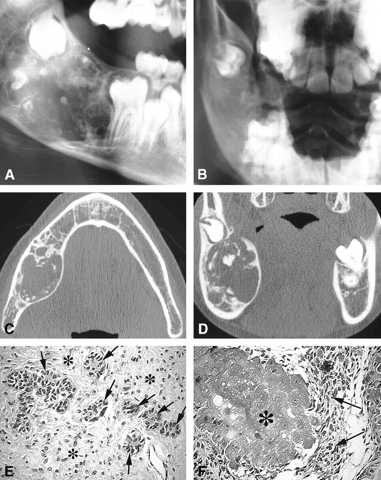
- fig 1.
Case 1.
A and B, Plain radiographs. Lateral plain radiograph (A) shows a large expansile mixed radiolucent/radiopaque lesion involving the right mandibular angle. Posteroanterior view of the right mandible (B) shows an anterior displacement of the first molar and a posterior displacement of the germ of the second molar. Scattered calcifications are seen.
C and D, CT scans. Axial view of the mandible (C) shows a large expansile radiolucent lesion of the mandibular angle containing scattered calcifications. Coronal view (D) shows a localized rupture of the thinned cortical plate with a posterosuperior displacement of the germ of the second molar.
E and F, Microscopic sections. These photomicrographs show the association of an ameloblastic fibroodontoma (E) with a CEOT (F) (hematoxylin-eosin, original magnification ×200). The ameloblastic fibroodontoma (E) has islands of epithelial cells that exhibit an enamel organlike arrangement (arrows) seen infiltrating the connective component of the tumor, similar to the dental pulp (asterisk). The CEOT (Pindborg tumor) (F) has epithelial cells (arrows) seen with foci of amyloidosis (asterisk)
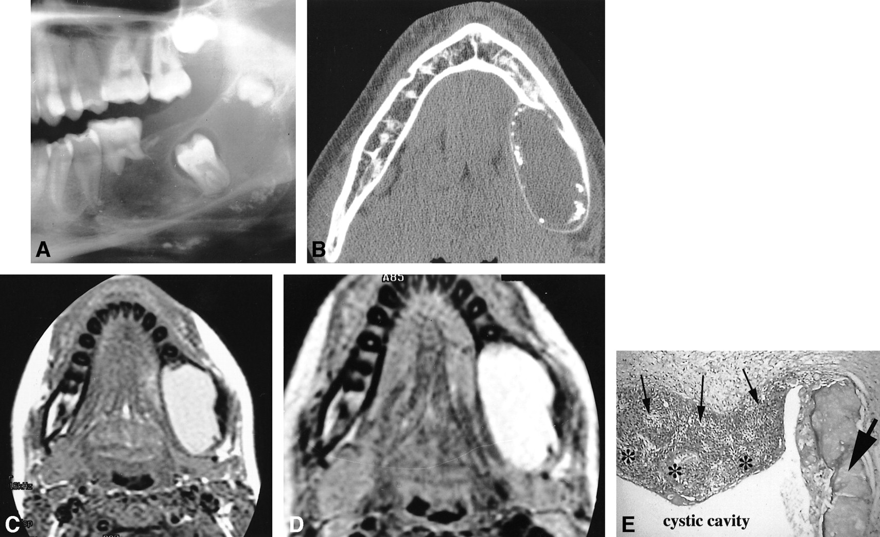
- fig 2.
Case 2.
A, Lateral plain radiograph of the mandible shows a large well-defined expansile radiolucency containing several small peripheral radiopacities. Focal resorption of the first molar with inferior displacement of the unerupted second molar is seen. Note the upper displacement of the germ of the third molar.
B, Axial CT view of the mandible shows a well-delineated expansile lesion with a large, smooth expansion of the thinned cortical plates. Small peripheral calcifications are seen.
C and D, MR images. Axial noncontrast spin-echo T1-weighted image (420/11/2) (C) shows a homogeneous, slightly hyperintense lesion. Axial fast spin-echo T2-weighted image (4000/104/1) (D) shows a markedly hyperintense lesion.
E, Photomicrograph shows the characteristic patterns of a COC close to a small odontoma (hematoxylin-eosin, original magnification ×200). Epithelial cyst lining (thin arrows) is seen with focal areas of necrosis (asterisks). Dentinoid component of the odontoma (thick arrow) is associated

Tables

Benign neoplasms related to the odontogenic apparatus


