Advertisement

Main menu

  • Home
  • Content
    • Current Issue
    • Accepted Manuscripts
    • Article Preview
    • Past Issue Archive
    • Video Articles
    • AJNR Case Collection
    • Case of the Week Archive
    • Case of the Month Archive
    • Classic Case Archive
  • Special Collections
    • AJNR Awards
    • ASNR Foundation Special Collection
    • Most Impactful AJNR Articles
    • Photon-Counting CT
    • Spinal CSF Leak Articles (Jan 2020-June 2024)
  • Multimedia
    • AJNR Podcasts
    • AJNR SCANtastic
    • Trainee Corner
    • MRI Safety Corner
    • Imaging Protocols
  • For Authors
    • Submit a Manuscript
    • Submit a Video Article
    • Submit an eLetter to the Editor/Response
    • Manuscript Submission Guidelines
    • Statistical Tips
    • Fast Publishing of Accepted Manuscripts
    • Graphical Abstract Preparation
    • Imaging Protocol Submission
    • Author Policies
  • About Us
    • About AJNR
    • Editorial Board
    • Editorial Board Alumni
  • More
    • Become a Reviewer/Academy of Reviewers
    • Subscribers
    • Permissions
    • Alerts
    • Feedback
    • Advertisers
    • ASNR Home

User menu

  • Alerts
  • Log in

Search

  • Advanced search
American Journal of Neuroradiology

American Journal of Neuroradiology

ASHNR American Society of Functional Neuroradiology ASHNR American Society of Pediatric Neuroradiology ASSR
  • Alerts
  • Log in

Advanced Search

  • Home
  • Content
    • Current Issue
    • Accepted Manuscripts
    • Article Preview
    • Past Issue Archive
    • Video Articles
    • AJNR Case Collection
    • Case of the Week Archive
    • Case of the Month Archive
    • Classic Case Archive
  • Special Collections
    • AJNR Awards
    • ASNR Foundation Special Collection
    • Most Impactful AJNR Articles
    • Photon-Counting CT
    • Spinal CSF Leak Articles (Jan 2020-June 2024)
  • Multimedia
    • AJNR Podcasts
    • AJNR SCANtastic
    • Trainee Corner
    • MRI Safety Corner
    • Imaging Protocols
  • For Authors
    • Submit a Manuscript
    • Submit a Video Article
    • Submit an eLetter to the Editor/Response
    • Manuscript Submission Guidelines
    • Statistical Tips
    • Fast Publishing of Accepted Manuscripts
    • Graphical Abstract Preparation
    • Imaging Protocol Submission
    • Author Policies
  • About Us
    • About AJNR
    • Editorial Board
    • Editorial Board Alumni
  • More
    • Become a Reviewer/Academy of Reviewers
    • Subscribers
    • Permissions
    • Alerts
    • Feedback
    • Advertisers
    • ASNR Home
  • Follow AJNR on Twitter
  • Visit AJNR on Facebook
  • Follow AJNR on Instagram
  • Join AJNR on LinkedIn
  • RSS Feeds

AJNR is seeking candidates for the AJNR Podcast Editor. Read the position description.

Research ArticleINTERVENTIONAL

Thrombus Formation at the Neck of Cerebral Aneurysms during Treatment with Guglielmi Detachable Coils

Michael J. Workman, Harry J. Cloft, Frank C. Tong, Jacques E. Dion, Mary E. Jensen, William F. Marx and David F. Kallmes
American Journal of Neuroradiology October 2002, 23 (9) 1568-1576;
Michael J. Workman
  • Find this author on Google Scholar
  • Find this author on PubMed
  • Search for this author on this site
Harry J. Cloft
  • Find this author on Google Scholar
  • Find this author on PubMed
  • Search for this author on this site
Frank C. Tong
  • Find this author on Google Scholar
  • Find this author on PubMed
  • Search for this author on this site
Jacques E. Dion
  • Find this author on Google Scholar
  • Find this author on PubMed
  • Search for this author on this site
Mary E. Jensen
  • Find this author on Google Scholar
  • Find this author on PubMed
  • Search for this author on this site
William F. Marx
  • Find this author on Google Scholar
  • Find this author on PubMed
  • Search for this author on this site
David F. Kallmes
  • Find this author on Google Scholar
  • Find this author on PubMed
  • Search for this author on this site
  • Article
  • Figures & Data
  • Info & Metrics
  • Responses
  • References
  • PDF
Loading

Article Figures & Data

Figures

  • Tables
  • Fig 1.
    • Download figure
    • Open in new tab
    • Download powerpoint
    Fig 1.

    Images from the case of a 54-year-old female patient with giant ophthalmic artery aneurysm remnant.

    A, Right internal carotid artery injection, oblique projection. Mask artifact outlines the margin of the dense coil pack filling the giant ophthalmic artery aneurysm. The first coil has been detached within the remnant at the aneurysm base (black arrow). Note GDCs within a previously embolized middle cerebral artery bifurcation aneurysm (arrowhead).

    B, After completion of the embolization procedure, control angiogram shows a small, poorly marginated, hazy opacity at the coil-parent artery interface, suggesting early thrombus formation (arrow).

    C, Repeat angiogram obtained 25 minutes later shows marked increase in thrombus burden at the coil surface and in the parent artery.

    D, Forty minutes after the administration of the abciximab bolus, near-complete resolution of thrombus at the coil surface can be seen.

  • Fig 2.
    • Download figure
    • Open in new tab
    • Download powerpoint
    Fig 2.

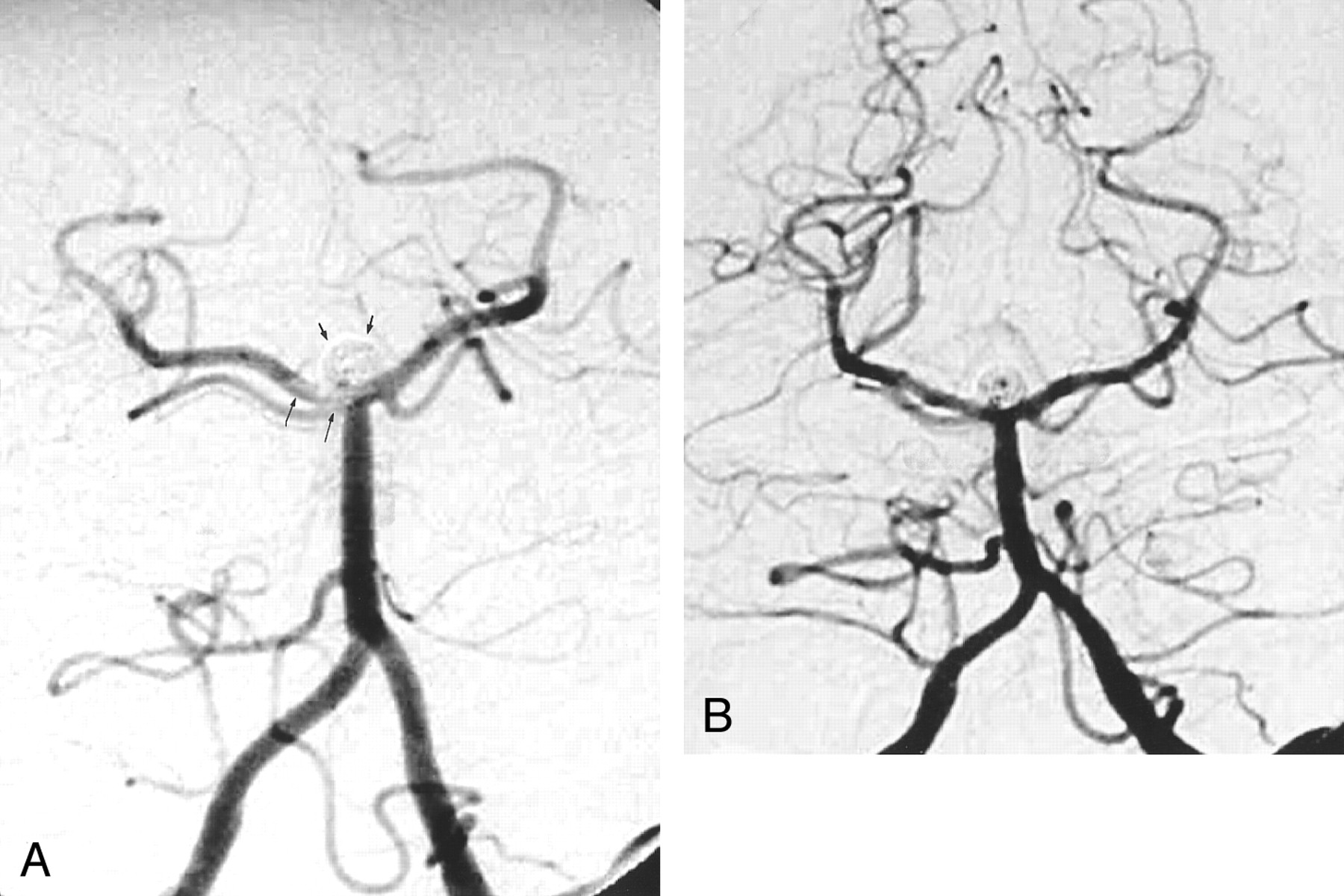
    Images from the case of a 49-year-old male patient with a basilar tip aneurysm.

    A, Angiogram of the left vertebral artery, transfacial projection. The basilar tip aneurysm is packed with GDCs (short arrows). A linear filling defect with hazy margins, consistent with thrombus, is present in the distal basilar artery and extends into the right P1 segment (long arrows).

    B, Angiogram of the left vertebral artery, transfacial projection. After 24 hours of heparin infusion, follow-up angiogram obtained after GDC treatment shows resolution of thrombus. However, there is partial recanalization of the aneurysm centrally.

  • Fig 3.
    • Download figure
    • Open in new tab
    • Download powerpoint
    Fig 3.

    Images from the case of a 57-year-old female patient with a superior hypophyseal artery aneurysm.

    A, Control angiogram of the right internal carotid artery, contralateral transorbital oblique projection. The superior hypophyseal artery aneurysm is tightly packed with GDCs (arrowheads). Thrombus has formed at the coil-parent artery interface, manifesting as a filling defect with a lobular contour with hazy margins (white arrow).

    B, Control angiogram obtained 10 minutes after the administration of the Integrilin bolus shows resolution of thrombus at the coil-parent artery interface.

Tables

  • Figures
    • View popup
    TABLE 1:

    Clinical data of GDC treatment cases with thrombus forming at coil-parent artery interface

    Case no.Age (yr)SexLocationAneurysm Diameter (mm)Aneurysm Neck (mm)RupturedProcedural TherapyFollow-up Therapy*Evidence of Stroke at Discharge
    154FOphthalmic6 × 35NoAbciximabAspirin, PlavixNo
    267FBasilar tip53.5NoAbciximabAspirinNo
    355FBasilar tip83.5NoAbciximabAspirin, PlavixNo
    445MBasilar tip6 × 85YesHeparinAspirinNo
    549MBasilar tip65YesHeparinAspirinNo
    643MPosterior communicating12 × 205YesHeparinAspirinNo
    739MPosterior communicating6 × 85YesHeparinAspirinNo
    857FSuperior hypophyseal106NoIntegrilin and heparinAspirinNo
    949FSuperior hypophyseal128NoAbciximabAspirin,PlavixNo
    • Note.—F indicates female; M, male.

    • * Aspirin, 325 mg/day; Plavix, 75 mg/day with 300 mg loading dose on day 1.

    • View popup
    TABLE 2:

    Case summary of intra-procedural anti-coagulation therapy

    Case No. Imaging FindingsActivated Clotting Time before Treatment (s)Initial TherapyActivated Clotting Time after Treatment (s)Findings after Anticoagulation TherapyAdditional Therapy*Technical Result
    1 Thrombus at coil-parent artery interface299Abciximab bolus and infusionNot measuredSmall residual thrombusAbciximab infusion for 12 hrNo follow-up angiography at 24 hr
    2 Thrombus at coil-parent artery interface238Heparin bolus, 3000 U318Unchanged after 30 minAbciximab bolus and infusionResolution of thrombus at 30 min after abciximab
    3 Coil loop, thrombus narrowing right P1 50%288Heparin infusion, 1000 U/hr400Resolved thrombus, coil protruding into left P1Abciximab bolus and infusionResolved thrombus
    4 Thrombus at coil-parent artery interface, extending into distal basilar and right P1250Heparin bolus, 2000 UNot measuredBasilar thrombus resolved after 20 min, nonocclusive thrombus right P2Heparin infusion for 24 hrNo follow-up angiography at 24 hr
    5 Thrombus at coil-parent artery interface, extending into left and right P1200Heparin bolus, 2000 U300Small residual, nonocclusive thrombus right P1Heparin infusion for 24 hrResolution of thrombus at 24 hr
    6 Thrombus at coil-parent artery interface155Heparin bolus, 3000 U300Small residual thrombusHeparin infusion for 24 hrNo follow-up angiography at 24 hr
    7 Thrombus at coil-parent artery interface220Heparin bolus, 4000 UNot measuredResolved thrombusHeparin infusion for 24 hrResolved thrombus
    8 Thrombus at coil-parent artery interfaceNot recordedHeparin bolus, 5000 U270Unchanged after 30 minIntegrilin bolus and infusionResolution of thrombus at 10 min after Integrilin
    9 Thrombus at coil-parent artery interface307Abciximab bolus and infusionNot measuredSmall residual thrombusAbciximab infusion for 12 hrNo follow-up angiography at 24 hr
    • Note.—P1 indicates P1 segment; P2, P2 segment.

    • * Integrilin dose: bolus, 180 μg/kg; infuse, 2 μg/kg/min for 20 hr. Abciximab dose: bolus, 0.25 mg/kg; infuse, 10 μg/min for 12 hr. Heparin infusion: heparin dosed to maintain a partial thromboplastin time of 50 to 70 s.

PreviousNext
Back to top

In this issue

American Journal of Neuroradiology: 23 (9)
American Journal of Neuroradiology
Vol. 23, Issue 9
1 Oct 2002
  • Table of Contents
  • Index by author
Advertisement
Print
Download PDF
Email Article

Thank you for your interest in spreading the word on American Journal of Neuroradiology.

NOTE: We only request your email address so that the person you are recommending the page to knows that you wanted them to see it, and that it is not junk mail. We do not capture any email address.

Enter multiple addresses on separate lines or separate them with commas.
Thrombus Formation at the Neck of Cerebral Aneurysms during Treatment with Guglielmi Detachable Coils
(Your Name) has sent you a message from American Journal of Neuroradiology
(Your Name) thought you would like to see the American Journal of Neuroradiology web site.
CAPTCHA
This question is for testing whether or not you are a human visitor and to prevent automated spam submissions.
Cite this article
Michael J. Workman, Harry J. Cloft, Frank C. Tong, Jacques E. Dion, Mary E. Jensen, William F. Marx, David F. Kallmes
Thrombus Formation at the Neck of Cerebral Aneurysms during Treatment with Guglielmi Detachable Coils
American Journal of Neuroradiology Oct 2002, 23 (9) 1568-1576;

Citation Manager Formats

  • BibTeX
  • Bookends
  • EasyBib
  • EndNote (tagged)
  • EndNote 8 (xml)
  • Medlars
  • Mendeley
  • Papers
  • RefWorks Tagged
  • Ref Manager
  • RIS
  • Zotero
0 Responses
Respond to this article
Share
Bookmark this article
Thrombus Formation at the Neck of Cerebral Aneurysms during Treatment with Guglielmi Detachable Coils
Michael J. Workman, Harry J. Cloft, Frank C. Tong, Jacques E. Dion, Mary E. Jensen, William F. Marx, David F. Kallmes
American Journal of Neuroradiology Oct 2002, 23 (9) 1568-1576;
del.icio.us logo Twitter logo Facebook logo Mendeley logo
  • Tweet Widget
  • Facebook Like
  • Google Plus One

Jump to section

  • Article
    • Abstract
    • Methods
    • Results
    • Representative Case Reports
    • Discussion
    • Footnotes
    • References
  • Figures & Data
  • Info & Metrics
  • Responses
  • References
  • PDF

Related Articles

  • No related articles found.
  • PubMed
  • Google Scholar

Cited By...

  • Intra-arterial versus intravenous abciximab therapy for thromboembolic complications of neuroendovascular procedures: case review and meta-analysis
  • Safety and efficacy of a new prophylactic tirofiban protocol without oral intraoperative antiplatelet therapy for endovascular treatment of ruptured intracranial aneurysms
  • Characterizing patterns of endothelialization following coil embolization: a whole-mount, dual immunostaining approach
  • Thromboembolic Complications in Patients with Clopidogrel Resistance after Coil Embolization for Unruptured Intracranial Aneurysms
  • Silent embolism after stent-assisted coiling of cerebral aneurysms: diffusion-weighted MRI study of 75 cases
  • Heparin dosing is associated with diffusion weighted imaging lesion load following aneurysm coiling
  • Attributing Hypodensities on CT to Angiographic Vasospasm Is Not Sensitive and Unreliable
  • Intra-arterial abciximab for the treatment of thrombus formation during coil embolization of intracranial aneurysms
  • Angiographic and Clinical Outcomes in 200 Consecutive Patients with Cerebral Aneurysm Treated with Hydrogel-Coated Coils
  • Abciximab Is a Safe Rescue Therapy in Thromboembolic Events Complicating Cerebral Aneurysm Coil Embolization: Single Center Experience in 42 Cases and Review of the Literature
  • Bailout Stent Deployment during Coil Embolization of Intracranial Aneurysms
  • Intravenous Administration of Acetylsalicylic Acid During Endovascular Treatment of Cerebral Aneurysms Reduces the Rate of Thromboembolic Events
  • Response to Letter by Wong et al
  • Thromboembolic Complications of Endovascular Aneurysm Occlusion Using Matrix Detachable Coils
  • Crossref
  • Google Scholar

This article has not yet been cited by articles in journals that are participating in Crossref Cited-by Linking.

More in this TOC Section

  • SAVE vs. Solumbra Techniques for Thrombectomy
  • Contrast-Induced Encephalopathy after NeuroIR
  • CT Perfusion&Reperfusion in Acute Ischemic Stroke
Show more Interventional

Similar Articles

Advertisement

Indexed Content

  • Current Issue
  • Accepted Manuscripts
  • Article Preview
  • Past Issues
  • Editorials
  • Editor's Choice
  • Fellows' Journal Club
  • Letters to the Editor
  • Video Articles

Cases

  • Case Collection
  • Archive - Case of the Week
  • Archive - Case of the Month
  • Archive - Classic Case

Special Collections

  • AJNR Awards
  • ASNR Foundation Special Collection
  • Most Impactful AJNR Articles
  • Photon-Counting CT
  • Spinal CSF Leak Articles (Jan 2020-June 2024)

More from AJNR

  • Trainee Corner
  • Imaging Protocols
  • MRI Safety Corner

Multimedia

  • AJNR Podcasts
  • AJNR Scantastics

Resources

  • Turnaround Time
  • Submit a Manuscript
  • Submit a Video Article
  • Submit an eLetter to the Editor/Response
  • Manuscript Submission Guidelines
  • Statistical Tips
  • Fast Publishing of Accepted Manuscripts
  • Graphical Abstract Preparation
  • Imaging Protocol Submission
  • Evidence-Based Medicine Level Guide
  • Publishing Checklists
  • Author Policies
  • Become a Reviewer/Academy of Reviewers
  • News and Updates

About Us

  • About AJNR
  • Editorial Board
  • Editorial Board Alumni
  • Alerts
  • Permissions
  • Not an AJNR Subscriber? Join Now
  • Advertise with Us
  • Librarian Resources
  • Feedback
  • Terms and Conditions
  • AJNR Editorial Board Alumni

American Society of Neuroradiology

  • Not an ASNR Member? Join Now

© 2025 by the American Society of Neuroradiology All rights, including for text and data mining, AI training, and similar technologies, are reserved.
Print ISSN: 0195-6108 Online ISSN: 1936-959X

Powered by HighWire