
Article Figures & Data
Figures
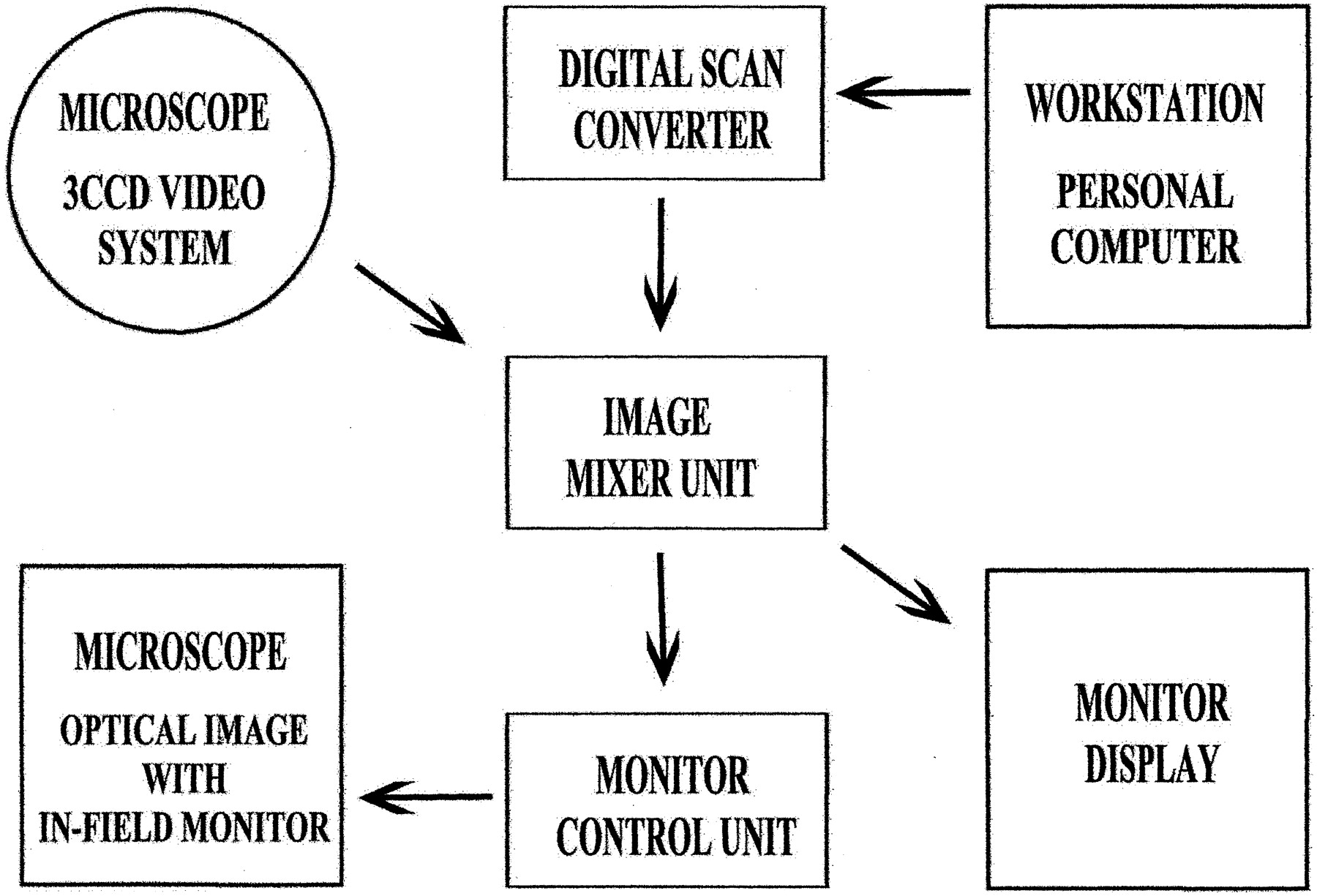
- Fig 1.
Schematic illustration of the setting for an intraoperative image reconstruction system.
- Fig 2.
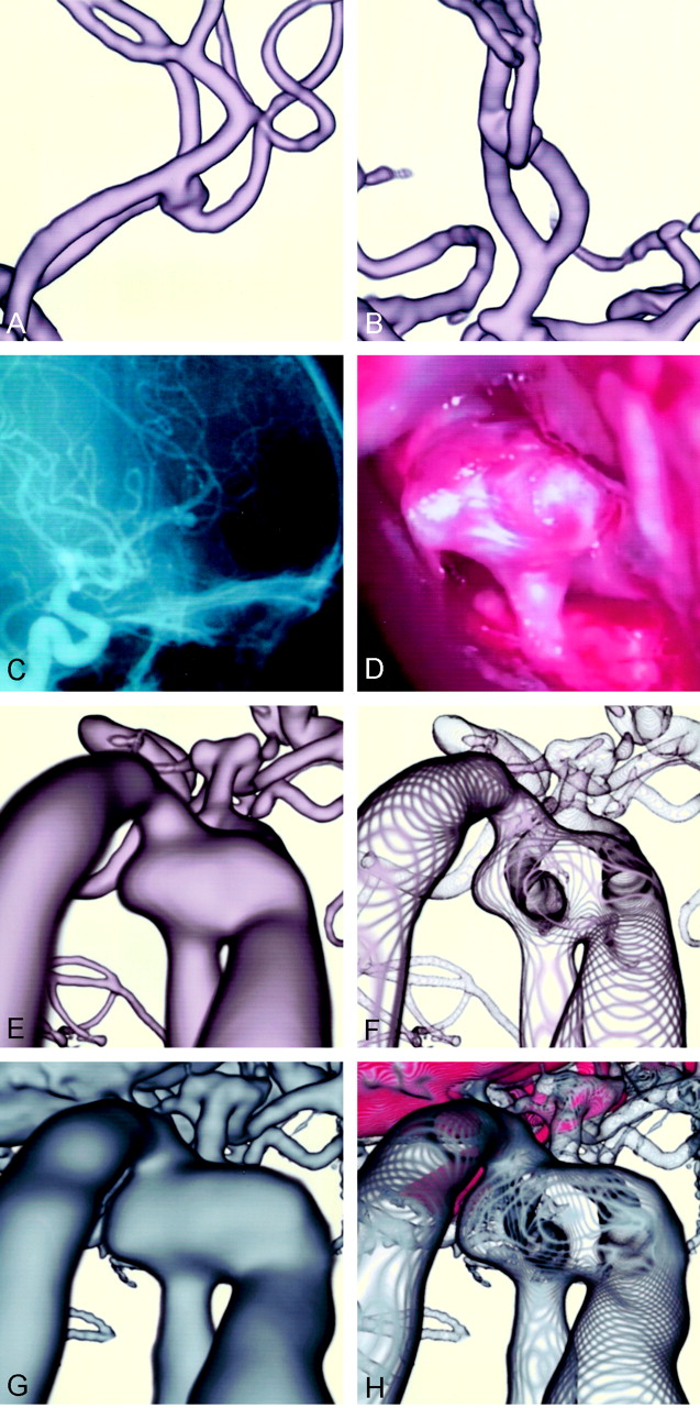
Case 1: A 74-year-old man with an unruptured right internal carotid-posterior communicating artery aneurysm.
A, Conventional parallel volume-rendered 3D MR angiogram, viewed from above.
B, Conventional parallel volume-rendered 3D CT angiogram, viewed from above.
C, Arteriogram, similar projection to the operative view in D.
D, Operative photograph before aneurysmal neck clipping shows the neck and dome of the aneurysm. A yellowish-colored atherosclerotic lesion of the wall is observed on the internal carotid artery and base of the aneurysmal neck.
E, Conventional perspective volume-rendered 3D MR angiogram shows the aneurysmal dome, internal carotid artery, and branching posterior communicating artery.
F, Transluminal 3D MR angiogram of the same projection shows the orifice of the posterior communicating artery at the neck.
G, Conventional 3D CT angiogram shows similar aneurysmal architecture, with the surrounding veins and bony structures.
H, Transluminal 3D CT angiogram shows the transparent view of the aneurysmal architecture, with calcified nodules at the neck enhanced in red.
- Fig 3.
Case 2: A 55-year-old woman with an unruptured distal anterior cerebral artery (A2–3) aneurysm.
A and B, Parallel volume-rendered 3D MR angiograms, right lateral (A) and anteroposterior (B) projections, show the aneurysm at the bifurcaton of the left A2 and A3, extending bilaterally, with the right side of the dome attached to the right A3.
C, Arteriogram, right lateral projection, shows the aneurysm.
D, Operative photograph through the right anterior interhemispheric approach, shows the superoposterior aspect of the aneurysmal dome with branching efferent artery. The right side of the dome is adhered firmly to the right A3, and the aneurysmal neck is hidden by the foreground dome.
E, Conventional perspective volume-rendered 3D MR angiogram shows the angioarchitecture of the aneurysm.
F, Transluminal 3D MR angiogram of the same projection shows the orifices of the afferent left A2 and efferent left A3 arteries transluminally through the vessel and aneurysmal walls.
G, Conventional 3D CT angiogram shows the aneurysmmal architecture with surrounding veins and frontal base bone.
H, Transluminal 3D CT anagiogram depicts similar findings as in G, with the frontal base bone color-coded in red.


In this issue


Jump to section
Related Articles
Cited By...
- No citing articles found.