
Article Figures & Data
Figures
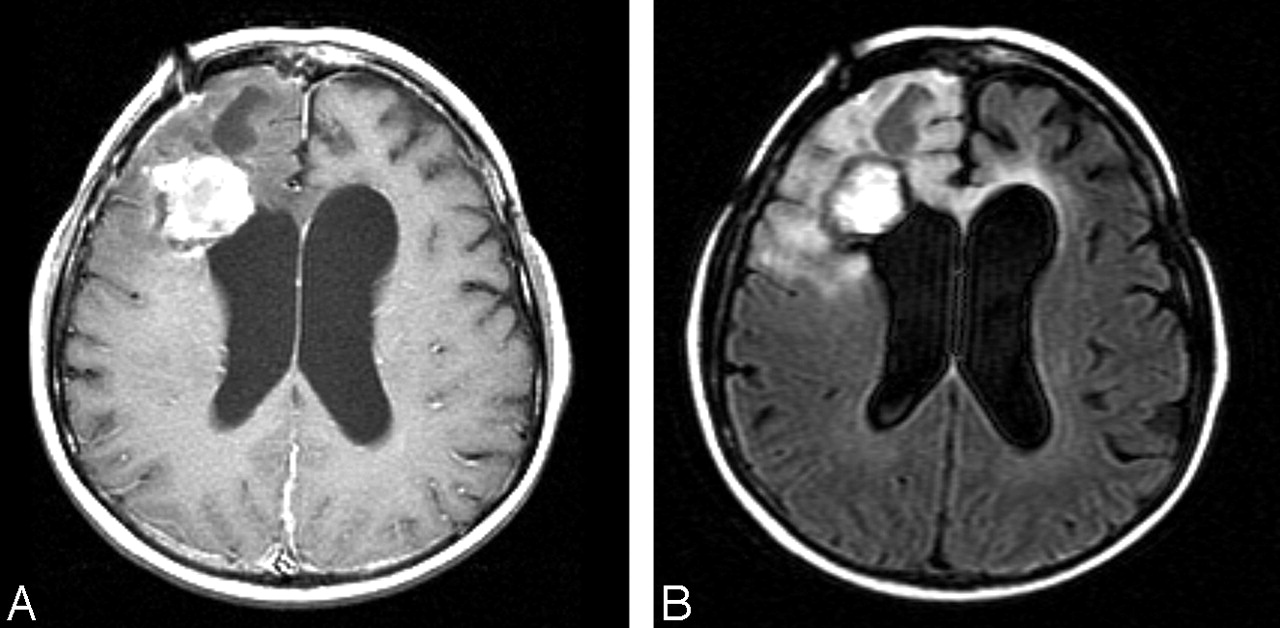
- Fig 1.
MR imaging shows a 5.1 × 6.4 cm heterogeneous mass in the right frontal lobe with areas of necrosis and surrounding edema.
A, Axial T1-weighted postgadolinium image (500/8/2; TR/TE/NEX).
B, Axial FLAIR image (8002/158/1; TI, 2000).
C, Axial FLAIR image (8002/158/1; TI, 2000) immediately after surgical resection.
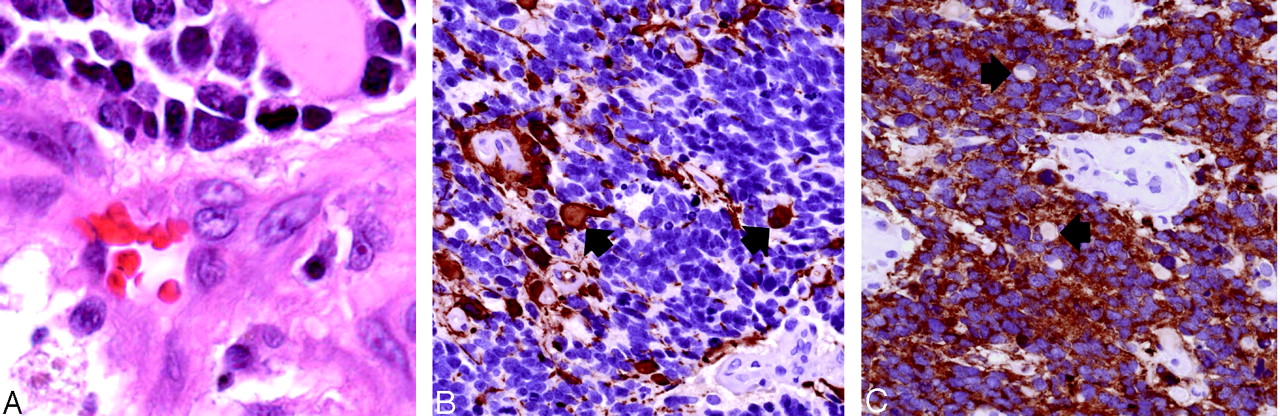
- Fig 2.
Histopathologic findings (original magnification ×400).
A, Hematoxylin-eosin stained sections show sheets of undifferentiated tumor with frequent apoptotic figures and endothelial proliferation. Malignant gemistocytes indicate astrocytic differentiation (arrows).
B, Immunostaining with anti-GFAP antibody shows positive staining of fibrillary tumor cell processes as well as of the rounded cell bodies of malignant gemistocytic astrocytes (arrows).
C, Scytplasmic staining of many tumor cells, indicating neuroblastic differentiation. Cells showing absent cytoplasmic staining (arrows) correspond to cells with astrocytic differentiation.
- Fig 3.
Follow-up MR imaging 4 months after resection shows a stable appearance of her right frontal tumor with decreased edema and mass effect.
A, Axial T1-weighted postgadolinium image (400/8/2).
B, Axial FLAIR image (8002/158/1; T1, 2000).


In this issue


Jump to section
Related Articles
Cited By...
- No citing articles found.