
Article Figures & Data
Figures
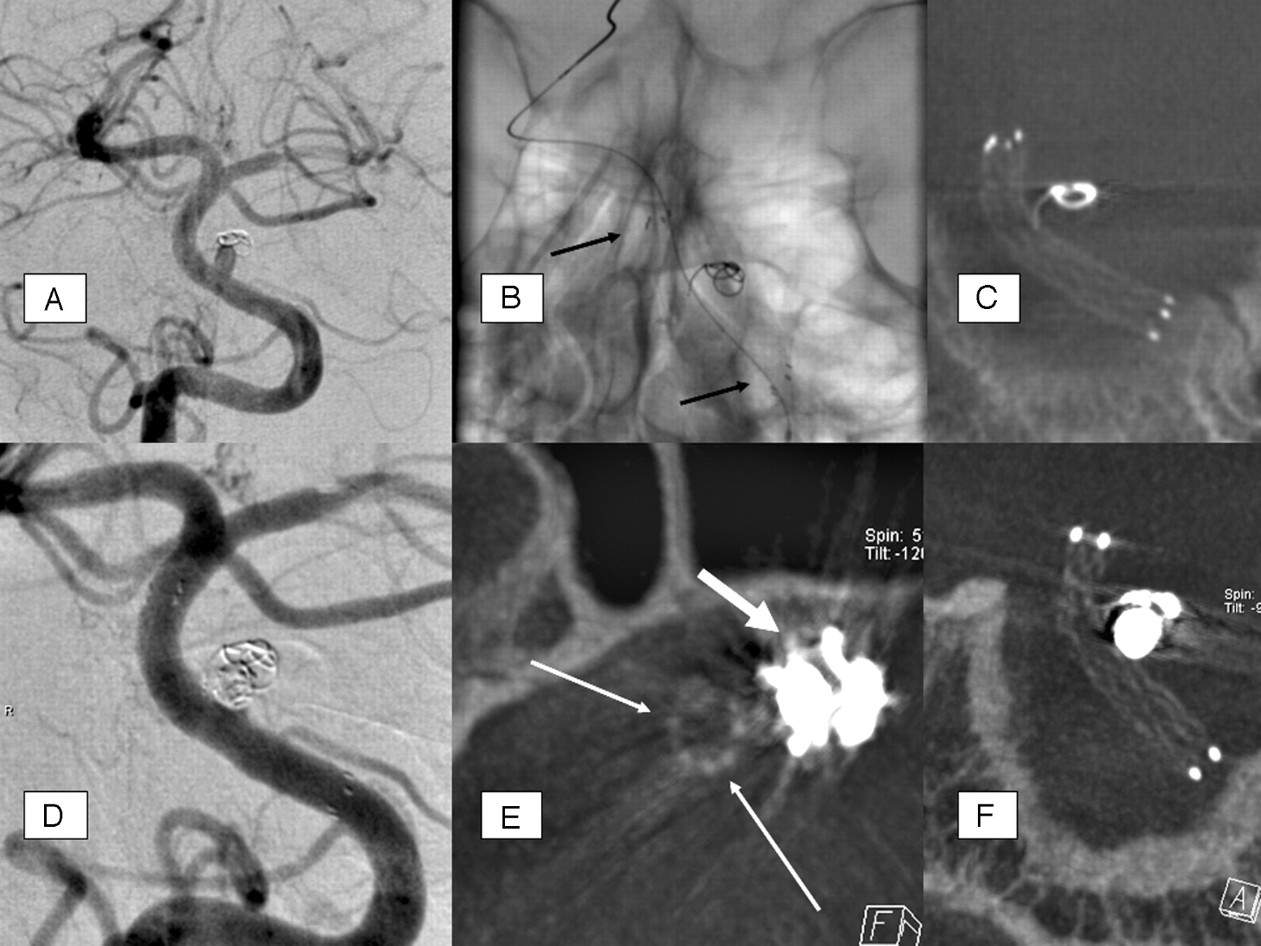
- Fig 1.
A, The basilar stem aneurysm showed complete occlusion initially after insertion of a single coil 2.5 mm in diameter: coil compaction and significant aneurysm growth in 6-month follow-up-DSA. B, Unsubtracted DSA image after insertion of a Neuroform stent (4 × 20 mm) shows the proximal and distal radiopaque markers of the stent (arrows); the stent itself is not visible. C, MIP reconstruction of ACT performed after stent deployment and before second coil embolization: excellent stent visibility and regular, complete stent deployment. The initially inserted single coil shows compaction and is distant to the parent (basilar) artery and to the stent struts as well, including one coil loop with position adjacent to stent wall, with definite extraluminal position. D, DSA. A total of 4 platinum microcoils (diameters, 2.5 and 2.0 mm, respectively) were additionally inserted. The basilar stem aneurysm shows satisfiable occlusion with minimal dog-ear remnant. E, Axial, thin (1 mm section thickness) MPR reconstruction of ACT performed after stent deployment and second coil embolization (same dataset as in F). This reconstruction allows definite exclusion of stent strut movement or coil protrusion: stent (thin arrows) without deformation adjacent to coil package (thick arrow), which causes some beam-hardening artifacts. F, MIP reconstruction (oblique coronal) of ACT performed after stent deployment and second coil embolization: excellent stent visibility without significant limitation by marginal beam-hardening artifacts through coil package; no change of stent configuration compared with the ACT imaging before the second coil embolization.
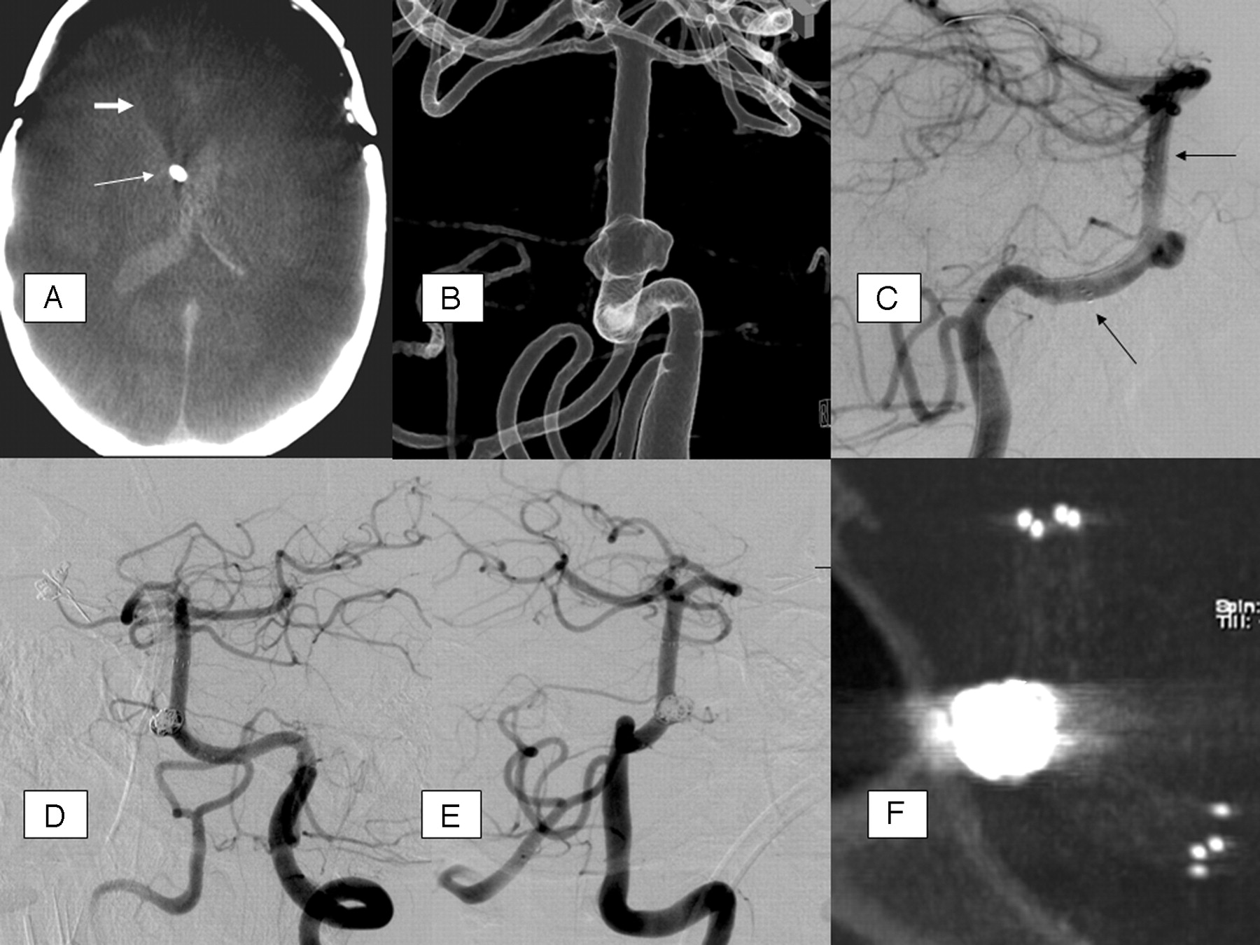
- Fig 2.
A, Native, axial ACT-reconstruction, 5 mm section thickness; extensive SAH with intraventricular, hyperattenuated blood; CSF drainage adjacent to the septum pellucidum (arrow); previous frontal trepanation in the course of aneurysm clipping (anterior communicating artery); hypoattenuated brain parenchyma defect (thick arrow) with dilation of the frontal of the right lateral ventricle due to previous aneurysm rupture and parenchymal hemorrhage. B, Rotational 3D angiography with intra-arterial contrast agent injection, transparent surface shaded reconstruction (posterior-anterior view); broad-based proximal basilar stem aneurysm, forward oriented. The aneurysm base surrounds the basilar artery at least as a semicircle. C, DSA, lateral view; guidewire in the left posterior cerebral artery; Neuroform stent with proximal and distal markers (arrows). D and E, DSA (oblique views) after stent-assisted aneurysm coiling. F, MIP reconstruction of ACT after stent-assisted aneurysm coiling, providing excellent overview of the stent and the adjacent coil package; moderate beam-hardening artifacts.
- Fig 3.
A, Follow-up DSA 3 months after initial coiling of the basilar artery tip aneurysm; partially reperfused aneurysm due to aneurysm growth and minimal coil compaction. B, Exchange microguidewire in the left posterior cerebral artery. C, Neuroform stent inserted, still undeployed, with radiopaque markers at the ends (arrows). Note the stretched vessel anatomy compared with B. D, Fully deployed stent, markers (arrows). E, ACT MIP reconstruction with impaired stent visibility on the level of the large coil package. F, ACT with MPR to exclude periprocedural bleeding; proximal stent marker (arrow).


Tables
Case-No. (Age, y) Sex Aneurysm Localization Presenting Symptoms Maximum Aneurysm Diameter, mm Maximum Aneurysm Neck Diameter, mm Head/Neck Ratio 1 (66) F ICA, extradural Progressive, ipsilateral visual impairment 18 6 3.0 2 (63) M Basilar stem Asymptomatic, elective procedure 4 3.5 1.1 3 (35) M Basilar stem SAH Hunt and Hess grade 4 4.5 3.5 1.3 4 (61) F Basilar tip Brain stem compression, hydrocephalus 13 7 1.3 5 (57) M Basilar tip SAH Hunt and Hess grade 4 at time of previous coiling 12 5.5 2.2 6 (24) F ICA Multilobulated No symptoms, elective procedure 10 6 1.7 7 (44) M Basilar stem SAH Hunt and Hess grade 3 at time of previous coiling 2.5 2 1.25 8 (45) F Basilar tip SAH Hunt and Hess grade 3 10 5.5 1.8 9 (46) F Basilar tip SAH Hunt and Hess grade 1 11 6 1.8 10 (58) M Fusiform vertebral artery aneurysm with saccular comp Asymptomatic, previous SAH from inversely located vertebral aneurysm 4 (saccular compartment) No neck, fusiform geometry – 11 (47) M Fusiform vertebral artery aneurysm Asymptomatic 11 No neck, fusiform geometry – Note:—ICA indicates internal carotid artery; M, male; F, female; SAH, subarachnoid hemorrhage; –, no data.
- Table 2:
Results and cases arbitrarily divided into 3 groups in terms of stent visibility with angiographic CT data reconstructions*
Case No. (Age, y) Stent (Neuroform 2/3) Maximum Aneurysm Diameter, mm No. of coils Aneurysm Occlusion Rate, I > 95% or II < 95% Stent Visibility, Grade 1, 2, or 3 1 (66) NF3, 4.5 × 20 mm 18 18 II 3 2 (63) NF3, 4.5 × 20 mm 4 2 II 1 3 (35) NF3, 4.5 × 20 mm 4.5 5 I 1 4 (61) NF3, 4.0 × 20 mm 13 4 II 3 5 (57) NF3, 4.0 × 20 mm 12 7 I 2 6 (24) NF2, 4.5 × 20 mm 10 10 I 2 7 (44) NF3, 4.0 × 20 mm 2.5 3 I 1 8 (45) NF3, 3.5 × 20 mm 10 8 I 2 9 (46) NF3, 4.0 × 20 mm 11 14 II 3 10 (58) NF3, 4.0 × 20 mm 4 (saccular compartment) 7 I 3 11(47) NF3, 4.5 × 30 mm 11 11 I 3 Note:—NF indicates Neuroform.
* Group 1, excellent; Group 2, favorable; Group 3, poor (see column on the right).
In this issue


Jump to section
Related Articles
Cited By...
- Clinical Impact of Flat Panel Volume CT Angiography in Evaluating the Accurate Intraoperative Deployment of Flow-Diverter Stents
- FRED Flow Diverter: A Study on Safety and Efficacy in a Consecutive Group of 50 Patients
- A novel reconstruction tool (syngo DynaCT Head Clear) in the post-processing of DynaCT images to reduce artefacts and improve image quality
- Low-Dose Volume-of-Interest C-Arm CT Imaging of Intracranial Stents and Flow Diverters
- Cone-beam CT angiography (Dyna CT) for intraoperative localization of cerebral arteriovenous malformations
- Role of C-Arm VasoCT in the Use of Endovascular WEB Flow Disruption in Intracranial Aneurysm Treatment
- In Vitro and In Vivo Imaging Characteristics Assessment of Polymeric Coils Compared with Standard Platinum Coils for the Treatment of Intracranial Aneurysms
- Angiographic CT for Intraprocedural Monitoring of Complex Neuroendovascular Procedures
- Applicability of Tableside Flat Panel Detector CT Parenchymal Cerebral Blood Volume Measurement in Neurovascular Interventions: Preliminary Clinical Experience
- Use of CT Angiography in Comparison with Other Imaging Techniques for the Determination of Embolus and Remnant Size in Experimental Aneurysms Embolized with Hydrogel Filaments
- Contrast-Enhanced Angiographic Cone-Beam CT of Cerebrovascular Stents: Experimental Optimization and Clinical Application
- Use of Angiographic CT Imaging in the Cardiac Catheterization Laboratory for Congenital Heart Disease
- Metal Artifact Reduction for Clipping and Coiling in Interventional C-Arm CT