Advertisement

Main menu

  • Home
  • Content
    • Current Issue
    • Accepted Manuscripts
    • Article Preview
    • Past Issue Archive
    • Video Articles
    • AJNR Case Collection
    • Case of the Week Archive
    • Case of the Month Archive
    • Classic Case Archive
  • Special Collections
    • AJNR Awards
    • ASNR Foundation Special Collection
    • Most Impactful AJNR Articles
    • Photon-Counting CT
    • Spinal CSF Leak Articles (Jan 2020-June 2024)
  • Multimedia
    • AJNR Podcasts
    • AJNR SCANtastic
    • Trainee Corner
    • MRI Safety Corner
    • Imaging Protocols
  • For Authors
    • Submit a Manuscript
    • Submit a Video Article
    • Submit an eLetter to the Editor/Response
    • Manuscript Submission Guidelines
    • Statistical Tips
    • Fast Publishing of Accepted Manuscripts
    • Graphical Abstract Preparation
    • Imaging Protocol Submission
    • Author Policies
  • About Us
    • About AJNR
    • Editorial Board
    • Editorial Board Alumni
  • More
    • Become a Reviewer/Academy of Reviewers
    • Subscribers
    • Permissions
    • Alerts
    • Feedback
    • Advertisers
    • ASNR Home

User menu

  • Alerts
  • Log in

Search

  • Advanced search
American Journal of Neuroradiology

American Journal of Neuroradiology

ASHNR American Society of Functional Neuroradiology ASHNR American Society of Pediatric Neuroradiology ASSR
  • Alerts
  • Log in

Advanced Search

  • Home
  • Content
    • Current Issue
    • Accepted Manuscripts
    • Article Preview
    • Past Issue Archive
    • Video Articles
    • AJNR Case Collection
    • Case of the Week Archive
    • Case of the Month Archive
    • Classic Case Archive
  • Special Collections
    • AJNR Awards
    • ASNR Foundation Special Collection
    • Most Impactful AJNR Articles
    • Photon-Counting CT
    • Spinal CSF Leak Articles (Jan 2020-June 2024)
  • Multimedia
    • AJNR Podcasts
    • AJNR SCANtastic
    • Trainee Corner
    • MRI Safety Corner
    • Imaging Protocols
  • For Authors
    • Submit a Manuscript
    • Submit a Video Article
    • Submit an eLetter to the Editor/Response
    • Manuscript Submission Guidelines
    • Statistical Tips
    • Fast Publishing of Accepted Manuscripts
    • Graphical Abstract Preparation
    • Imaging Protocol Submission
    • Author Policies
  • About Us
    • About AJNR
    • Editorial Board
    • Editorial Board Alumni
  • More
    • Become a Reviewer/Academy of Reviewers
    • Subscribers
    • Permissions
    • Alerts
    • Feedback
    • Advertisers
    • ASNR Home
  • Follow AJNR on Twitter
  • Visit AJNR on Facebook
  • Follow AJNR on Instagram
  • Join AJNR on LinkedIn
  • RSS Feeds

AJNR is seeking candidates for the AJNR Podcast Editor. Read the position description.

Research ArticleSPINE

Quantitative and Diffusion MR Imaging as a New Method To Assess Osteoporosis

H.G. Hatipoglu, A. Selvi, D. Ciliz and E. Yuksel
American Journal of Neuroradiology November 2007, 28 (10) 1934-1937; DOI: https://doi.org/10.3174/ajnr.A0704
H.G. Hatipoglu
  • Find this author on Google Scholar
  • Find this author on PubMed
  • Search for this author on this site
A. Selvi
  • Find this author on Google Scholar
  • Find this author on PubMed
  • Search for this author on this site
D. Ciliz
  • Find this author on Google Scholar
  • Find this author on PubMed
  • Search for this author on this site
E. Yuksel
  • Find this author on Google Scholar
  • Find this author on PubMed
  • Search for this author on this site
  • Article
  • Figures & Data
  • Info & Metrics
  • Responses
  • References
  • PDF
Loading

Article Figures & Data

Figures

  • Fig 1.
    • Download figure
    • Open in new tab
    • Download powerpoint
    Fig 1.

    A, Graph of ADC values for normal, osteopenic, and osteoporotic subjects. The differences between groups are significant (P < .0001, Kruskal-Wallis Test). Normal versus osteopenia, P = .026; normal versus osteoporosis, P < .0001; osteopenia versus osteoporosis, P = .077 (Mann-Whitney U test). B, Graph of diffusion signal-intensity values for normal, osteopenic, and osteoporotic subjects. The differences between groups are significant (P < .0001, Kruskal-Wallis test). Normal versus osteopenia, P = .004; normal versus osteoporosis, P < .0001; osteopenia versus osteoporosis, P < .0001 (Mann-Whitney U test).
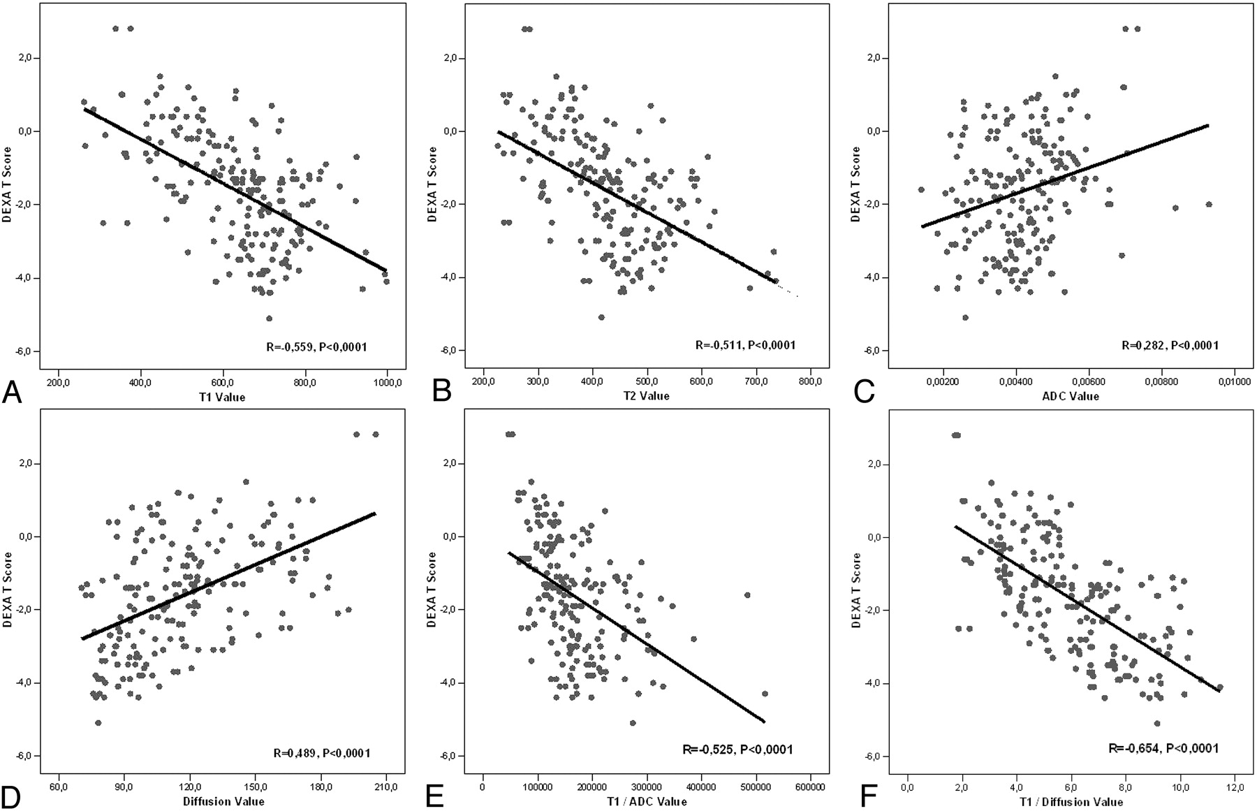
  • Fig 2.
    • Download figure
    • Open in new tab
    • Download powerpoint
    Fig 2.

    A, The correlations graphics of DEXA t-scores and T1 signal-intensity values (R = −0.559, P < .0001). B, The correlations graphics of DEXA t-scores and T2 signal intensity values (R = −0.511, P < .0001). C, The correlations graphics of DEXA t-scores and ADC values (R = 0.282, P < .0001). D, The correlations graphics of DEXA t-scores and diffusion signal-intensity values (R = 0.489, P < .0001). E, The correlations graphics of DEXA t-scores and T1/ADC values (R = −0.525, P < .0001). F, The correlations graphics of DEXA t-scores and T1/diffusion values (R = −0.654, P < .0001).

PreviousNext
Back to top

In this issue

American Journal of Neuroradiology: 28 (10)
American Journal of Neuroradiology
Vol. 28, Issue 10
November 2007
  • Table of Contents
  • Index by author
Advertisement
Print
Download PDF
Email Article

Thank you for your interest in spreading the word on American Journal of Neuroradiology.

NOTE: We only request your email address so that the person you are recommending the page to knows that you wanted them to see it, and that it is not junk mail. We do not capture any email address.

Enter multiple addresses on separate lines or separate them with commas.
Quantitative and Diffusion MR Imaging as a New Method To Assess Osteoporosis
(Your Name) has sent you a message from American Journal of Neuroradiology
(Your Name) thought you would like to see the American Journal of Neuroradiology web site.
CAPTCHA
This question is for testing whether or not you are a human visitor and to prevent automated spam submissions.
Cite this article
H.G. Hatipoglu, A. Selvi, D. Ciliz, E. Yuksel
Quantitative and Diffusion MR Imaging as a New Method To Assess Osteoporosis
American Journal of Neuroradiology Nov 2007, 28 (10) 1934-1937; DOI: 10.3174/ajnr.A0704

Citation Manager Formats

  • BibTeX
  • Bookends
  • EasyBib
  • EndNote (tagged)
  • EndNote 8 (xml)
  • Medlars
  • Mendeley
  • Papers
  • RefWorks Tagged
  • Ref Manager
  • RIS
  • Zotero
0 Responses
Respond to this article
Share
Bookmark this article
Quantitative and Diffusion MR Imaging as a New Method To Assess Osteoporosis
H.G. Hatipoglu, A. Selvi, D. Ciliz, E. Yuksel
American Journal of Neuroradiology Nov 2007, 28 (10) 1934-1937; DOI: 10.3174/ajnr.A0704
del.icio.us logo Twitter logo Facebook logo Mendeley logo
  • Tweet Widget
  • Facebook Like
  • Google Plus One
Purchase

Jump to section

  • Article
    • Abstract
    • Methods
    • Results
    • Discussion
    • Conclusion
    • References
  • Figures & Data
  • Info & Metrics
  • Responses
  • References
  • PDF

Related Articles

  • No related articles found.
  • PubMed
  • Google Scholar

Cited By...

  • No citing articles found.
  • Crossref (51)
  • Google Scholar

This article has been cited by the following articles in journals that are participating in Crossref Cited-by Linking.

  • Quantitative MRI and spectroscopy of bone marrow
    Dimitrios C. Karampinos, Stefan Ruschke, Michael Dieckmeyer, Maximilian Diefenbach, Daniela Franz, Alexandra S. Gersing, Roland Krug, Thomas Baum
    Journal of Magnetic Resonance Imaging 2018 47 2
  • Optimising diffusion weighted MRI for imaging metastatic and myeloma bone disease and assessing reproducibility
    C. Messiou, D. J. Collins, V. A. Morgan, N. M. deSouza
    European Radiology 2011 21 8
  • Diffusion and perfusion imaging of bone marrow
    Andreas Biffar, Olaf Dietrich, Steven Sourbron, Hans-Roland Duerr, Maximilian F. Reiser, Andrea Baur-Melnyk
    European Journal of Radiology 2010 76 3
  • Evaluation of MR spectroscopy and diffusion-weighted MRI in detecting bone marrow changes in postmenopausal women with osteoporosis
    G.Y. Tang, Z.W. Lv, R.B. Tang, Y. Liu, Y.F. Peng, W. Li, Y.S. Cheng
    Clinical Radiology 2010 65 5
  • Diffusion imaging of the vertebral bone marrow
    Olaf Dietrich, Tobias Geith, Maximilian F. Reiser, Andrea Baur‐Melnyk
    NMR in Biomedicine 2017 30 3
  • Diffusion Magnetic Resonance Imaging to Differentiate Degenerative From Infectious Endplate Abnormalities in the Lumbar Spine
    Yawara Eguchi, Seiji Ohtori, Masaomi Yamashita, Kazuyo Yamauchi, Munetaka Suzuki, Sumihisa Orita, Hiroto Kamoda, Gen Arai, Tetsuhiro Ishikawa, Masayuki Miyagi, Nobuyasu Ochiai, Shunji Kishida, Yoshitada Masuda, Shigehiro Ochi, Takashi Kikawa, Masashi Takaso, Yasuchika Aoki, Gen Inoue, Tomoaki Toyone, Kazuhisa Takahashi
    Spine 2011 36 3
  • The Relationship of Fat Distribution and Insulin Resistance with Lumbar Spine Bone Mass in Women
    Francisco J. A. de Paula, Iana M. de Araújo, Adriana L. Carvalho, Jorge Elias, Carlos E. G. Salmon, Marcello H. Nogueira-Barbosa, Victor Sanchez-Margalet
    PLOS ONE 2015 10 6
  • Combined diffusion-weighted and dynamic contrast-enhanced imaging of patients with acute osteoporotic vertebral fractures
    Andreas Biffar, Steven Sourbron, Olaf Dietrich, Gerwin Schmidt, Michael Ingrisch, Maximilian F. Reiser, Andrea Baur-Melnyk
    European Journal of Radiology 2010 76 3
  • In vitroandin vivoMR evaluation of internal gradient to assess trabecular bone density
    S De Santis, M Rebuzzi, G Di Pietro, F Fasano, B Maraviglia, S Capuani
    Physics in Medicine and Biology 2010 55 19
  • Diffusion-weighted magnetic resonance imaging in differentiating acute infectious spondylitis from degenerative Modic type 1 change; the role ofb-value, apparent diffusion coefficient, claw sign and amorphous increased signal
    Mohammad Hossein Daghighi, Masoud Poureisa, Mohsen Safarpour, Razieh Behzadmehr, Daniel F Fouladi, Ali Meshkini, Mojtaba Varshochi, Ali Kiani Nazarlou
    The British Journal of Radiology 2016 89 1066

More in this TOC Section

  • MP2RAGE 7T in MS Lesions of the Cervical Spine
  • Bern Score Validity for SIH
  • Resisted Inspiration for CSF-Venous Fistula
Show more Spine

Similar Articles

Advertisement

Indexed Content

  • Current Issue
  • Accepted Manuscripts
  • Article Preview
  • Past Issues
  • Editorials
  • Editor's Choice
  • Fellows' Journal Club
  • Letters to the Editor
  • Video Articles

Cases

  • Case Collection
  • Archive - Case of the Week
  • Archive - Case of the Month
  • Archive - Classic Case

Special Collections

  • AJNR Awards
  • ASNR Foundation Special Collection
  • Most Impactful AJNR Articles
  • Photon-Counting CT
  • Spinal CSF Leak Articles (Jan 2020-June 2024)

More from AJNR

  • Trainee Corner
  • Imaging Protocols
  • MRI Safety Corner

Multimedia

  • AJNR Podcasts
  • AJNR Scantastics

Resources

  • Turnaround Time
  • Submit a Manuscript
  • Submit a Video Article
  • Submit an eLetter to the Editor/Response
  • Manuscript Submission Guidelines
  • Statistical Tips
  • Fast Publishing of Accepted Manuscripts
  • Graphical Abstract Preparation
  • Imaging Protocol Submission
  • Evidence-Based Medicine Level Guide
  • Publishing Checklists
  • Author Policies
  • Become a Reviewer/Academy of Reviewers
  • News and Updates

About Us

  • About AJNR
  • Editorial Board
  • Editorial Board Alumni
  • Alerts
  • Permissions
  • Not an AJNR Subscriber? Join Now
  • Advertise with Us
  • Librarian Resources
  • Feedback
  • Terms and Conditions
  • AJNR Editorial Board Alumni

American Society of Neuroradiology

  • Not an ASNR Member? Join Now

© 2025 by the American Society of Neuroradiology All rights, including for text and data mining, AI training, and similar technologies, are reserved.
Print ISSN: 0195-6108 Online ISSN: 1936-959X

Powered by HighWire