
Article Figures & Data
Figures
- Fig 1.
Signal intensity of thrombosed venous segments without susceptibility effect on GRE T2*- weighted sequences. Black, hypointense relative to gray matter; gray, isointense to gray matter; white, hyperintense to gray matter.
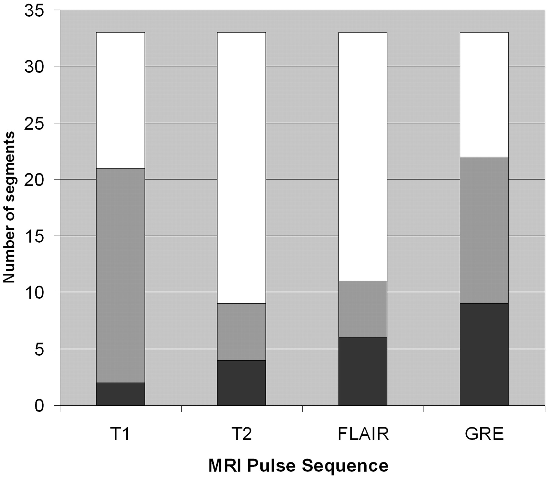
- Fig 2.
Signal intensity of thrombosed venous segments with susceptibility effect on GRE T2*- weighted sequences. Black, hypointense relative to gray matter; gray, isointense to gray matter; white, hyperintense to gray matter. GRE*, all segments exhibiting susceptibility effect were hypointense on GRE T2*- weighted sequences.
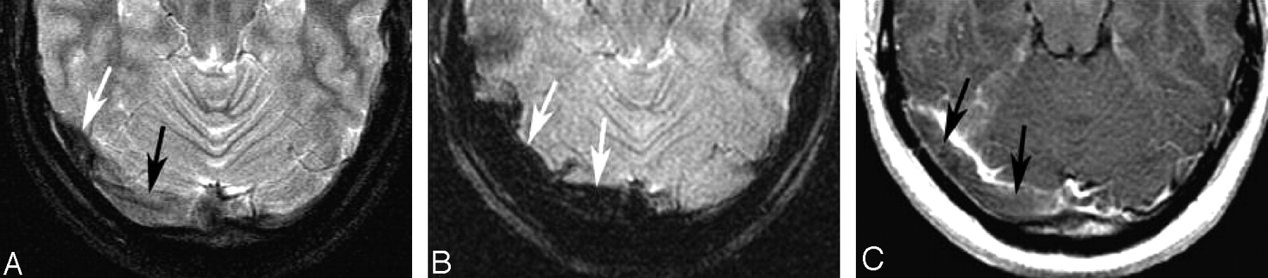
- Fig 3.
A 22-year-old woman with a 6-day history of headache. Note the subtle abnormal signal intensity in the right transverse sinus on T2-weighting (A). The medial sinus is isointense (black arrow, A) and the lateral transverse sinus is hypointense (white arrow, A). The signal intensity on GRE sequences is very dark and enlarged compared with the size of the sinus on other sequences, consistent with susceptibility effect (arrows, B). On the postcontrast T1-weighted image, an isointense filling defect is noted consistent with thrombus (arrows, C).
A, FSE T2-weighted image.
B, GRE T2*-weighted image.
C, T1WI after the administration of contrast.
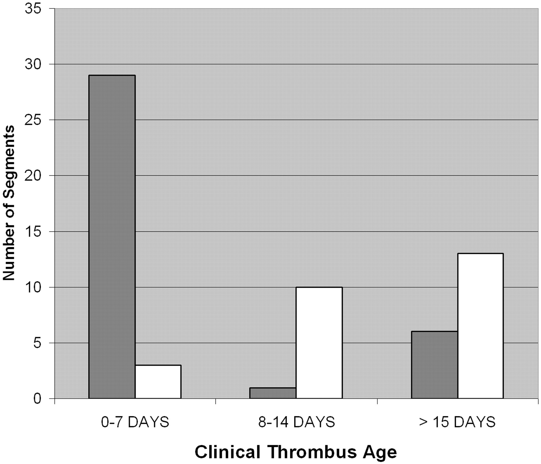
- Fig 4.
Clinical thrombus age and susceptibility effect. Gray, susceptibility effect present; white, susceptibility effect absent.
- Fig 5.
A 20-year-old woman with 2-day history of severe headache. Thrombosis of the superior sagittal sinus and multiple cortical veins is present. Note the prominent hypointensity of the thrombosed superior sagittal sinus and cortical veins in B (arrows), much larger than the thrombosed venous structures on other sequences (A and C, arrows). Graded as 3+ susceptibility effect. Signal intensity of the thrombosed segments is hypointense on T2WI and isointense on T1WI. Note the normal parietal cortical vein on the left (arrowheads).
A, FSE T2-weighted image.
B, GRE T2*-weighted image.
C, T1WI after the administration of contrast.


Tables
- Table 1:
Susceptibility effect on gradient recalled-echo MR images and clinical thrombus age
Clinical Thrombus Age SE+ n (%) SE− n (%) Total 0–7 days 29 (80.5%) 3 (11.5%) 32 8–14 days 1 (2.8%) 10 (38.5%) 11 >15 days 6 (16.7%) 13 (50%) 19 Total 36 26 62 Note:—SE+ indicates susceptibility effect present; SE−, susceptibility effect absent. Accurate time from onset of symptoms to initial imaging (clinical thrombus age) was available in 16 patients (62 thrombosed venous segments).
- Table 2:
Signal intensity of venographically normal venous segments on gradient recalled-echo images
Venous Segment Hyperintense n (%) Isointense n (%) Hypointense n (%) SSS 10 (76.9) 3 (23.1) 0 (0) MTS 6 (46.1) 4 (30.8) 3 (23.1) LTS 6 (46.1) 3 (23.1) 4 (30.8) SS 12 (92.3) 1 (7.7) 0 (0) CV 0 (0) 0 (0) 13 (100) Note:—SSS indicates superior sagittal sinus; MTS, medial transverse sinus; LTS, lateral transverse sinus; SS, sigmoid sinus; CV, cerebral cortical veins. Signal intensity is most prominent signal intensity graded relative to normal gray matter.
In this issue


Jump to section
Related Articles
Cited By...
- Child Neurology: Mimics of cerebral sinovenous thrombosis: A pediatric case series
- Pediatric Cortical Vein Thrombosis: Frequency and Association With Venous Infarction
- Cerebral Venous Thrombosis with Subarachnoid Hemorrhage: a Case Report
- Simultaneous Arteriovenous Shunting and Venous Congestion Identification in Dural Arteriovenous Fistulas Using Susceptibility-Weighted Imaging: Initial Experience
- Diagnosis and Management of Cerebral Venous Thrombosis: A Statement for Healthcare Professionals From the American Heart Association/American Stroke Association
- Cerebral Venous Thrombosis: Diagnostic Accuracy of Combined, Dynamic and Static, Contrast-Enhanced 4D MR Venography
- T2* Signal Hyperintensity in Subacute Cerebral Vein Thrombosis
- MR Imaging Features of Isolated Cortical Vein Thrombosis: Diagnosis and Follow-Up