Advertisement

Main menu

  • Home
  • Content
    • Current Issue
    • Accepted Manuscripts
    • Article Preview
    • Past Issue Archive
    • Video Articles
    • AJNR Case Collection
    • Case of the Week Archive
    • Case of the Month Archive
    • Classic Case Archive
  • Special Collections
    • AJNR Awards
    • ASNR Foundation Special Collection
    • Most Impactful AJNR Articles
    • Photon-Counting CT
    • Spinal CSF Leak Articles (Jan 2020-June 2024)
  • Multimedia
    • AJNR Podcasts
    • AJNR SCANtastic
    • Trainee Corner
    • MRI Safety Corner
    • Imaging Protocols
  • For Authors
    • Submit a Manuscript
    • Submit a Video Article
    • Submit an eLetter to the Editor/Response
    • Manuscript Submission Guidelines
    • Statistical Tips
    • Fast Publishing of Accepted Manuscripts
    • Graphical Abstract Preparation
    • Imaging Protocol Submission
    • Author Policies
  • About Us
    • About AJNR
    • Editorial Board
    • Editorial Board Alumni
  • More
    • Become a Reviewer/Academy of Reviewers
    • Subscribers
    • Permissions
    • Alerts
    • Feedback
    • Advertisers
    • ASNR Home

User menu

  • Alerts
  • Log in

Search

  • Advanced search
American Journal of Neuroradiology

American Journal of Neuroradiology

ASHNR American Society of Functional Neuroradiology ASHNR American Society of Pediatric Neuroradiology ASSR
  • Alerts
  • Log in

Advanced Search

  • Home
  • Content
    • Current Issue
    • Accepted Manuscripts
    • Article Preview
    • Past Issue Archive
    • Video Articles
    • AJNR Case Collection
    • Case of the Week Archive
    • Case of the Month Archive
    • Classic Case Archive
  • Special Collections
    • AJNR Awards
    • ASNR Foundation Special Collection
    • Most Impactful AJNR Articles
    • Photon-Counting CT
    • Spinal CSF Leak Articles (Jan 2020-June 2024)
  • Multimedia
    • AJNR Podcasts
    • AJNR SCANtastic
    • Trainee Corner
    • MRI Safety Corner
    • Imaging Protocols
  • For Authors
    • Submit a Manuscript
    • Submit a Video Article
    • Submit an eLetter to the Editor/Response
    • Manuscript Submission Guidelines
    • Statistical Tips
    • Fast Publishing of Accepted Manuscripts
    • Graphical Abstract Preparation
    • Imaging Protocol Submission
    • Author Policies
  • About Us
    • About AJNR
    • Editorial Board
    • Editorial Board Alumni
  • More
    • Become a Reviewer/Academy of Reviewers
    • Subscribers
    • Permissions
    • Alerts
    • Feedback
    • Advertisers
    • ASNR Home
  • Follow AJNR on Twitter
  • Visit AJNR on Facebook
  • Follow AJNR on Instagram
  • Join AJNR on LinkedIn
  • RSS Feeds

AJNR is seeking candidates for the AJNR Podcast Editor. Read the position description.

Research ArticleHEAD & NECK

Diagnostic Accuracy of the Constructive Interference in Steady State Sequence Alone for Follow-Up Imaging of Vestibular Schwannomas

B. Ozgen, B. Oguz and A. Dolgun
American Journal of Neuroradiology May 2009, 30 (5) 985-991; DOI: https://doi.org/10.3174/ajnr.A1472
B. Ozgen
  • Find this author on Google Scholar
  • Find this author on PubMed
  • Search for this author on this site
B. Oguz
  • Find this author on Google Scholar
  • Find this author on PubMed
  • Search for this author on this site
A. Dolgun
  • Find this author on Google Scholar
  • Find this author on PubMed
  • Search for this author on this site
  • Article
  • Figures & Data
  • Supplemental
  • Info & Metrics
  • Responses
  • References
  • PDF
Loading

Article Figures & Data

Figures

  • Tables
  • Fig 1.
    • Download figure
    • Open in new tab
    • Download powerpoint
    Fig 1.

    A and B, Axial CISS (A) and postcontrast T1WI (B) of the left ear in a patient with Koos stage 3 VS. C and D, The follow-up imaging with CISS (C) and postcontrast T1WI (D) performed 1 year later demonstrates interval growth of the lesion with new brain stem compression (arrow), consistent with progression to Koos stage 4. Note that loss of internal heterogeneity is less well appreciated on the CISS sequence (C) than on the postcontrast image (D).
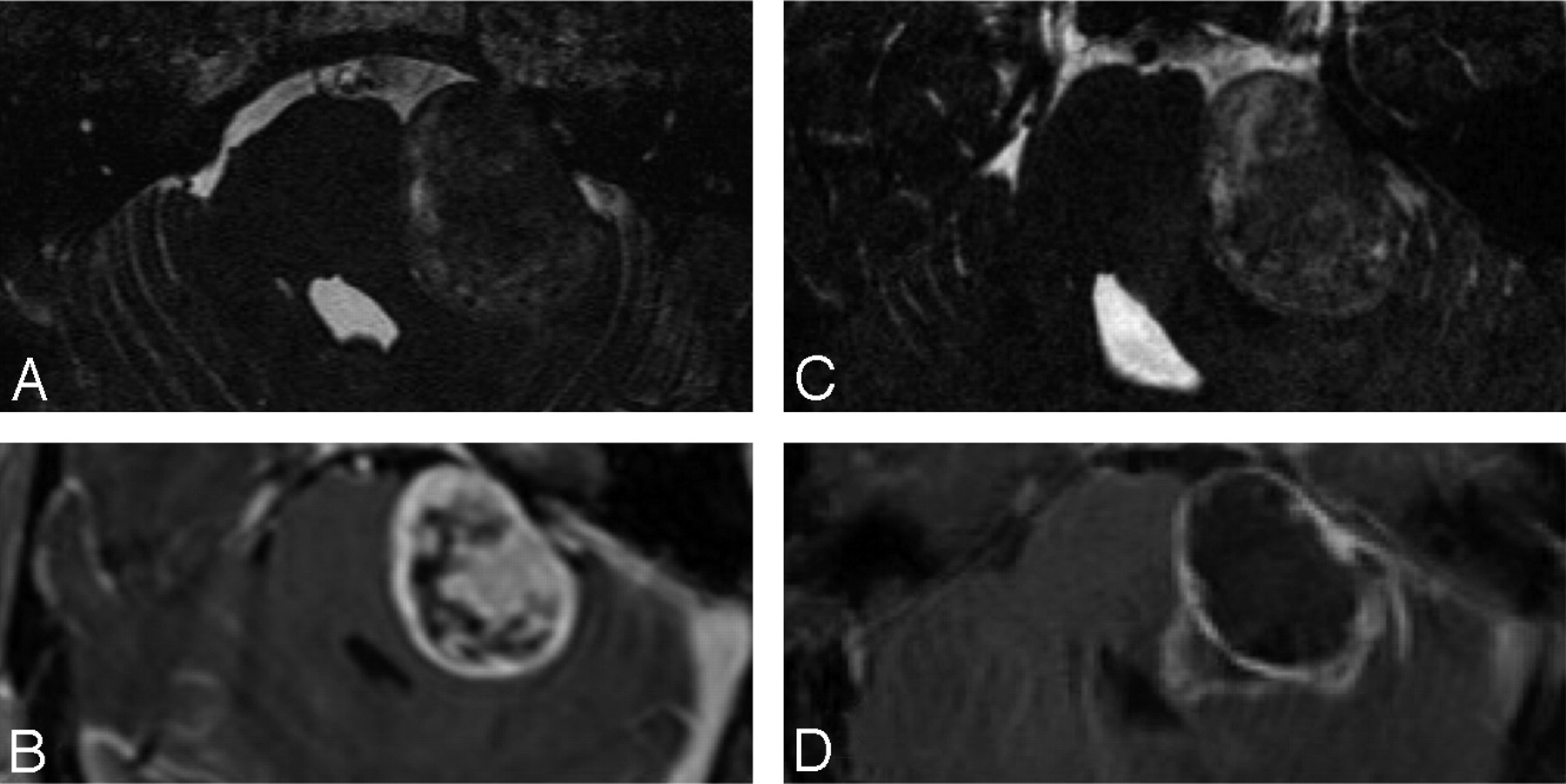
  • Fig 2.
    • Download figure
    • Open in new tab
    • Download powerpoint
    Fig 2.

    A and B, Koos stage 2 VS on the axial CISS image (A) and postcontrast T1WI (B). C and D, The follow-up imaging reveals minimal progression of the lesion with increased protrusion into the cerebellopontine cistern, both on the CISS (C) and postcontrast T1WI (D).
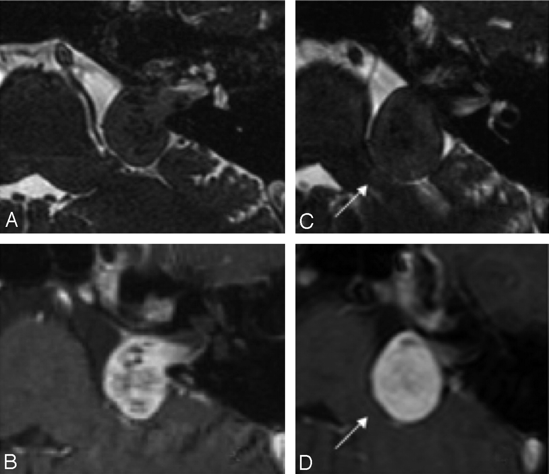
  • Fig 3.
    • Download figure
    • Open in new tab
    • Download powerpoint
    Fig 3.

    A−C, Postoperative MR imaging of a patient with right-sided VS. The axial T1WI (A), postcontrast T1WI (B), and CISS (C) image demonstrate postoperative changes on the right and a small residual lesion (arrow) within the cerebellopontine angle. D−F, The residual mass (arrow) is stable in the follow-up T1WI (D), postcontrast T1WI (E), and CISS imaging (F) performed 2 years later.
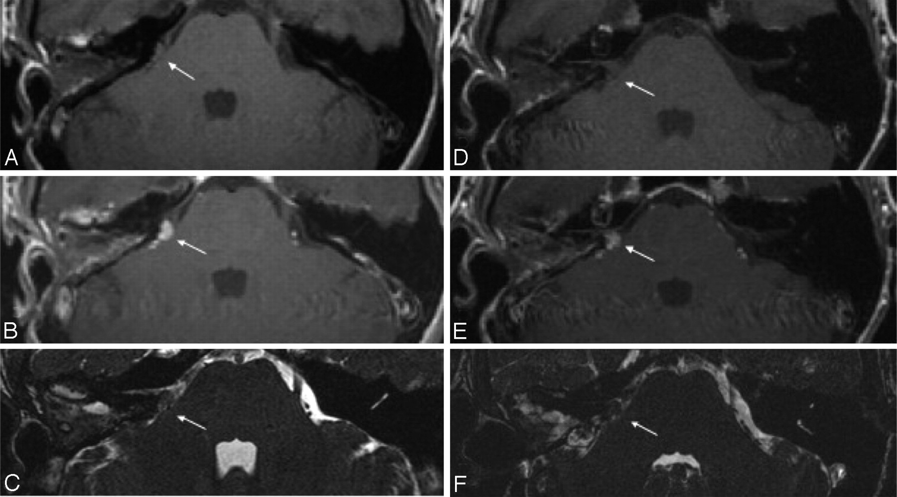
  • Fig 4.
    • Download figure
    • Open in new tab
    • Download powerpoint
    Fig 4.

    Imaging appearance of a Koos stage 4 VS treated with CyberKnife. The CISS image (A) and postcontrast T1WI (B) demonstrate a large heterogeneous mass compressing the brain stem. The CISS image (C) and postcontrast T1WI (D) of the follow-up study reveal an interval increase in the size of the mass. The increased internal heterogeneity is not well reflected in the CISS image (C).

Tables

  • Figures
    • View popup
    Table 1:

    Patient data and imaging information

    PatientAge (yr)/SexClinical HistoryKoos StageNo. of StudiesFollow-Up Interval (Months)Progression (Observer 1)Progression (Observer 2)
    171/FHearing loss1450––
    278/FHearing loss2226––
    351/FHearing loss1443––
    459/FFollow-up after surgery2442––
    558/FHearing loss2530––
    631/FHearing loss2655+–
    754/FFollow-up after CK426+–
    850/FHearing loss1314––
    962/MHearing loss128––
    1025/FHearing loss127––
    1171/MHearing loss4212––
    1270/FHearing loss2212––
    1370/FFollow-up after surgery2224––
    1457/MHearing loss2224++
    1566/MHearing loss226––
    1628/MHearing loss3212++
    1759/MHearing loss1214––
    1834/MHearing loss1224+–
    • Note:—+ indicates increase in size; –, no progression; CK, CyberKnife.

    • View popup
    Table 2:

    Comparison of measurements performed on CISS and Post Gad images

    MeasurementsMean (mm)SDP Value
    Observer 1
        CISS AP8.06.5.048
        Post Gad AP8.26.7
        CISS TRA10.55.1.123
        Post Gad TRA10.85.1
        CISS SI7.15.6.007
        Post Gad SI7.66.3
    Observer 2
        CISS AP7.55.9.030
        Post Gad AP7.86.4
        CISS TRA10.85.1.214
        Post Gad TRA10.54.8
        CISS SI6.95.1.983
        Post Gad SI6.95.7
    • Note:—Post Gad indicates postcontrast; CISS, constructive interference in steady state; AP, anteroposterior diameter; TRA, transverse diameter; SI, superoinferior diameter.

    • View popup
    Table 3:

    Interobserver variations between the measurements performed by 2 observers

    MeasurementsMean (mm)SDP Value
    CISS
        AP.009
            Observer 18.06.5
            Observer 27.55.9
        TRA.244
            Observer 110.55.1
            Observer 210.85.1
        SI.602
            Observer 17.185.6
            Observer 27.05.3
    Post Gad
        AP.009
            Observer 18.26.7
            Observer 27.86.4
        TRA.067
            Observer 110.85.1
            Observer 210.54.8
        SI.011
            Observer 17.46.2
            Observer 26.95.7
PreviousNext
Back to top

In this issue

American Journal of Neuroradiology: 30 (5)
American Journal of Neuroradiology
Vol. 30, Issue 5
May 2009
  • Table of Contents
  • Index by author
Advertisement
Print
Download PDF
Email Article

Thank you for your interest in spreading the word on American Journal of Neuroradiology.

NOTE: We only request your email address so that the person you are recommending the page to knows that you wanted them to see it, and that it is not junk mail. We do not capture any email address.

Enter multiple addresses on separate lines or separate them with commas.
Diagnostic Accuracy of the Constructive Interference in Steady State Sequence Alone for Follow-Up Imaging of Vestibular Schwannomas
(Your Name) has sent you a message from American Journal of Neuroradiology
(Your Name) thought you would like to see the American Journal of Neuroradiology web site.
CAPTCHA
This question is for testing whether or not you are a human visitor and to prevent automated spam submissions.
Cite this article
B. Ozgen, B. Oguz, A. Dolgun
Diagnostic Accuracy of the Constructive Interference in Steady State Sequence Alone for Follow-Up Imaging of Vestibular Schwannomas
American Journal of Neuroradiology May 2009, 30 (5) 985-991; DOI: 10.3174/ajnr.A1472

Citation Manager Formats

  • BibTeX
  • Bookends
  • EasyBib
  • EndNote (tagged)
  • EndNote 8 (xml)
  • Medlars
  • Mendeley
  • Papers
  • RefWorks Tagged
  • Ref Manager
  • RIS
  • Zotero
0 Responses
Respond to this article
Share
Bookmark this article
Diagnostic Accuracy of the Constructive Interference in Steady State Sequence Alone for Follow-Up Imaging of Vestibular Schwannomas
B. Ozgen, B. Oguz, A. Dolgun
American Journal of Neuroradiology May 2009, 30 (5) 985-991; DOI: 10.3174/ajnr.A1472
del.icio.us logo Twitter logo Facebook logo Mendeley logo
  • Tweet Widget
  • Facebook Like
  • Google Plus One
Purchase

Jump to section

  • Article
    • Abstract
    • Materials and Methods
    • Results
    • Discussion
    • Conclusions
    • Footnotes
    • References
  • Figures & Data
  • Supplemental
  • Info & Metrics
  • Responses
  • References
  • PDF

Related Articles

  • PubMed
  • Google Scholar

Cited By...

  • Noncontrast MRI Surveillance of Craniopharyngiomas Using a Balanced Steady-state Free Precession (bSSFP) Sequence
  • Balanced Steady-State Free Precession Sequence (CISS/FIESTA/3D Driven Equilibrium Radiofrequency Reset Pulse) Increases the Diagnostic Yield for Spinal Drop Metastases in Children with Brain Tumors
  • Crossref (21)
  • Google Scholar

This article has been cited by the following articles in journals that are participating in Crossref Cited-by Linking.

  • Clinical Practice Guideline: Sudden Hearing Loss (Update)
    Sujana S. Chandrasekhar, Betty S. Tsai Do, Seth R. Schwartz, Laura J. Bontempo, Erynne A. Faucett, Sandra A. Finestone, Deena B. Hollingsworth, David M. Kelley, Steven T. Kmucha, Gul Moonis, Gayla L. Poling, J. Kirk Roberts, Robert J. Stachler, Daniel M. Zeitler, Maureen D. Corrigan, Lorraine C. Nnacheta, Lisa Satterfield
    Otolaryngology–Head and Neck Surgery 2019 161 S1
  • Surgical management for large vestibular schwannomas: a systematic review, meta-analysis, and consensus statement on behalf of the EANS skull base section
    Daniele Starnoni, Lorenzo Giammattei, Giulia Cossu, Michael J. Link, Pierre-Hugues Roche, Ari G. Chacko, Kenji Ohata, Majid Samii, Ashish Suri, Michael Bruneau, Jan F. Cornelius, Luigi Cavallo, Torstein R. Meling, Sebastien Froelich, Marcos Tatagiba, Albert Sufianov, Dimitrios Paraskevopoulos, Idoya Zazpe, Moncef Berhouma, Emmanuel Jouanneau, Jeroen B. Verheul, Constantin Tuleasca, Mercy George, Marc Levivier, Mahmoud Messerer, Roy Thomas Daniel
    Acta Neurochirurgica 2020 162 11
  • European Society for Paediatric Oncology (SIOPE) MRI guidelines for imaging patients with central nervous system tumours
    Shivaram Avula, Andrew Peet, Giovanni Morana, Paul Morgan, Monika Warmuth-Metz, Tim Jaspan
    Child's Nervous System 2021 37 8
  • “Wait and scan” management of patients with vestibular schwannoma and the relevance of non-contrast MRI in the follow-up
    Jing Zou, Timo Hirvonen
    Journal of Otology 2017 12 4
  • Balanced Steady-State Free Precession Sequence (CISS/FIESTA/3D Driven Equilibrium Radiofrequency Reset Pulse) Increases the Diagnostic Yield for Spinal Drop Metastases in Children with Brain Tumors
    K. Buch, P. Caruso, D. Ebb, S. Rincon
    American Journal of Neuroradiology 2018 39 7
  • Imaging of the Vestibular Schwannoma
    Steve E.J. Connor
    Neuroimaging Clinics of North America 2021 31 4
  • Hearing Preservation in Retrosigmoid Approach of Small Vestibular Schwannomas
    Stéphane Tringali, Chantal Ferber-Viart, Carine Fuchsmann, Guillaume Buiret, Sandra Zaouche, Christian Dubreuil
    Otology & Neurotology 2010 31 9
  • Repeat stereotactic radiosurgery for progressive vestibular schwannomas after previous radiosurgery: a systematic review and meta-analysis
    Anne Balossier, Jean Régis, Nicolas Reyns, Pierre-Hugues Roche, Roy Thomas Daniel, Mercy George, Mohamed Faouzi, Marc Levivier, Constantin Tuleasca
    Neurosurgical Review 2021 44 6
  • Noncontrast vestibular schwannoma surveillance imaging including an MR cisternographic sequence: is there a need for postcontrast imaging?
    Karen Buch, Amy Juliano, Konstantina M. Stankovic, Hugh D. Curtin, Mary Beth Cunnane
    Journal of Neurosurgery 2019 131 2
  • Automated Koos Classification of Vestibular Schwannoma
    Aaron Kujawa, Reuben Dorent, Steve Connor, Anna Oviedova, Mohamed Okasha, Diana Grishchuk, Sebastien Ourselin, Ian Paddick, Neil Kitchen, Tom Vercauteren, Jonathan Shapey
    Frontiers in Radiology 2022 2

More in this TOC Section

  • Peritumoral Signal in Vestibular Schwannomas
  • NI-RADS for HEAD&NECK Cancer Recurrence
  • Chondrosarcoma vs Synovial Chondromatosis: Imaging
Show more HEAD & NECK

Similar Articles

Advertisement

Indexed Content

  • Current Issue
  • Accepted Manuscripts
  • Article Preview
  • Past Issues
  • Editorials
  • Editor's Choice
  • Fellows' Journal Club
  • Letters to the Editor
  • Video Articles

Cases

  • Case Collection
  • Archive - Case of the Week
  • Archive - Case of the Month
  • Archive - Classic Case

Special Collections

  • AJNR Awards
  • ASNR Foundation Special Collection
  • Most Impactful AJNR Articles
  • Photon-Counting CT
  • Spinal CSF Leak Articles (Jan 2020-June 2024)

More from AJNR

  • Trainee Corner
  • Imaging Protocols
  • MRI Safety Corner

Multimedia

  • AJNR Podcasts
  • AJNR Scantastics

Resources

  • Turnaround Time
  • Submit a Manuscript
  • Submit a Video Article
  • Submit an eLetter to the Editor/Response
  • Manuscript Submission Guidelines
  • Statistical Tips
  • Fast Publishing of Accepted Manuscripts
  • Graphical Abstract Preparation
  • Imaging Protocol Submission
  • Evidence-Based Medicine Level Guide
  • Publishing Checklists
  • Author Policies
  • Become a Reviewer/Academy of Reviewers
  • News and Updates

About Us

  • About AJNR
  • Editorial Board
  • Editorial Board Alumni
  • Alerts
  • Permissions
  • Not an AJNR Subscriber? Join Now
  • Advertise with Us
  • Librarian Resources
  • Feedback
  • Terms and Conditions
  • AJNR Editorial Board Alumni

American Society of Neuroradiology

  • Not an ASNR Member? Join Now

© 2025 by the American Society of Neuroradiology All rights, including for text and data mining, AI training, and similar technologies, are reserved.
Print ISSN: 0195-6108 Online ISSN: 1936-959X

Powered by HighWire