Advertisement

Main menu

  • Home
  • Content
    • Current Issue
    • Accepted Manuscripts
    • Article Preview
    • Past Issue Archive
    • Video Articles
    • AJNR Case Collection
    • Case of the Week Archive
    • Case of the Month Archive
    • Classic Case Archive
  • Special Collections
    • AJNR Awards
    • ASNR Foundation Special Collection
    • Most Impactful AJNR Articles
    • Photon-Counting CT
    • Spinal CSF Leak Articles (Jan 2020-June 2024)
  • Multimedia
    • AJNR Podcasts
    • AJNR SCANtastic
    • Trainee Corner
    • MRI Safety Corner
    • Imaging Protocols
  • For Authors
    • Submit a Manuscript
    • Submit a Video Article
    • Submit an eLetter to the Editor/Response
    • Manuscript Submission Guidelines
    • Statistical Tips
    • Fast Publishing of Accepted Manuscripts
    • Graphical Abstract Preparation
    • Imaging Protocol Submission
    • Author Policies
  • About Us
    • About AJNR
    • Editorial Board
    • Editorial Board Alumni
  • More
    • Become a Reviewer/Academy of Reviewers
    • Subscribers
    • Permissions
    • Alerts
    • Feedback
    • Advertisers
    • ASNR Home

User menu

  • Alerts
  • Log in

Search

  • Advanced search
American Journal of Neuroradiology

American Journal of Neuroradiology

ASHNR American Society of Functional Neuroradiology ASHNR American Society of Pediatric Neuroradiology ASSR
  • Alerts
  • Log in

Advanced Search

  • Home
  • Content
    • Current Issue
    • Accepted Manuscripts
    • Article Preview
    • Past Issue Archive
    • Video Articles
    • AJNR Case Collection
    • Case of the Week Archive
    • Case of the Month Archive
    • Classic Case Archive
  • Special Collections
    • AJNR Awards
    • ASNR Foundation Special Collection
    • Most Impactful AJNR Articles
    • Photon-Counting CT
    • Spinal CSF Leak Articles (Jan 2020-June 2024)
  • Multimedia
    • AJNR Podcasts
    • AJNR SCANtastic
    • Trainee Corner
    • MRI Safety Corner
    • Imaging Protocols
  • For Authors
    • Submit a Manuscript
    • Submit a Video Article
    • Submit an eLetter to the Editor/Response
    • Manuscript Submission Guidelines
    • Statistical Tips
    • Fast Publishing of Accepted Manuscripts
    • Graphical Abstract Preparation
    • Imaging Protocol Submission
    • Author Policies
  • About Us
    • About AJNR
    • Editorial Board
    • Editorial Board Alumni
  • More
    • Become a Reviewer/Academy of Reviewers
    • Subscribers
    • Permissions
    • Alerts
    • Feedback
    • Advertisers
    • ASNR Home
  • Follow AJNR on Twitter
  • Visit AJNR on Facebook
  • Follow AJNR on Instagram
  • Join AJNR on LinkedIn
  • RSS Feeds

AJNR is seeking candidates for the AJNR Podcast Editor. Read the position description.

Research ArticleBrain

Corpus Callosum Morphology and Microstructure Assessed Using Structural MR Imaging and Diffusion Tensor Imaging: Initial Findings in Adults with Neurofibromatosis Type 1

E.L. Wignall, P.D. Griffiths, N.G. Papadakis, I.D. Wilkinson, L.I. Wallis, O. Bandmann, P.E.E. Cowell and N. Hoggard
American Journal of Neuroradiology May 2010, 31 (5) 856-861; DOI: https://doi.org/10.3174/ajnr.A2005
E.L. Wignall
  • Find this author on Google Scholar
  • Find this author on PubMed
  • Search for this author on this site
P.D. Griffiths
  • Find this author on Google Scholar
  • Find this author on PubMed
  • Search for this author on this site
N.G. Papadakis
  • Find this author on Google Scholar
  • Find this author on PubMed
  • Search for this author on this site
I.D. Wilkinson
  • Find this author on Google Scholar
  • Find this author on PubMed
  • Search for this author on this site
L.I. Wallis
  • Find this author on Google Scholar
  • Find this author on PubMed
  • Search for this author on this site
O. Bandmann
  • Find this author on Google Scholar
  • Find this author on PubMed
  • Search for this author on this site
P.E.E. Cowell
  • Find this author on Google Scholar
  • Find this author on PubMed
  • Search for this author on this site
N. Hoggard
  • Find this author on Google Scholar
  • Find this author on PubMed
  • Search for this author on this site
  • Article
  • Figures & Data
  • Info & Metrics
  • Responses
  • References
  • PDF
Loading

Article Figures & Data

Figures

  • Tables
  • Fig 1.
    • Download figure
    • Open in new tab
    • Download powerpoint
    Fig 1.

    Line diagram of the divisions of the CC used for regional width measurements and factor structure by using the method of Denenberg et al (1991)13 and Denenberg et al (1991).14 See text for details.

  • Fig 2.
    • Download figure
    • Open in new tab
    • Download powerpoint
    Fig 2.

    A, The mean widths (±95% CIs) of the individual factors for both subjects with NF-1 and controls. B, Schematic representation of the regions of the CC that are statistically significantly wider in subjects with NF-1 compared with controls (white indicates not significant; light gray, significant to P < .05; and dark gray, significant to P < .005). C and D, Midsagittal images from the T1-weighted volume dataset from a person with NF-1 (C) and a matched control (D). The CC is visibly larger in the person with NF-1, particularly in the posterior aspect.
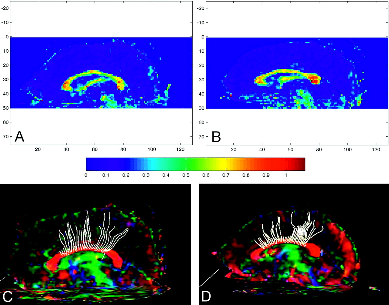
  • Fig 3.
    • Download figure
    • Open in new tab
    • Download powerpoint
    Fig 3.

    A and B, Sagittally reformatted color FA maps for a subject with NF-1 (A) and a matched control (B), with color-scale guide below. C and D, Tractography for the subject with NF-1 (C) and the control (D).

Tables

  • Figures
    • View popup
    Table 1:

    Mean (± SD) global and regional CC measures for patients with NF-1 and matched controlsa

    Patients with NF-1 (n= 10)Matched Controls (n= 10)t Statistic (df = 9)
    Global CC measures
        Central length (mm)94.1 (5.7)85.4 (5.7)3.46b
        Area (mm2)708.4 (74.9)587.2 (53.8)4.15b
        Total brain volume (cm3)1050 (110)1010 (90)0.89
    Factor groupings (mm)
        F1 (widths 3–18)13.0 (3.2)14.5 (1.6)1.33
        F2 (widths 22–39)9.2 (1.7)7.3 (0.7)3.21b
        F3 (widths 49–62)7.9 (1.4)6.6 (0.7)2.58c
        F4 (widths 65–74)7.9 (0.8)6.2 (0.7)5.09b
        F5 (widths 77–85)12.7 (1.9)10.8 (1.2)2.67c
        F6 (widths 89–94)14.3 (1.7)13.2 (1.9)1.40
        F7 (widths 95–99)5.1 (0.4)4.6 (0.3)2.55c
    • a There is no significant difference between the total brain volumes of the subjects with NF-1 and controls, but the area and length of the CC are larger in NF-1. The factor groupings are also presented pictorially in Fig 1.

    • b P <.05 after the Bonferroni correction.

    • c P < .05 before the Bonferroni correction (.05/10) only.

    • View popup
    Table 2:

    Mean (± SD) diffusion measures for subjects with NF-1 and matched controlsa

    Patients with NF-1 (n = 10)Matched Controls (n = 10)t Statistic (df = 18)
    Genu
        FA0.68 (0.06)0.77 (0.05)3.644b
        λ∥1.92 (0.35)1.91 (0.14)0.084
        λ⊥c0.52 (0.07)0.39 (0.06)4.46b
        Trace0.99 (0.16)0.89 (0.04)0.36
    Body
        FA0.70 (0.05)0.77 (0.04)3.46b
        λ∥1.66 (0.08)1.77 (0.13)2.28d
        λ⊥c0.42 (0.07)0.35 (0.06)2.40d
        Trace0.83 (0.06)0.82 (0.06)0.37
    • a See text for discussion.

    • b P < .05 after Bonferroni correction.

    • c Diffusion trace, λ∥, and λ⊥ are expressed in 10−3 mm2/s.

    • d P < .05 before Bonferroni correction only (.05/8).

PreviousNext
Back to top

In this issue

American Journal of Neuroradiology: 31 (5)
American Journal of Neuroradiology
Vol. 31, Issue 5
1 May 2010
  • Table of Contents
  • Index by author
Advertisement
Print
Download PDF
Email Article

Thank you for your interest in spreading the word on American Journal of Neuroradiology.

NOTE: We only request your email address so that the person you are recommending the page to knows that you wanted them to see it, and that it is not junk mail. We do not capture any email address.

Enter multiple addresses on separate lines or separate them with commas.
Corpus Callosum Morphology and Microstructure Assessed Using Structural MR Imaging and Diffusion Tensor Imaging: Initial Findings in Adults with Neurofibromatosis Type 1
(Your Name) has sent you a message from American Journal of Neuroradiology
(Your Name) thought you would like to see the American Journal of Neuroradiology web site.
CAPTCHA
This question is for testing whether or not you are a human visitor and to prevent automated spam submissions.
Cite this article
E.L. Wignall, P.D. Griffiths, N.G. Papadakis, I.D. Wilkinson, L.I. Wallis, O. Bandmann, P.E.E. Cowell, N. Hoggard
Corpus Callosum Morphology and Microstructure Assessed Using Structural MR Imaging and Diffusion Tensor Imaging: Initial Findings in Adults with Neurofibromatosis Type 1
American Journal of Neuroradiology May 2010, 31 (5) 856-861; DOI: 10.3174/ajnr.A2005

Citation Manager Formats

  • BibTeX
  • Bookends
  • EasyBib
  • EndNote (tagged)
  • EndNote 8 (xml)
  • Medlars
  • Mendeley
  • Papers
  • RefWorks Tagged
  • Ref Manager
  • RIS
  • Zotero
0 Responses
Respond to this article
Share
Bookmark this article
Corpus Callosum Morphology and Microstructure Assessed Using Structural MR Imaging and Diffusion Tensor Imaging: Initial Findings in Adults with Neurofibromatosis Type 1
E.L. Wignall, P.D. Griffiths, N.G. Papadakis, I.D. Wilkinson, L.I. Wallis, O. Bandmann, P.E.E. Cowell, N. Hoggard
American Journal of Neuroradiology May 2010, 31 (5) 856-861; DOI: 10.3174/ajnr.A2005
del.icio.us logo Twitter logo Facebook logo Mendeley logo
  • Tweet Widget
  • Facebook Like
  • Google Plus One
Purchase

Jump to section

  • Article
    • Abstract
    • Abbreviations
    • Materials and Methods
    • Results
    • Discussion
    • Acknowledgments
    • Footnotes
    • References
  • Figures & Data
  • Info & Metrics
  • Responses
  • References
  • PDF

Related Articles

  • No related articles found.
  • PubMed
  • Google Scholar

Cited By...

  • Impact of Pathogenic Variants of the Ras-MAPK Pathway on Major White Matter Tracts in the Human Brain
  • Alterations in Brain Morphology by MRI in Adults with Neurofibromatosis 1
  • White Matter is Increased in the Brains of Adults with Neurofibromatosis 1
  • Neurofibromatosis Type 1: Modeling CNS Dysfunction
  • Crossref (25)
  • Google Scholar

This article has been cited by the following articles in journals that are participating in Crossref Cited-by Linking.

  • Neurofibromatosis Type 1: Modeling CNS Dysfunction
    David H. Gutmann, Luis F. Parada, Alcino J. Silva, Nancy Ratner
    The Journal of Neuroscience 2012 32 41
  • Gyrification, cortical and subcortical morphometry in neurofibromatosis type 1: an uneven profile of developmental abnormalities
    Inês R Violante, Maria J Ribeiro, Eduardo D Silva, Miguel Castelo-Branco
    Journal of Neurodevelopmental Disorders 2013 5 1
  • Multivariate pattern analysis reveals subtle brain anomalies relevant to the cognitive phenotype in neurofibromatosis type 1
    João V. Duarte, Maria J. Ribeiro, Inês R. Violante, Gil Cunha, Eduardo Silva, Miguel Castelo-Branco
    Human Brain Mapping 2014 35 1
  • Resting state functional MRI reveals abnormal network connectivity in neurofibromatosis 1
    Steffie N. Tomson, Matthew J. Schreiner, Manjari Narayan, Tena Rosser, Nicole Enrique, Alcino J. Silva, Genevera I. Allen, Susan Y. Bookheimer, Carrie E. Bearden
    Human Brain Mapping 2015 36 11
  • MR imaging of neoplastic and non-neoplastic lesions of the brain and spine in neurofibromatosis type I
    Ahmed Abdel Khalek Abdel Razek
    Neurological Sciences 2018 39 5
  • Brain and behaviour phenotyping of a mouse model of neurofibromatosis type‐1: an MRI/DTI study on social cognition
    L. I. Petrella, Y. Cai, J. V. Sereno, S. I. Gonçalves, A. J. Silva, M. Castelo‐Branco
    Genes, Brain and Behavior 2016 15 7
  • Diffusion-Tensor Imaging Derived Metrics of the Corpus Callosum in Children With Neurofibromatosis Type I
    Christopher G. Filippi, Richard Watts, Lindsay A. N. Duy, Keith A. Cauley
    American Journal of Roentgenology 2013 200 1
  • Handedness and the Corpus Callosum: A Review and Further Analyses of Discordant Twins
    Patricia Cowell, Jennifer Gurd
    Neuroscience 2018 388
  • fMRI and corpus callosum relationships in monozygotic twins discordant for handedness
    J. M. Gurd, P. E. Cowell, S. Lux, R. Rezai, L. Cherkas, G. C. Ebers
    Brain Structure and Function 2013 218 2
  • Imaging genetics in neurodevelopmental psychopathology
    Marieke Klein, Marjolein van Donkelaar, Ellen Verhoef, Barbara Franke
    American Journal of Medical Genetics Part B: Neuropsychiatric Genetics 2017 174 5

More in this TOC Section

  • Usefulness of Quantitative Susceptibility Mapping for the Diagnosis of Parkinson Disease
  • White Matter Alterations in the Brains of Patients with Active, Remitted, and Cured Cushing Syndrome: A DTI Study
  • Qualitative and Quantitative Analysis of MR Imaging Findings in Patients with Middle Cerebral Artery Stroke Implanted with Mesenchymal Stem Cells
Show more BRAIN

Similar Articles

Advertisement

Indexed Content

  • Current Issue
  • Accepted Manuscripts
  • Article Preview
  • Past Issues
  • Editorials
  • Editor's Choice
  • Fellows' Journal Club
  • Letters to the Editor
  • Video Articles

Cases

  • Case Collection
  • Archive - Case of the Week
  • Archive - Case of the Month
  • Archive - Classic Case

Special Collections

  • AJNR Awards
  • ASNR Foundation Special Collection
  • Most Impactful AJNR Articles
  • Photon-Counting CT
  • Spinal CSF Leak Articles (Jan 2020-June 2024)

More from AJNR

  • Trainee Corner
  • Imaging Protocols
  • MRI Safety Corner

Multimedia

  • AJNR Podcasts
  • AJNR Scantastics

Resources

  • Turnaround Time
  • Submit a Manuscript
  • Submit a Video Article
  • Submit an eLetter to the Editor/Response
  • Manuscript Submission Guidelines
  • Statistical Tips
  • Fast Publishing of Accepted Manuscripts
  • Graphical Abstract Preparation
  • Imaging Protocol Submission
  • Evidence-Based Medicine Level Guide
  • Publishing Checklists
  • Author Policies
  • Become a Reviewer/Academy of Reviewers
  • News and Updates

About Us

  • About AJNR
  • Editorial Board
  • Editorial Board Alumni
  • Alerts
  • Permissions
  • Not an AJNR Subscriber? Join Now
  • Advertise with Us
  • Librarian Resources
  • Feedback
  • Terms and Conditions
  • AJNR Editorial Board Alumni

American Society of Neuroradiology

  • Not an ASNR Member? Join Now

© 2025 by the American Society of Neuroradiology All rights, including for text and data mining, AI training, and similar technologies, are reserved.
Print ISSN: 0195-6108 Online ISSN: 1936-959X

Powered by HighWire