Advertisement

Main menu

  • Home
  • Content
    • Current Issue
    • Accepted Manuscripts
    • Article Preview
    • Past Issue Archive
    • Video Articles
    • AJNR Case Collection
    • Case of the Week Archive
    • Case of the Month Archive
    • Classic Case Archive
  • Special Collections
    • AJNR Awards
    • ASNR Foundation Special Collection
    • Most Impactful AJNR Articles
    • Photon-Counting CT
    • Spinal CSF Leak Articles (Jan 2020-June 2024)
  • Multimedia
    • AJNR Podcasts
    • AJNR SCANtastic
    • Trainee Corner
    • MRI Safety Corner
    • Imaging Protocols
  • For Authors
    • Submit a Manuscript
    • Submit a Video Article
    • Submit an eLetter to the Editor/Response
    • Manuscript Submission Guidelines
    • Statistical Tips
    • Fast Publishing of Accepted Manuscripts
    • Graphical Abstract Preparation
    • Imaging Protocol Submission
    • Author Policies
  • About Us
    • About AJNR
    • Editorial Board
    • Editorial Board Alumni
  • More
    • Become a Reviewer/Academy of Reviewers
    • Subscribers
    • Permissions
    • Alerts
    • Feedback
    • Advertisers
    • ASNR Home

User menu

  • Alerts
  • Log in

Search

  • Advanced search
American Journal of Neuroradiology

American Journal of Neuroradiology

ASHNR American Society of Functional Neuroradiology ASHNR American Society of Pediatric Neuroradiology ASSR
  • Alerts
  • Log in

Advanced Search

  • Home
  • Content
    • Current Issue
    • Accepted Manuscripts
    • Article Preview
    • Past Issue Archive
    • Video Articles
    • AJNR Case Collection
    • Case of the Week Archive
    • Case of the Month Archive
    • Classic Case Archive
  • Special Collections
    • AJNR Awards
    • ASNR Foundation Special Collection
    • Most Impactful AJNR Articles
    • Photon-Counting CT
    • Spinal CSF Leak Articles (Jan 2020-June 2024)
  • Multimedia
    • AJNR Podcasts
    • AJNR SCANtastic
    • Trainee Corner
    • MRI Safety Corner
    • Imaging Protocols
  • For Authors
    • Submit a Manuscript
    • Submit a Video Article
    • Submit an eLetter to the Editor/Response
    • Manuscript Submission Guidelines
    • Statistical Tips
    • Fast Publishing of Accepted Manuscripts
    • Graphical Abstract Preparation
    • Imaging Protocol Submission
    • Author Policies
  • About Us
    • About AJNR
    • Editorial Board
    • Editorial Board Alumni
  • More
    • Become a Reviewer/Academy of Reviewers
    • Subscribers
    • Permissions
    • Alerts
    • Feedback
    • Advertisers
    • ASNR Home
  • Follow AJNR on Twitter
  • Visit AJNR on Facebook
  • Follow AJNR on Instagram
  • Join AJNR on LinkedIn
  • RSS Feeds

AJNR is seeking candidates for the AJNR Podcast Editor. Read the position description.

Research ArticlePediatrics
Open Access

Intracranial Meningeal Hemangiopericytomas in Children and Adolescents: CT and MR Imaging Findings

Q. Chen, X.-Z. Chen, J.-M. Wang, S.-W. Li, T. Jiang and J.-P. Dai
American Journal of Neuroradiology January 2012, 33 (1) 195-199; DOI: https://doi.org/10.3174/ajnr.A2721
Q. Chen
  • Find this author on Google Scholar
  • Find this author on PubMed
  • Search for this author on this site
X.-Z. Chen
  • Find this author on Google Scholar
  • Find this author on PubMed
  • Search for this author on this site
J.-M. Wang
  • Find this author on Google Scholar
  • Find this author on PubMed
  • Search for this author on this site
S.-W. Li
  • Find this author on Google Scholar
  • Find this author on PubMed
  • Search for this author on this site
T. Jiang
  • Find this author on Google Scholar
  • Find this author on PubMed
  • Search for this author on this site
J.-P. Dai
  • Find this author on Google Scholar
  • Find this author on PubMed
  • Search for this author on this site
  • Article
  • Figures & Data
  • Info & Metrics
  • Responses
  • References
  • PDF
Loading

Article Figures & Data

Figures

  • Tables
  • Fig 1.
    • Download figure
    • Open in new tab
    • Download powerpoint
    Fig 1.

    Patient 3. Hemangiopericytoma with peripheral calcification and bone erosion. A, Axial CT examination shows a mass over the right middle cranial fossa with upward extension to the temporal lobe. The mass shows mixed low, iso-, and high attenuation with peripheral calcification. B, Axial CT (bone window) shows destruction of right temporal bone and lateral wall of right orbit.
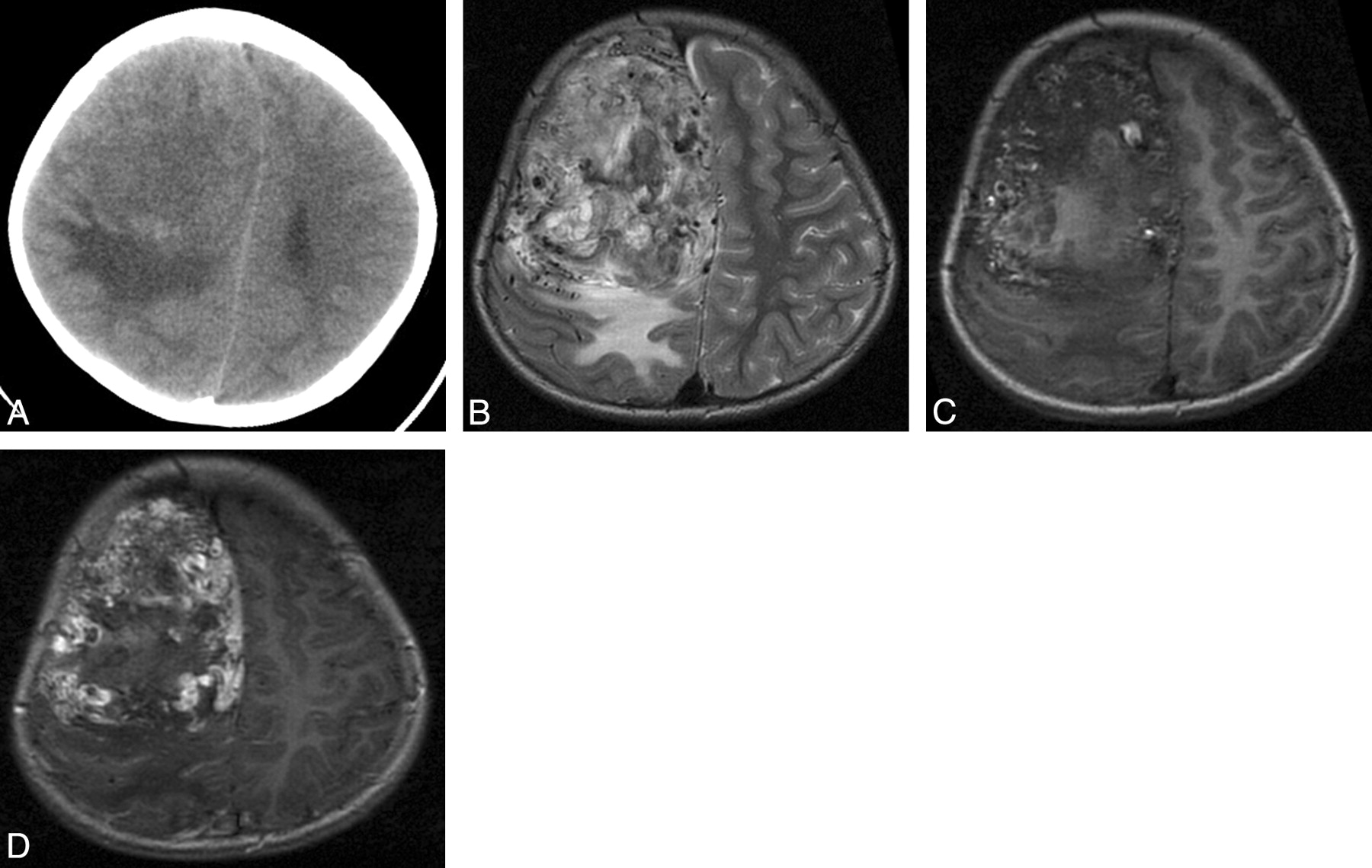
  • Fig 2.
    • Download figure
    • Open in new tab
    • Download powerpoint
    Fig 2.

    Patient 6. Anaplastic hemangiopericytoma with narrow-based dural attachment and AVM-like features. A, Axial CT examination shows a mass located in the right frontal parasagittal convexity. The mass shows mixed low and isoattenuation with moderate surrounding edema. B and C, Mass shows mixed hypo-, iso-, and hyperintensity with AVM-like signal intensity flow voids on precontrast axial T2 and T1-weighted images. D, Mass shows marked and heterogeneous enhancement with narrow-based dural attachment on postcontrast axial T1-weighted images but lacks the “dural tail” sign.

  • Fig 3.
    • Download figure
    • Open in new tab
    • Download powerpoint
    Fig 3.

    Patient 9. Hemangiopericytoma with the “dural tail” sign. A, Mass shows mixed hypo-, iso-, and hyperintensity on precontrast axial T2-weighted images. B, Mass shows marked and heterogeneous enhancement with narrow-based dural attachment on postcontrast axial T1-weighted images and shows the “dural tail” sign.

Tables

  • Figures
    • View popup
    Table 1:

    Demographics, surgery, follow-up, and pathology of 9 patients with hemangiopericytoma

    PatientSexAge (yr)Blood Loss (mL)Duration of Symptoms (mo)SymptomsExtent of Tumor RemovalPostoperative Radiotherapy (Gy)Follow-Up (mo)Local RecurrenceMetastasesPathology
    1M34000.5SeizuresCRNone95/aNoNoAH
    2F1810000.5HeadacheCR6039/aNoNoAH
    3F1040006HeadacheCR6036/aNoNoH
    4F1710000.5HeadacheCR6029/aNoNoAH
    5M33500.3HeadacheCRNone27/aNoNoAH
    6M7200012SeizuresCR6016/aNoNoAH
    7M1713,0003HeadacheCR6012/aNoNoAH
    8M53001.5HeadacheCR608/dYesNoAH
    9F18900012HeadacheSR602/aNoNoH
    • a indicates alive; d, dead; CR, complete resection; SR, subtotal resection; AH, anaplastic hemangiopericytoma; H, hemangiopericytoma

    • View popup
    Table 2:

    General imaging features of 9 patients with hemangiopericytoma

    PatientLocationShapeSizea (mm)NecrosisEdemaHyperostosis or Bone Erosion
    1Right middle cranial fossaMultilobular55 × 48 × 41YesMildIndeterminate
    2Left frontal-temporal convexityMultilobular60 × 58 × 55YesMildBone erosion of inner table
    3Right middle cranial fossaMultilobular67 × 73 × 72YesNoneBone erosion
    4Left frontal convexityMultilobular50 × 47 × 51YesMildIndeterminate, bone erosion
    5Right occipital-parietal parasagittalMultilobular54 × 41 × 46YesMildNo
    6Right frontal parasagittalOval78 × 76 × 87YesModerateNo
    7Bifrontal, anterior cranial fossaMultilobular102 × 72 × 61YesMildIndeterminate
    8Right occipital-parietal convexityMultilobular80 × 80 × 70YesModerateIndeterminate, bone erosion
    9Bioccipital-parietal parasagittalMultilobular116 × 66 × 50YesModerateNo
    • ↵a Length × width × height.

PreviousNext
Back to top

In this issue

American Journal of Neuroradiology: 33 (1)
American Journal of Neuroradiology
Vol. 33, Issue 1
1 Jan 2012
  • Table of Contents
  • Index by author
Advertisement
Print
Download PDF
Email Article

Thank you for your interest in spreading the word on American Journal of Neuroradiology.

NOTE: We only request your email address so that the person you are recommending the page to knows that you wanted them to see it, and that it is not junk mail. We do not capture any email address.

Enter multiple addresses on separate lines or separate them with commas.
Intracranial Meningeal Hemangiopericytomas in Children and Adolescents: CT and MR Imaging Findings
(Your Name) has sent you a message from American Journal of Neuroradiology
(Your Name) thought you would like to see the American Journal of Neuroradiology web site.
CAPTCHA
This question is for testing whether or not you are a human visitor and to prevent automated spam submissions.
Cite this article
Q. Chen, X.-Z. Chen, J.-M. Wang, S.-W. Li, T. Jiang, J.-P. Dai
Intracranial Meningeal Hemangiopericytomas in Children and Adolescents: CT and MR Imaging Findings
American Journal of Neuroradiology Jan 2012, 33 (1) 195-199; DOI: 10.3174/ajnr.A2721

Citation Manager Formats

  • BibTeX
  • Bookends
  • EasyBib
  • EndNote (tagged)
  • EndNote 8 (xml)
  • Medlars
  • Mendeley
  • Papers
  • RefWorks Tagged
  • Ref Manager
  • RIS
  • Zotero
0 Responses
Respond to this article
Share
Bookmark this article
Intracranial Meningeal Hemangiopericytomas in Children and Adolescents: CT and MR Imaging Findings
Q. Chen, X.-Z. Chen, J.-M. Wang, S.-W. Li, T. Jiang, J.-P. Dai
American Journal of Neuroradiology Jan 2012, 33 (1) 195-199; DOI: 10.3174/ajnr.A2721
del.icio.us logo Twitter logo Facebook logo Mendeley logo
  • Tweet Widget
  • Facebook Like
  • Google Plus One
Purchase

Jump to section

  • Article
    • Abstract
    • Case Series
    • Discussion
    • Conclusions
    • Footnotes
    • References
  • Figures & Data
  • Info & Metrics
  • Responses
  • References
  • PDF

Related Articles

  • No related articles found.
  • PubMed
  • Google Scholar

Cited By...

  • No citing articles found.
  • Crossref (23)
  • Google Scholar

This article has been cited by the following articles in journals that are participating in Crossref Cited-by Linking.

  • Presurgical differentiation between malignant haemangiopericytoma and angiomatous meningioma by a radiomics approach based on texture analysis
    Xuanxuan Li, Yiping Lu, Ji Xiong, Dongdong Wang, Dejun She, Xinping Kuai, Daoying Geng, Bo Yin
    Journal of Neuroradiology 2019 46 5
  • Preoperative Prediction of Solitary Fibrous Tumor/Hemangiopericytoma and Angiomatous Meningioma Using Magnetic Resonance Imaging Texture Analysis
    Tokunori Kanazawa, Yasuhiro Minami, Masahiro Jinzaki, Masahiro Toda, Kazunari Yoshida, Hikaru Sasaki
    World Neurosurgery 2018 120
  • Accurate Preoperative Distinction of Intracranial Hemangiopericytoma From Meningioma Using a Multihabitat and Multisequence-Based Radiomics Diagnostic Technique
    Jingwei Wei, Lianwang Li, Yuqi Han, Dongsheng Gu, Qian Chen, Junmei Wang, Runting Li, Jiong Zhan, Jie Tian, Dabiao Zhou
    Frontiers in Oncology 2020 10
  • Comparison of ADC values of intracranial hemangiopericytomas and angiomatous and anaplastic meningiomas
    Li Liu, Bo Yin, Dao-ying Geng, Yuan Li, Bi-yun Zhang, Wei-jun Peng
    Journal of Neuroradiology 2014 41 3
  • Role of intratumoral flow void signs in the differential diagnosis of intracranial solitary fibrous tumors and meningiomas
    Chunhong Wang, Yikai Xu, Xiang Xiao, Jing Zhang, Fang Zhou, Xixi Zhao
    Journal of Neuroradiology 2016 43 5
  • C11orf95‐RELA fusion present in a primary intracranial extra‐axial ependymoma: Report of a case with literature review
    Aruna Nambirajan, Prit Benny Malgulwar, Mehar C. Sharma, Anutosh Singh, Pankaj Pathak, Guru Dutta Satyarthee, Ajay Garg
    Neuropathology 2016 36 5
  • Intracranial meningeal hemangiopericytoma: Recurrences at the initial and distant intracranial sites and extraneural metastases to multiple organs
    GUANGQUAN WEI, XIAOWEI KANG, XIANPING LIU, XING TANG, QINLONG LI, JUNTAO HAN, HONG YIN
    Molecular and Clinical Oncology 2015 3 4
  • Preoperative Assessment of Pathologic Subtypes of Meningioma and Solitary Fibrous Tumor/Hemangiopericytoma Using Dynamic Computed Tomography: A Clinical Research Study
    Nobuhiko Arai, Katsuhiro Mizutani, Satoshi Takahashi, Yukina Morimoto, Takenori Akiyama, Takashi Horiguchi, Hatano Mami, Kazunari Yoshida
    World Neurosurgery 2018 115
  • Apparent diffusion coefficient values in differential diagnosis and prognostic prediction of solitary of fibrous tumor/hemangiopericytoma (WHOII) and atypical meningioma
    Ziwei Lu, Zhiqun You, Daohai Xie, Zhongling Wang
    Technology and Health Care 2019 27 2
  • Endovascular embolization of complex hypervascular skull base tumors
    Aaron A. Cho, Michael Annen
    Operative Techniques in Otolaryngology-Head and Neck Surgery 2014 25 1

More in this TOC Section

  • SyMRI & MR Fingerprinting in Brainstem Myelination
  • Comparison of Image Quality and Radiation Dose in Pediatric Temporal Bone CT Using Photon-Counting Detector CT and Energy-Integrating Detector CT
  • Dual-Layer Detector CT for PEDS Image Quality
Show more Pediatrics

Similar Articles

Advertisement

Indexed Content

  • Current Issue
  • Accepted Manuscripts
  • Article Preview
  • Past Issues
  • Editorials
  • Editor's Choice
  • Fellows' Journal Club
  • Letters to the Editor
  • Video Articles

Cases

  • Case Collection
  • Archive - Case of the Week
  • Archive - Case of the Month
  • Archive - Classic Case

Special Collections

  • AJNR Awards
  • ASNR Foundation Special Collection
  • Most Impactful AJNR Articles
  • Photon-Counting CT
  • Spinal CSF Leak Articles (Jan 2020-June 2024)

More from AJNR

  • Trainee Corner
  • Imaging Protocols
  • MRI Safety Corner

Multimedia

  • AJNR Podcasts
  • AJNR Scantastics

Resources

  • Turnaround Time
  • Submit a Manuscript
  • Submit a Video Article
  • Submit an eLetter to the Editor/Response
  • Manuscript Submission Guidelines
  • Statistical Tips
  • Fast Publishing of Accepted Manuscripts
  • Graphical Abstract Preparation
  • Imaging Protocol Submission
  • Evidence-Based Medicine Level Guide
  • Publishing Checklists
  • Author Policies
  • Become a Reviewer/Academy of Reviewers
  • News and Updates

About Us

  • About AJNR
  • Editorial Board
  • Editorial Board Alumni
  • Alerts
  • Permissions
  • Not an AJNR Subscriber? Join Now
  • Advertise with Us
  • Librarian Resources
  • Feedback
  • Terms and Conditions
  • AJNR Editorial Board Alumni

American Society of Neuroradiology

  • Not an ASNR Member? Join Now

© 2025 by the American Society of Neuroradiology All rights, including for text and data mining, AI training, and similar technologies, are reserved.
Print ISSN: 0195-6108 Online ISSN: 1936-959X

Powered by HighWire