
Article Figures & Data
Figures
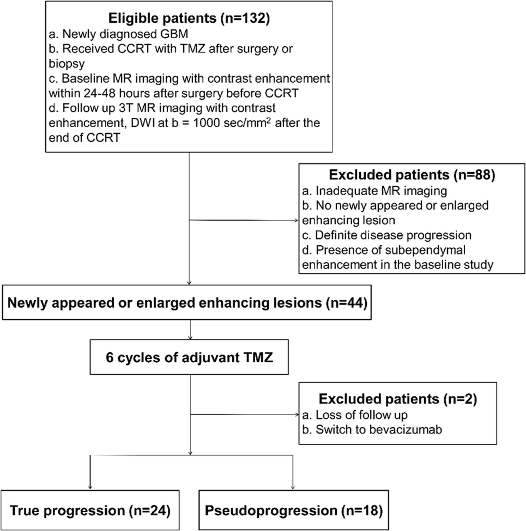
- Fig 1.
Flow diagram of patient selection, with inclusion and exclusion criteria.
- Fig 2.
Pseudoprogression in a 40-year-old man with glioblastoma who had undergone surgical resection. A, Axial contrast-enhanced T1-weighted MR image obtained within 1 month after the end of concurrent chemotherapy and radiation therapy shows a newly developed enhancing lesion (arrow) in the right frontal periventricular white matter. The ventricular margin adjacent to the enhancing lesion (arrow) also shows linear enhancement (arrowhead), with the distance of extension measuring ≤1 cm. B, On the ADC map, a decrease in ADC value is not apparent at the lesion (arrow) (mean, 1264 × 10−6 mm2/s; fifth percentile, 1059 × 10−6 mm2/s). C, Follow-up MR image after a 6-month continuation of temozolomide reveals resolution of the enhancing lesion.
- Fig 3.
True progression in a 36-year-old man with glioblastoma who had undergone surgical resection. A, On the axial contrast-enhanced T1-weighted MR image obtained within 1 month after the end of concurrent chemotherapy and radiation therapy, a newly developed enhancing lesion (arrow) is noted in the left occipital lobe. B, On the ADC map, the ADC value is decreased in some portion of the lesion (arrow) (mean, 1292 × 10−6 mm2/s; fifth percentile, 991 × 10−6 mm2/s). C, Axial contrast-enhanced T1-weighted MR image at a higher level reveals linear enhancement (arrowheads) along the ventricular margin, with the distance of extension measuring >1 cm. D, Follow-up MR image after a 6-month continuation of temozolomide demonstrates aggravation of both the left occipital lobe lesion (arrows) and subependymal enhancement (arrowheads) in the left lateral ventricle.


Tables
Parameters 3D MPRAGE Axial TSE T2WI FLAIR DWI TR (ms) 1500 4500–5160 9000–9902 6900–10,000 TE (ms) 1.9 91–106 97–163 55–67 TI (ms) 900 NA 2500 NA Echo-train length 1 16–19 0–11 1 Flip angle (degree) 9 90–130 90–130 90 Section thickness (mm) 1 5 5 3–5 Intersection gap (mm) 0 1 1 0.9–1 FOV (mm) 250 × 250 199–220 × 220 199–220 × 220 240 × 240 Matrix 256 × 256 448–640 × 256–290 320–384 × 192–209 160 × 160 No. of signals acquired 1 0–2 0–1 0–3 No. of sections 192 25 25 50–70 Note:—NA indicates not available.
Characteristic True Progression (n = 24) Pseudoprogression (n = 18) P Value Age (yr) 60.50 ± 11.58 48.22 ± 12.54 .002 Sex .347 Male 17 10 Female 7 8 Karnofsky performance score .371 <70 4 1 ≥70 20 17 Surgery .623 Biopsy 3 1 Resection 21 17 Radiation dose (Gy) 55.52 ± 8.17 57.83 ± 7.52 .376 Methylated MGMT promotera 1.000 Negative 6 5 Positive 15 13 Note:—MGMT indicates O6-methylguanine DNA methyltransferase.
↵a The promoter methylation status of MGMT, which was investigated by using the methylation-specific polymerase chain reaction technique, was documented whenever available.
True Progression (n = 24) Pseudoprogression (n = 18) P Value Subependymal enhancement 19 (79) 8 (44) .027 Local 9 (38) 6 (33) >.99 Type I 8 (33) 4 (22) Type II 1 (4) 2 (11) Distant 10 (42) 2 (11) .042 Type III 5 (21) 0 (0) Type IV 5 (21) 2 (11) ADC (×10−6 mm2/s) Mean 1247 ± 197 1310 ± 182 .298 Fifth percentile 895 ± 136 998 ± 120 .014 ↵a Numbers in parentheses are percentages.


