Advertisement

Main menu

  • Home
  • Content
    • Current Issue
    • Accepted Manuscripts
    • Article Preview
    • Past Issue Archive
    • Video Articles
    • AJNR Case Collection
    • Case of the Week Archive
    • Case of the Month Archive
    • Classic Case Archive
  • Special Collections
    • AJNR Awards
    • ASNR Foundation Special Collection
    • Most Impactful AJNR Articles
    • Photon-Counting CT
    • Spinal CSF Leak Articles (Jan 2020-June 2024)
  • Multimedia
    • AJNR Podcasts
    • AJNR SCANtastic
    • Trainee Corner
    • MRI Safety Corner
    • Imaging Protocols
  • For Authors
    • Submit a Manuscript
    • Submit a Video Article
    • Submit an eLetter to the Editor/Response
    • Manuscript Submission Guidelines
    • Statistical Tips
    • Fast Publishing of Accepted Manuscripts
    • Graphical Abstract Preparation
    • Imaging Protocol Submission
    • Author Policies
  • About Us
    • About AJNR
    • Editorial Board
    • Editorial Board Alumni
  • More
    • Become a Reviewer/Academy of Reviewers
    • Subscribers
    • Permissions
    • Alerts
    • Feedback
    • Advertisers
    • ASNR Home

User menu

  • Alerts
  • Log in

Search

  • Advanced search
American Journal of Neuroradiology

American Journal of Neuroradiology

ASHNR American Society of Functional Neuroradiology ASHNR American Society of Pediatric Neuroradiology ASSR
  • Alerts
  • Log in

Advanced Search

  • Home
  • Content
    • Current Issue
    • Accepted Manuscripts
    • Article Preview
    • Past Issue Archive
    • Video Articles
    • AJNR Case Collection
    • Case of the Week Archive
    • Case of the Month Archive
    • Classic Case Archive
  • Special Collections
    • AJNR Awards
    • ASNR Foundation Special Collection
    • Most Impactful AJNR Articles
    • Photon-Counting CT
    • Spinal CSF Leak Articles (Jan 2020-June 2024)
  • Multimedia
    • AJNR Podcasts
    • AJNR SCANtastic
    • Trainee Corner
    • MRI Safety Corner
    • Imaging Protocols
  • For Authors
    • Submit a Manuscript
    • Submit a Video Article
    • Submit an eLetter to the Editor/Response
    • Manuscript Submission Guidelines
    • Statistical Tips
    • Fast Publishing of Accepted Manuscripts
    • Graphical Abstract Preparation
    • Imaging Protocol Submission
    • Author Policies
  • About Us
    • About AJNR
    • Editorial Board
    • Editorial Board Alumni
  • More
    • Become a Reviewer/Academy of Reviewers
    • Subscribers
    • Permissions
    • Alerts
    • Feedback
    • Advertisers
    • ASNR Home
  • Follow AJNR on Twitter
  • Visit AJNR on Facebook
  • Follow AJNR on Instagram
  • Join AJNR on LinkedIn
  • RSS Feeds

AJNR is seeking candidates for the AJNR Podcast Editor. Read the position description.

Research ArticleAdult Brain

Chordoid Meningioma: Differentiating a Rare World Health Organization Grade II Tumor from Other Meningioma Histologic Subtypes Using MRI

J.B. Pond, T.G. Morgan, K.J. Hatanpaa, Z.F. Yetkin, B.E. Mickey and D.B. Mendelsohn
American Journal of Neuroradiology July 2015, 36 (7) 1253-1258; DOI: https://doi.org/10.3174/ajnr.A4309
J.B. Pond
aFrom the Departments of Neuroradiology (J.B.P., T.G.M., Z.F.Y., D.B.M.)
  • Find this author on Google Scholar
  • Find this author on PubMed
  • Search for this author on this site
  • ORCID record for J.B. Pond
T.G. Morgan
aFrom the Departments of Neuroradiology (J.B.P., T.G.M., Z.F.Y., D.B.M.)
  • Find this author on Google Scholar
  • Find this author on PubMed
  • Search for this author on this site
K.J. Hatanpaa
bNeuropathology (K.J.H.)
  • Find this author on Google Scholar
  • Find this author on PubMed
  • Search for this author on this site
Z.F. Yetkin
aFrom the Departments of Neuroradiology (J.B.P., T.G.M., Z.F.Y., D.B.M.)
  • Find this author on Google Scholar
  • Find this author on PubMed
  • Search for this author on this site
B.E. Mickey
cNeurosurgery (B.E.M.), University of Texas Southwestern Medical Center, Dallas, Texas.
  • Find this author on Google Scholar
  • Find this author on PubMed
  • Search for this author on this site
D.B. Mendelsohn
aFrom the Departments of Neuroradiology (J.B.P., T.G.M., Z.F.Y., D.B.M.)
  • Find this author on Google Scholar
  • Find this author on PubMed
  • Search for this author on this site
  • Article
  • Figures & Data
  • Supplemental
  • Info & Metrics
  • Responses
  • References
  • PDF
Loading

Article Figures & Data

Figures

  • Fig 1.
    • Download figure
    • Open in new tab
    • Download powerpoint
    Fig 1.

    Three different cases of chordoid meningioma show the characteristic T1, T2 FLAIR, and postcontrast T1 appearances, which are similar to those of other meningioma subtypes. A, T2-weighted FLAIR image shows slight hyperintensity to gray matter with mild surrounding edema. B, T1-weighted image shows isointensity of the chordoid meningioma (arrow) to gray matter. C, Contrast-enhanced T1-weighted image shows avid homogeneous enhancement of the meningioma.
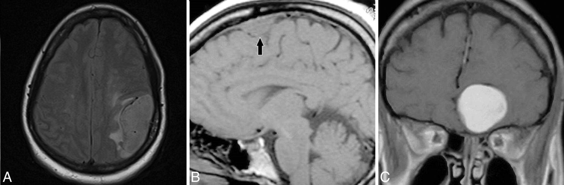
  • Fig 2.
    • Download figure
    • Open in new tab
    • Download powerpoint
    Fig 2.

    Chordoid meningioma shows facilitated diffusion. A, Postcontrast T1-weighted image showing avid homogeneous enhancement of a left temporal chordoid meningioma. B, DWI shows hypointensity of this chordoid meningioma. C, ADC map corresponding to the tumor shown in B depicting hyperintensity of the meningioma with an ADC value of 2.11 and an NADC ratio of 2.93, which represent increased diffusion. D, Postcontrast T1-weighted image of a different patient shows avid homogeneous enhancement of a left frontal convexity chordoid meningioma. E, DWI of this tumor shows isointensity to slight hypointensity of the tumor. F, ADC map corresponding to tumor shown in E shows increased signal of the meningioma with an ADC value of 1.47 and an NADC ratio of 1.99, which are consistent with facilitated diffusion.

  • Fig 3.
    • Download figure
    • Open in new tab
    • Download powerpoint
    Fig 3.

    Boxplot of the ADC values of all WHO grade I, II, and III meningiomas shows that chordoid meningiomas have significantly higher ADC values than all other subtypes, and there is a clear gap between the lowest chordoid meningioma ADC and the highest ADC from all other subtypes.
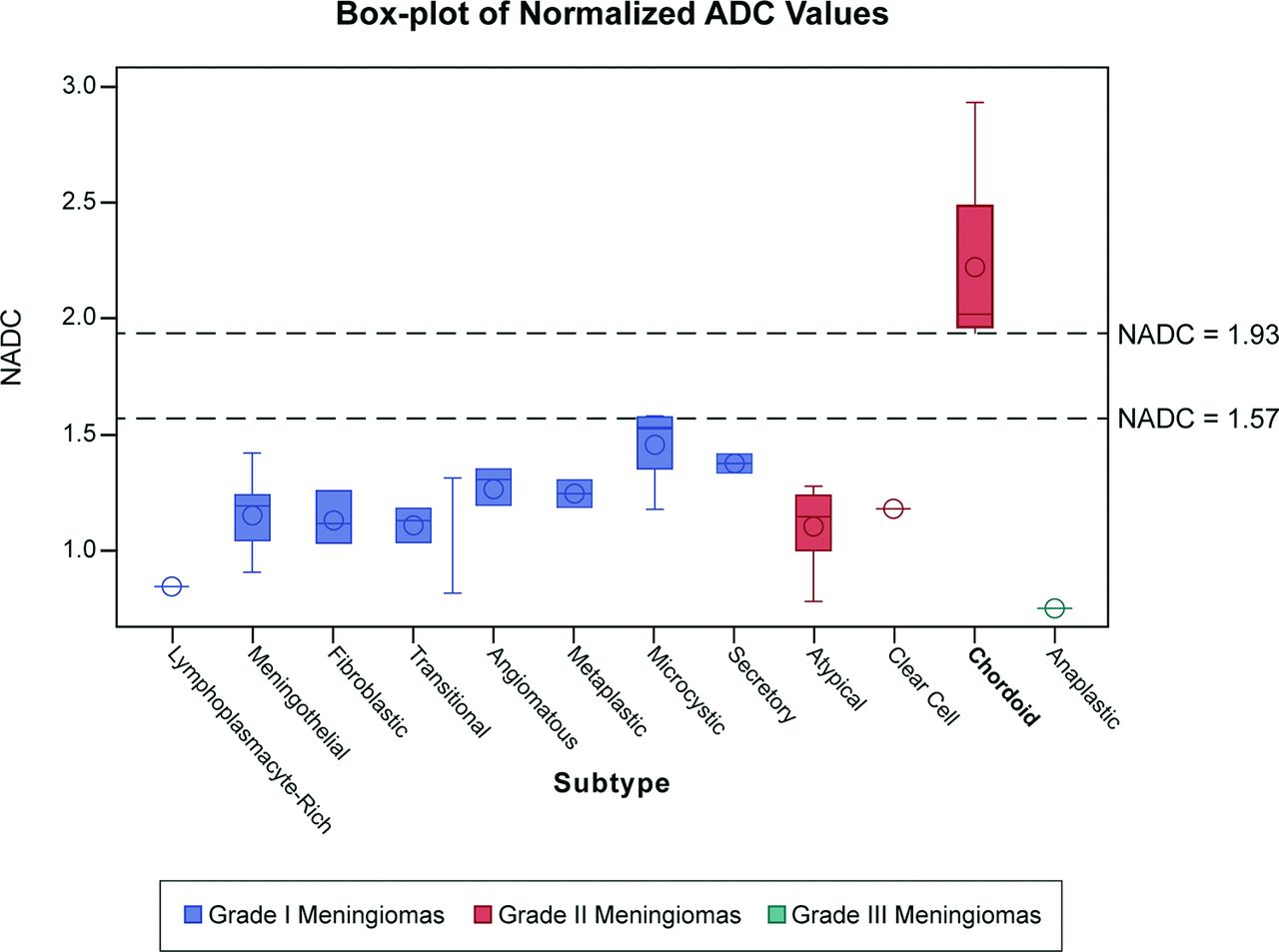
  • Fig 4.
    • Download figure
    • Open in new tab
    • Download powerpoint
    Fig 4.

    Boxplot of the NADC ratios of all WHO grade I, II, and III meningiomas shows that chordoid meningioma tumors have significantly higher NADC ratios than all other subtypes, and there is a clear gap between the lowest chordoid meningioma NADC ratio and the highest NADC ratio from all other types.

PreviousNext
Back to top

In this issue

American Journal of Neuroradiology: 36 (7)
American Journal of Neuroradiology
Vol. 36, Issue 7
1 Jul 2015
  • Table of Contents
  • Index by author
  • Complete Issue (PDF)
Advertisement
Print
Download PDF
Email Article

Thank you for your interest in spreading the word on American Journal of Neuroradiology.

NOTE: We only request your email address so that the person you are recommending the page to knows that you wanted them to see it, and that it is not junk mail. We do not capture any email address.

Enter multiple addresses on separate lines or separate them with commas.
Chordoid Meningioma: Differentiating a Rare World Health Organization Grade II Tumor from Other Meningioma Histologic Subtypes Using MRI
(Your Name) has sent you a message from American Journal of Neuroradiology
(Your Name) thought you would like to see the American Journal of Neuroradiology web site.
CAPTCHA
This question is for testing whether or not you are a human visitor and to prevent automated spam submissions.
Cite this article
J.B. Pond, T.G. Morgan, K.J. Hatanpaa, Z.F. Yetkin, B.E. Mickey, D.B. Mendelsohn
Chordoid Meningioma: Differentiating a Rare World Health Organization Grade II Tumor from Other Meningioma Histologic Subtypes Using MRI
American Journal of Neuroradiology Jul 2015, 36 (7) 1253-1258; DOI: 10.3174/ajnr.A4309

Citation Manager Formats

  • BibTeX
  • Bookends
  • EasyBib
  • EndNote (tagged)
  • EndNote 8 (xml)
  • Medlars
  • Mendeley
  • Papers
  • RefWorks Tagged
  • Ref Manager
  • RIS
  • Zotero
0 Responses
Respond to this article
Share
Bookmark this article
Chordoid Meningioma: Differentiating a Rare World Health Organization Grade II Tumor from Other Meningioma Histologic Subtypes Using MRI
J.B. Pond, T.G. Morgan, K.J. Hatanpaa, Z.F. Yetkin, B.E. Mickey, D.B. Mendelsohn
American Journal of Neuroradiology Jul 2015, 36 (7) 1253-1258; DOI: 10.3174/ajnr.A4309
del.icio.us logo Twitter logo Facebook logo Mendeley logo
  • Tweet Widget
  • Facebook Like
  • Google Plus One
Purchase

Jump to section

  • Article
    • Abstract
    • ABBREVIATIONS:
    • Materials and Methods
    • Results
    • Discussion
    • Conclusions
    • Acknowledgments
    • Footnotes
    • REFERENCES
  • Figures & Data
  • Supplemental
  • Info & Metrics
  • Responses
  • References
  • PDF

Related Articles

  • PubMed
  • Google Scholar

Cited By...

  • Preoperative MR Imaging to Differentiate Chordoid Meningiomas from Other Meningioma Histologic Subtypes
  • Crossref (21)
  • Google Scholar

This article has been cited by the following articles in journals that are participating in Crossref Cited-by Linking.

  • Variants of meningiomas: a review of imaging findings and clinical features
    Akira Kunimatsu, Natsuko Kunimatsu, Kouhei Kamiya, Masaki Katsura, Harushi Mori, Kuni Ohtomo
    Japanese Journal of Radiology 2016 34 7
  • Prediction of High-Grade Histology and Recurrence in Meningiomas Using Routine Preoperative Magnetic Resonance Imaging: A Systematic Review
    Dorothee Cäcilia Spille, Peter B. Sporns, Katharina Heß, Walter Stummer, Benjamin Brokinkel
    World Neurosurgery 2019 128
  • Histological Grade of Meningioma: Prediction by Intravoxel Incoherent Motion Histogram Parameters
    Manisha Bohara, Masanori Nakajo, Kiyohisa Kamimura, Tomohide Yoneyama, Yoshihiko Fukukura, Yutaro Kiyao, Hajime Yonezawa, Nayuta Higa, Mari Kirishima, Takashi Yoshiura
    Academic Radiology 2020 27 3
  • Uncommon Cranial Meningioma: Key Imaging Features on Conventional and Advanced Imaging
    Nader Zakhari, Carlos Torres, Mauricio Castillo, Thanh B. Nguyen
    Clinical Neuroradiology 2017 27 2
  • T1 and ADC histogram parameters may be an in vivo biomarker for predicting the grade, subtype, and proliferative activity of meningioma
    Tiexin Cao, Rifeng Jiang, Lingmin Zheng, Rufei Zhang, Xiaodan Chen, Zongmeng Wang, Peirong Jiang, Yilin Chen, Tianjin Zhong, Hu Chen, PuYeh Wu, Yunjing Xue, Lin Lin
    European Radiology 2022 33 1
  • Imaging spectrum of meningiomas: a review of uncommon imaging appearances and their histopathological and prognostic significance
    Venkatram Krishnan, Mahesh K. Mittal, Mukul Sinha
    Polish Journal of Radiology 2019 84
  • Clinical Features, Treatment, and Prognostic Factors of Chordoid Meningioma: Radiological and Pathological Features in 60 Cases of Chordoid Meningioma
    Yang Yang, Da Li, Xiao-Yu Cao, Shu-Yu Hao, Liang Wang, Zhen Wu, Jun-Ting Zhang
    World Neurosurgery 2016 93
  • Preoperative Apparent Diffusion Coefficient Values for Differentiation between Low and High Grade Meningiomas: An Updated Systematic Review and Meta-Analysis
    Yueh-Ting Tsai, Kuo-Chuan Hung, Yun-Ju Shih, Sher-Wei Lim, Cheng-Chun Yang, Yu-Ting Kuo, Jeon-Hor Chen, Ching-Chung Ko
    Diagnostics 2022 12 3
  • Preoperative MR Imaging to Differentiate Chordoid Meningiomas from Other Meningioma Histologic Subtypes
    J.D. Baal, W.C. Chen, D.A. Solomon, J.S. Pai, C.-H. Lucas, J.H. Hara, N.A. Oberheim Bush, M.W. McDermott, D.R. Raleigh, J.E. Villanueva-Meyer
    American Journal of Neuroradiology 2019
  • Histogram analysis in predicting the grade and histological subtype of meningiomas based on diffusion kurtosis imaging
    Xiaodan Chen, Lin Lin, Jie Wu, Guang Yang, Tianjin Zhong, Xiaoqiang Du, Zhiyong Chen, Ganggang Xu, Yang Song, Yunjing Xue, Qing Duan
    Acta Radiologica 2020 61 9

More in this TOC Section

  • Diagnostic Neuroradiology of Monoclonal Antibodies
  • Clinical Outcomes After Chiari I Decompression
  • Segmentation of Brain Metastases with BLAST
Show more Adult Brain

Similar Articles

Advertisement

Indexed Content

  • Current Issue
  • Accepted Manuscripts
  • Article Preview
  • Past Issues
  • Editorials
  • Editor's Choice
  • Fellows' Journal Club
  • Letters to the Editor
  • Video Articles

Cases

  • Case Collection
  • Archive - Case of the Week
  • Archive - Case of the Month
  • Archive - Classic Case

Special Collections

  • AJNR Awards
  • ASNR Foundation Special Collection
  • Most Impactful AJNR Articles
  • Photon-Counting CT
  • Spinal CSF Leak Articles (Jan 2020-June 2024)

More from AJNR

  • Trainee Corner
  • Imaging Protocols
  • MRI Safety Corner

Multimedia

  • AJNR Podcasts
  • AJNR Scantastics

Resources

  • Turnaround Time
  • Submit a Manuscript
  • Submit a Video Article
  • Submit an eLetter to the Editor/Response
  • Manuscript Submission Guidelines
  • Statistical Tips
  • Fast Publishing of Accepted Manuscripts
  • Graphical Abstract Preparation
  • Imaging Protocol Submission
  • Evidence-Based Medicine Level Guide
  • Publishing Checklists
  • Author Policies
  • Become a Reviewer/Academy of Reviewers
  • News and Updates

About Us

  • About AJNR
  • Editorial Board
  • Editorial Board Alumni
  • Alerts
  • Permissions
  • Not an AJNR Subscriber? Join Now
  • Advertise with Us
  • Librarian Resources
  • Feedback
  • Terms and Conditions
  • AJNR Editorial Board Alumni

American Society of Neuroradiology

  • Not an ASNR Member? Join Now

© 2025 by the American Society of Neuroradiology All rights, including for text and data mining, AI training, and similar technologies, are reserved.
Print ISSN: 0195-6108 Online ISSN: 1936-959X

Powered by HighWire