Advertisement

Main menu

  • Home
  • Content
    • Current Issue
    • Accepted Manuscripts
    • Article Preview
    • Past Issue Archive
    • Video Articles
    • AJNR Case Collection
    • Case of the Week Archive
    • Case of the Month Archive
    • Classic Case Archive
  • Special Collections
    • AJNR Awards
    • ASNR Foundation Special Collection
    • Most Impactful AJNR Articles
    • Photon-Counting CT
    • Spinal CSF Leak Articles (Jan 2020-June 2024)
  • Multimedia
    • AJNR Podcasts
    • AJNR SCANtastic
    • Trainee Corner
    • MRI Safety Corner
    • Imaging Protocols
  • For Authors
    • Submit a Manuscript
    • Submit a Video Article
    • Submit an eLetter to the Editor/Response
    • Manuscript Submission Guidelines
    • Statistical Tips
    • Fast Publishing of Accepted Manuscripts
    • Graphical Abstract Preparation
    • Imaging Protocol Submission
    • Author Policies
  • About Us
    • About AJNR
    • Editorial Board
    • Editorial Board Alumni
  • More
    • Become a Reviewer/Academy of Reviewers
    • Subscribers
    • Permissions
    • Alerts
    • Feedback
    • Advertisers
    • ASNR Home

User menu

  • Alerts
  • Log in

Search

  • Advanced search
American Journal of Neuroradiology

American Journal of Neuroradiology

ASHNR American Society of Functional Neuroradiology ASHNR American Society of Pediatric Neuroradiology ASSR
  • Alerts
  • Log in

Advanced Search

  • Home
  • Content
    • Current Issue
    • Accepted Manuscripts
    • Article Preview
    • Past Issue Archive
    • Video Articles
    • AJNR Case Collection
    • Case of the Week Archive
    • Case of the Month Archive
    • Classic Case Archive
  • Special Collections
    • AJNR Awards
    • ASNR Foundation Special Collection
    • Most Impactful AJNR Articles
    • Photon-Counting CT
    • Spinal CSF Leak Articles (Jan 2020-June 2024)
  • Multimedia
    • AJNR Podcasts
    • AJNR SCANtastic
    • Trainee Corner
    • MRI Safety Corner
    • Imaging Protocols
  • For Authors
    • Submit a Manuscript
    • Submit a Video Article
    • Submit an eLetter to the Editor/Response
    • Manuscript Submission Guidelines
    • Statistical Tips
    • Fast Publishing of Accepted Manuscripts
    • Graphical Abstract Preparation
    • Imaging Protocol Submission
    • Author Policies
  • About Us
    • About AJNR
    • Editorial Board
    • Editorial Board Alumni
  • More
    • Become a Reviewer/Academy of Reviewers
    • Subscribers
    • Permissions
    • Alerts
    • Feedback
    • Advertisers
    • ASNR Home
  • Follow AJNR on Twitter
  • Visit AJNR on Facebook
  • Follow AJNR on Instagram
  • Join AJNR on LinkedIn
  • RSS Feeds

AJNR is seeking candidates for the AJNR Podcast Editor. Read the position description.

Research ArticleADULT BRAIN

Memory Part 3: The Role of the Fornix and Clinical Cases

F.D. Raslau, J.C. Augustinack, A.P. Klein, J.L. Ulmer, V.P. Mathews and L.P. Mark
American Journal of Neuroradiology September 2015, 36 (9) 1604-1608; DOI: https://doi.org/10.3174/ajnr.A4371
F.D. Raslau
aFrom the Department of Radiology (F.D.R.), University of Kentucky, Lexington, Kentucky
  • Find this author on Google Scholar
  • Find this author on PubMed
  • Search for this author on this site
J.C. Augustinack
bDepartment of Radiology (J.C.A.), Harvard Medical School, Boston, Massachusetts
cAthinoula A. Martinos Center for Biomedical Imaging (J.C.A.), Massachusetts General Hospital and Harvard Medical School, Boston, Massachusetts
  • Find this author on Google Scholar
  • Find this author on PubMed
  • Search for this author on this site
A.P. Klein
dDepartment of Radiology (A.P.K., J.L.U., V.P.M., L.P.M.), Medical College of Wisconsin, Milwaukee, Wisconsin.
  • Find this author on Google Scholar
  • Find this author on PubMed
  • Search for this author on this site
J.L. Ulmer
dDepartment of Radiology (A.P.K., J.L.U., V.P.M., L.P.M.), Medical College of Wisconsin, Milwaukee, Wisconsin.
  • Find this author on Google Scholar
  • Find this author on PubMed
  • Search for this author on this site
  • ORCID record for J.L. Ulmer
V.P. Mathews
dDepartment of Radiology (A.P.K., J.L.U., V.P.M., L.P.M.), Medical College of Wisconsin, Milwaukee, Wisconsin.
  • Find this author on Google Scholar
  • Find this author on PubMed
  • Search for this author on this site
L.P. Mark
dDepartment of Radiology (A.P.K., J.L.U., V.P.M., L.P.M.), Medical College of Wisconsin, Milwaukee, Wisconsin.
  • Find this author on Google Scholar
  • Find this author on PubMed
  • Search for this author on this site
  • ORCID record for L.P. Mark
  • Article
  • Figures & Data
  • Info & Metrics
  • Responses
  • References
  • PDF
Loading

Article Figures & Data

Figures

  • Fig 1.
    • Download figure
    • Open in new tab
    • Download powerpoint
    Fig 1.

    Sagittal T1-weighted image showing the archlike configuration of the fornix (yellow arrow) at the upper margin of the velum interpositum (VI), which is situated between the lateral (LV) and third (3V) ventricles. The fornix is attached to the septum pellucidum, which in turn is attached to the inner curving surface of the corpus callosum (CC). The fornix bifurcates at the level of the anterior commissure (red arrow), with the postcommissural fibers projecting to the mammillary bodies (green arrow).

  • Fig 2.
    • Download figure
    • Open in new tab
    • Download powerpoint
    Fig 2.

    High-resolution coronal T2-weighted image of the right hippocampal formation showing the dark signal of the white matter fibers of the alveus (red arrow) positioned on top of Ammon's horn (CA) of the hippocampus and below the CSF of the temporal horn of the lateral ventricle, which also contains the choroid plexus (not labeled). The dentate gyrus (DG) is shown. The alveus thickens at its medial margin to form the fimbria (yellow arrow).
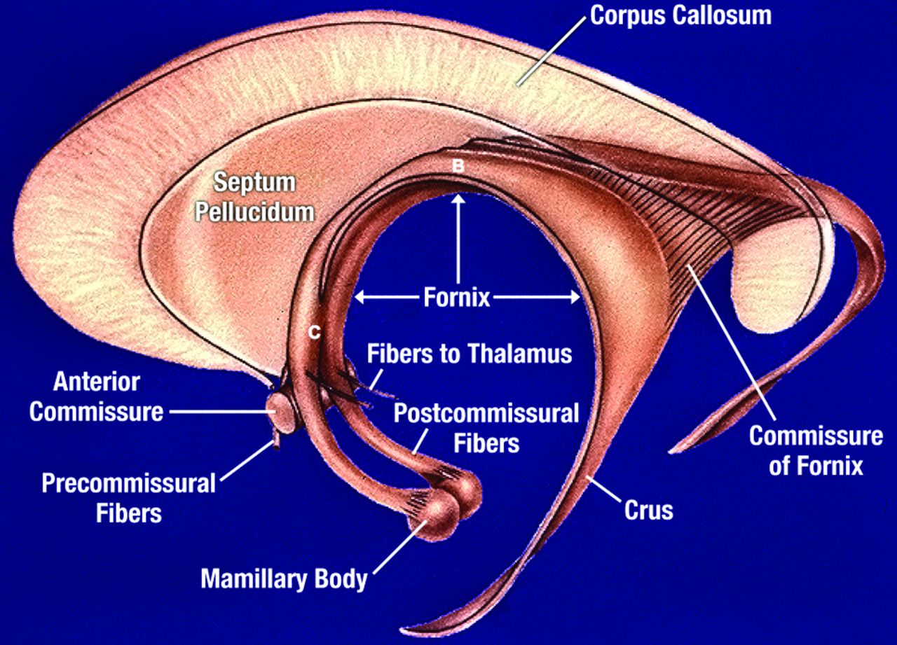
  • Fig 3.
    • Download figure
    • Open in new tab
    • Download powerpoint
    Fig 3.

    Schematic overview of the fornix from the side. The fimbria forms the crus of the fornix, which is joined to the opposite fornix across the midline by the commissure of the fornix (hippocampal commissure, psalterium). The body of the fornix (B) at the top of the fornical arch is connected to the inferior surface of the body of the corpus callosum by the septum pellucidum. The column of the fornix (C) has a more superoinferior orientation and is connected to the posterior margin of the genu of the corpus callosum by the septum pellucidum. The columns of the fornix are split at the level of the anterior commissure with most the fibers (postcommissural fibers) projecting to the mammillary bodies. Published with permission from the estate of David L. Daniels.
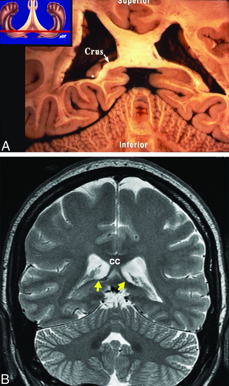
  • Fig 4.
    • Download figure
    • Open in new tab
    • Download powerpoint
    Fig 4.

    A, Coronal anatomic specimen with a schematic reference image. The white matter fibers of the crus of the fornix can be seen forming the inferior medial margin of the posterior aspect of the body of the lateral ventricles at the level of the splenium of the corpus callosum. B, Coronal T2-weighted image shows the dark signal of the crura of the fornices (yellow arrows) forming the inferior medial margin of the posterior lateral ventricles at the level of the posterior aspect of the body of the corpus callosum (CC).
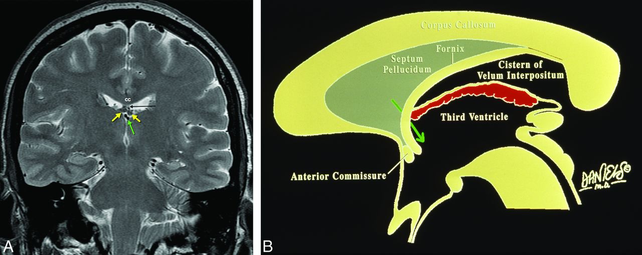
  • Fig 5.
    • Download figure
    • Open in new tab
    • Download powerpoint
    Fig 5.

    A, Coronal T2-weighted image shows the body of the fornices (yellow arrows) attached to the inferior margin of the body of the corpus callosum (CC) via the septum pellucidum (black arrow). The body of the fornices are at the upper aspect of the velum interpositum, which is traversed by the internal cerebral veins (green arrow), situated above the third ventricle but inferomedial to the body of the lateral ventricles. B, Sagittal schematic shows the position of the fornix relative to the velum interpositum. The foramen of Monro (green arrow) forms the anterior border of the velum interpositum, while the posterior border opens between the splenium of the corpus callosum above and the pineal gland below into the quadrigeminal plate cistern. The choroid plexus (red) is indicated at the roof of the third ventricle below but not within the velum interpositum. Published with permission from the estate of David L. Daniels.

  • Fig 6.
    • Download figure
    • Open in new tab
    • Download powerpoint
    Fig 6.

    A, Axial T2-weighted image shows the postcommissural fibers of the fornix (yellow arrows) coursing posterior to the anterior commissure (green arrow). B, Coronal T2-weighted image shows the superoinferior orientation of the columns of the fornix (yellow arrows).

  • Fig 7.
    • Download figure
    • Open in new tab
    • Download powerpoint
    Fig 7.

    Drawing showing verbal memory information carried primarily by the left fornix (orange), while the visuospatial memory information is primarily transmitted by the right fornix (light purple).
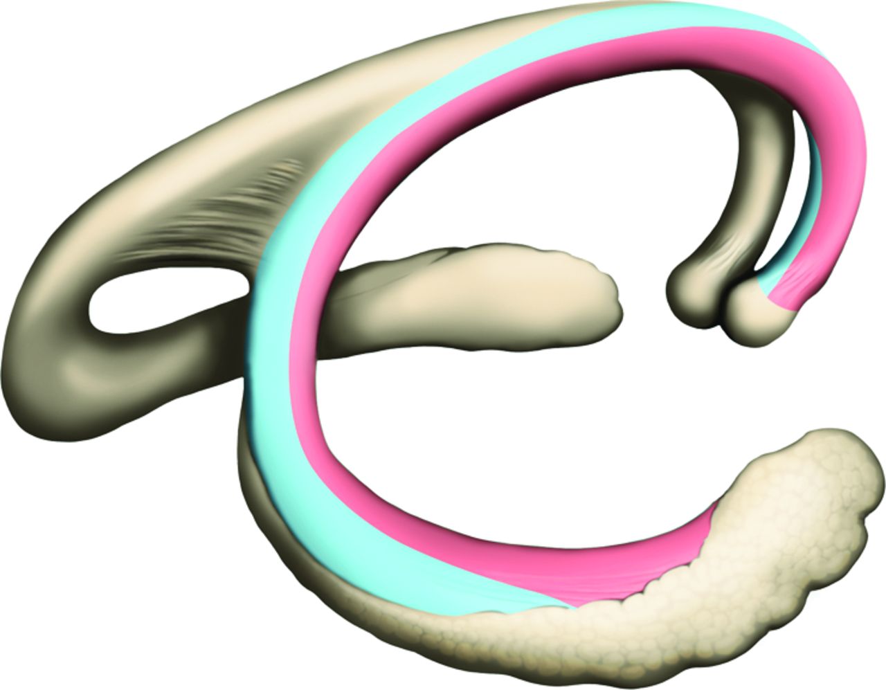
  • Fig 8.
    • Download figure
    • Open in new tab
    • Download powerpoint
    Fig 8.

    Drawing shows the medial aspect of the fornix (light blue) carrying fibers from the posterior hippocampus for processing of exteroceptive information for scene learning. The more lateral fibers (light red), meanwhile, carry fibers from the more rostral portions of the hippocampus for processing of interceptive signals for emotional and motivational memory and learning.

  • Fig 9.
    • Download figure
    • Open in new tab
    • Download powerpoint
    Fig 9.

    A, T2-weighted axial image shows a glioneuronal tumor (red arrow) within the right lateral ventricle near the foramen of Monro displacing and distorting the septum pellucidum and columns of the fornices (yellow arrow). B, The intraventricular glioneuronal tumor (red arrow) is better visualized on the axial FLAIR image. C, Postoperative coronal T2-weighted image shows the intraventricular neoplasm still distorting the columns of the fornix and septum pellucidum.

  • Fig 10.
    • Download figure
    • Open in new tab
    • Download powerpoint
    Fig 10.

    A, Gradient recalled-echo image shows hemorrhage (red arrow) in the cistern of the velum interpositum involving the bodies of the fornices. B, Coronal T1-weighted image 5 months later without treatment shows atrophy of the mammillary bodies, worse on the left (yellow arrow).

  • Fig 11.
    • Download figure
    • Open in new tab
    • Download powerpoint
    Fig 11.

    A, Sagittal T1 postcontrast image shows an enhancing mass involving the floor of the hypothalamus and mammillary body with extension into the third ventricle. B, Coronal FLAIR image shows extensive abnormal signal involving the hypothalamus on either side of the midline. Pathology (not shown) showed this to be Langerhans cell histiocytosis.

PreviousNext
Back to top

In this issue

American Journal of Neuroradiology: 36 (9)
American Journal of Neuroradiology
Vol. 36, Issue 9
1 Sep 2015
  • Table of Contents
  • Index by author
  • Complete Issue (PDF)
Advertisement
Print
Download PDF
Email Article

Thank you for your interest in spreading the word on American Journal of Neuroradiology.

NOTE: We only request your email address so that the person you are recommending the page to knows that you wanted them to see it, and that it is not junk mail. We do not capture any email address.

Enter multiple addresses on separate lines or separate them with commas.
Memory Part 3: The Role of the Fornix and Clinical Cases
(Your Name) has sent you a message from American Journal of Neuroradiology
(Your Name) thought you would like to see the American Journal of Neuroradiology web site.
CAPTCHA
This question is for testing whether or not you are a human visitor and to prevent automated spam submissions.
Cite this article
F.D. Raslau, J.C. Augustinack, A.P. Klein, J.L. Ulmer, V.P. Mathews, L.P. Mark
Memory Part 3: The Role of the Fornix and Clinical Cases
American Journal of Neuroradiology Sep 2015, 36 (9) 1604-1608; DOI: 10.3174/ajnr.A4371

Citation Manager Formats

  • BibTeX
  • Bookends
  • EasyBib
  • EndNote (tagged)
  • EndNote 8 (xml)
  • Medlars
  • Mendeley
  • Papers
  • RefWorks Tagged
  • Ref Manager
  • RIS
  • Zotero
0 Responses
Respond to this article
Share
Bookmark this article
Memory Part 3: The Role of the Fornix and Clinical Cases
F.D. Raslau, J.C. Augustinack, A.P. Klein, J.L. Ulmer, V.P. Mathews, L.P. Mark
American Journal of Neuroradiology Sep 2015, 36 (9) 1604-1608; DOI: 10.3174/ajnr.A4371
del.icio.us logo Twitter logo Facebook logo Mendeley logo
  • Tweet Widget
  • Facebook Like
  • Google Plus One
Purchase

Jump to section

  • Article
    • Acknowledgments
    • Footnotes
    • References
  • Figures & Data
  • Info & Metrics
  • Responses
  • References
  • PDF

Related Articles

  • No related articles found.
  • PubMed
  • Google Scholar

Cited By...

  • Long-term cognitive recovery following isolated bilateral infarction of the fornix presenting with amnesia
  • Brain connectivity changes underlying depression and fatigue in relapsing-remitting multiple sclerosis: a systematic review
  • Uncovering a role for the dorsal hippocampal commissure in episodic memory
  • Crossref (21)
  • Google Scholar

This article has been cited by the following articles in journals that are participating in Crossref Cited-by Linking.

  • Volumetric MRI Findings in Mild Traumatic Brain Injury (mTBI) and Neuropsychological Outcome
    Erin D. Bigler
    Neuropsychology Review 2023 33 1
  • Are Anesthesia and Surgery during Infancy Associated with Decreased White Matter Integrity and Volume during Childhood?
    Robert I. Block, Vincent A. Magnotta, Emine O. Bayman, James Y. Choi, Joss J. Thomas, Karolie K. Kimble
    Anesthesiology 2017 127 5
  • The Surgical White Matter Chassis: A Practical 3-Dimensional Atlas for Planning Subcortical Surgical Trajectories
    Jonathan E Jennings, Amin B Kassam, Melanie B Fukui, Alejandro Monroy-Sosa, Srikant Chakravarthi, Nathan Kojis, Richard A Rovin
    Operative Neurosurgery 2018 14 5
  • Microstructural white matter alterations in borderline personality disorder: A minireview
    M. Grottaroli, G. Delvecchio, C. Bressi, C. Moltrasio, J.C. Soares, P. Brambilla
    Journal of Affective Disorders 2020 264
  • The Fornix: Functional Anatomy, Normal Neuroimaging, and Various Pathological Conditions
    Young Jae Choi, Eun Ja Lee, Jung Eun Lee
    Investigative Magnetic Resonance Imaging 2021 25 2
  • Uncovering a Role for the Dorsal Hippocampal Commissure in Recognition Memory
    M Postans, G D Parker, H Lundell, M Ptito, K Hamandi, W P Gray, J P Aggleton, T B Dyrby, D K Jones, M Winter
    Cerebral Cortex 2020 30 3
  • Awake Surgical Management of Third Ventricular Tumors: A Preliminary Safety, Feasibility, and Clinical Applications Study
    Srikant S Chakravarthi, Amin B Kassam, Melanie B Fukui, Alejandro Monroy-Sosa, Nichelle Rothong, Joseph Cunningham, Jonathan E Jennings, Neil Guenther, Jeremy Connelly, Tobias Kaemmerer, Kenneth C Nash, Mark Lindsay, Janie Rissell, Juanita M Celix, Richard A Rovin
    Operative Neurosurgery 2019 17 2
  • Diffusion properties of the fornix assessed by deterministic tractography shows age, sex, volume, cognitive, hemispheric, and twin relationships in young adults from the Human Connectome Project
    Ariana J. Cahn, Graham Little, Christian Beaulieu, Pascal Tétreault
    Brain Structure and Function 2021 226 2
  • Diffusion imaging of fornix and interconnected limbic deep grey matter is linked to cognitive impairment in multiple sclerosis
    Diana Valdés Cabrera, Penelope Smyth, Gregg Blevins, Derek Emery, Christian Beaulieu
    European Journal of Neuroscience 2022 55 1
  • Fornix Stimulation Induces Metabolic Activity and Dopaminergic Response in the Nucleus Accumbens
    Hojin Shin, Sang-Yoon Lee, Hyun-U Cho, Yoonbae Oh, In Young Kim, Kendall H. Lee, Dong Pyo Jang, Hoon-Ki Min
    Frontiers in Neuroscience 2019 13

More in this TOC Section

ADULT BRAIN

  • Diagnostic Neuroradiology of Monoclonal Antibodies
  • Clinical Outcomes After Chiari I Decompression
  • Segmentation of Brain Metastases with BLAST
Show more ADULT BRAIN

PEDIATRICS

  • SyMRI & MR Fingerprinting in Brainstem Myelination
  • Comparison of Image Quality and Radiation Dose in Pediatric Temporal Bone CT Using Photon-Counting Detector CT and Energy-Integrating Detector CT
  • Axenfeld-Rieger Syndrome: Neuroimaging Findings
Show more PEDIATRICS

Similar Articles

Advertisement

Indexed Content

  • Current Issue
  • Accepted Manuscripts
  • Article Preview
  • Past Issues
  • Editorials
  • Editor's Choice
  • Fellows' Journal Club
  • Letters to the Editor
  • Video Articles

Cases

  • Case Collection
  • Archive - Case of the Week
  • Archive - Case of the Month
  • Archive - Classic Case

Special Collections

  • AJNR Awards
  • ASNR Foundation Special Collection
  • Most Impactful AJNR Articles
  • Photon-Counting CT
  • Spinal CSF Leak Articles (Jan 2020-June 2024)

More from AJNR

  • Trainee Corner
  • Imaging Protocols
  • MRI Safety Corner

Multimedia

  • AJNR Podcasts
  • AJNR Scantastics

Resources

  • Turnaround Time
  • Submit a Manuscript
  • Submit a Video Article
  • Submit an eLetter to the Editor/Response
  • Manuscript Submission Guidelines
  • Statistical Tips
  • Fast Publishing of Accepted Manuscripts
  • Graphical Abstract Preparation
  • Imaging Protocol Submission
  • Evidence-Based Medicine Level Guide
  • Publishing Checklists
  • Author Policies
  • Become a Reviewer/Academy of Reviewers
  • News and Updates

About Us

  • About AJNR
  • Editorial Board
  • Editorial Board Alumni
  • Alerts
  • Permissions
  • Not an AJNR Subscriber? Join Now
  • Advertise with Us
  • Librarian Resources
  • Feedback
  • Terms and Conditions
  • AJNR Editorial Board Alumni

American Society of Neuroradiology

  • Not an ASNR Member? Join Now

© 2025 by the American Society of Neuroradiology All rights, including for text and data mining, AI training, and similar technologies, are reserved.
Print ISSN: 0195-6108 Online ISSN: 1936-959X

Powered by HighWire