Advertisement

Main menu

  • Home
  • Content
    • Current Issue
    • Accepted Manuscripts
    • Article Preview
    • Past Issue Archive
    • Video Articles
    • AJNR Case Collection
    • Case of the Week Archive
    • Case of the Month Archive
    • Classic Case Archive
  • Special Collections
    • AJNR Awards
    • ASNR Foundation Special Collection
    • Most Impactful AJNR Articles
    • Photon-Counting CT
    • Spinal CSF Leak Articles (Jan 2020-June 2024)
  • Multimedia
    • AJNR Podcasts
    • AJNR SCANtastic
    • Trainee Corner
    • MRI Safety Corner
    • Imaging Protocols
  • For Authors
    • Submit a Manuscript
    • Submit a Video Article
    • Submit an eLetter to the Editor/Response
    • Manuscript Submission Guidelines
    • Statistical Tips
    • Fast Publishing of Accepted Manuscripts
    • Graphical Abstract Preparation
    • Imaging Protocol Submission
    • Author Policies
  • About Us
    • About AJNR
    • Editorial Board
    • Editorial Board Alumni
  • More
    • Become a Reviewer/Academy of Reviewers
    • Subscribers
    • Permissions
    • Alerts
    • Feedback
    • Advertisers
    • ASNR Home

User menu

  • Alerts
  • Log in

Search

  • Advanced search
American Journal of Neuroradiology

American Journal of Neuroradiology

ASHNR American Society of Functional Neuroradiology ASHNR American Society of Pediatric Neuroradiology ASSR
  • Alerts
  • Log in

Advanced Search

  • Home
  • Content
    • Current Issue
    • Accepted Manuscripts
    • Article Preview
    • Past Issue Archive
    • Video Articles
    • AJNR Case Collection
    • Case of the Week Archive
    • Case of the Month Archive
    • Classic Case Archive
  • Special Collections
    • AJNR Awards
    • ASNR Foundation Special Collection
    • Most Impactful AJNR Articles
    • Photon-Counting CT
    • Spinal CSF Leak Articles (Jan 2020-June 2024)
  • Multimedia
    • AJNR Podcasts
    • AJNR SCANtastic
    • Trainee Corner
    • MRI Safety Corner
    • Imaging Protocols
  • For Authors
    • Submit a Manuscript
    • Submit a Video Article
    • Submit an eLetter to the Editor/Response
    • Manuscript Submission Guidelines
    • Statistical Tips
    • Fast Publishing of Accepted Manuscripts
    • Graphical Abstract Preparation
    • Imaging Protocol Submission
    • Author Policies
  • About Us
    • About AJNR
    • Editorial Board
    • Editorial Board Alumni
  • More
    • Become a Reviewer/Academy of Reviewers
    • Subscribers
    • Permissions
    • Alerts
    • Feedback
    • Advertisers
    • ASNR Home
  • Follow AJNR on Twitter
  • Visit AJNR on Facebook
  • Follow AJNR on Instagram
  • Join AJNR on LinkedIn
  • RSS Feeds

AJNR is seeking candidates for the AJNR Podcast Editor. Read the position description.

Research ArticleADULT BRAIN

Neuroradiologic Diagnosis of Minor Leak prior to Major SAH: Diagnosis by T1-FLAIR Mismatch

S. Oda, M. Shimoda, A. Hirayama, M. Imai, F. Komatsu, H. Shigematsu, J. Nishiyama and M. Matsumae
American Journal of Neuroradiology September 2015, 36 (9) 1616-1622; DOI: https://doi.org/10.3174/ajnr.A4325
S. Oda
aFrom the Department of Neurosurgery (S.O., M.S., A.H., M.I., F.K., H.S., J.N.), Tokai University Hachioji Hospital, Tokyo, Japan
  • Find this author on Google Scholar
  • Find this author on PubMed
  • Search for this author on this site
M. Shimoda
aFrom the Department of Neurosurgery (S.O., M.S., A.H., M.I., F.K., H.S., J.N.), Tokai University Hachioji Hospital, Tokyo, Japan
  • Find this author on Google Scholar
  • Find this author on PubMed
  • Search for this author on this site
A. Hirayama
aFrom the Department of Neurosurgery (S.O., M.S., A.H., M.I., F.K., H.S., J.N.), Tokai University Hachioji Hospital, Tokyo, Japan
  • Find this author on Google Scholar
  • Find this author on PubMed
  • Search for this author on this site
M. Imai
aFrom the Department of Neurosurgery (S.O., M.S., A.H., M.I., F.K., H.S., J.N.), Tokai University Hachioji Hospital, Tokyo, Japan
  • Find this author on Google Scholar
  • Find this author on PubMed
  • Search for this author on this site
F. Komatsu
aFrom the Department of Neurosurgery (S.O., M.S., A.H., M.I., F.K., H.S., J.N.), Tokai University Hachioji Hospital, Tokyo, Japan
  • Find this author on Google Scholar
  • Find this author on PubMed
  • Search for this author on this site
H. Shigematsu
aFrom the Department of Neurosurgery (S.O., M.S., A.H., M.I., F.K., H.S., J.N.), Tokai University Hachioji Hospital, Tokyo, Japan
  • Find this author on Google Scholar
  • Find this author on PubMed
  • Search for this author on this site
J. Nishiyama
aFrom the Department of Neurosurgery (S.O., M.S., A.H., M.I., F.K., H.S., J.N.), Tokai University Hachioji Hospital, Tokyo, Japan
  • Find this author on Google Scholar
  • Find this author on PubMed
  • Search for this author on this site
M. Matsumae
bDepartment of Neurosurgery (M.M.), Tokai University School of Medicine, Kanagawa, Japan.
  • Find this author on Google Scholar
  • Find this author on PubMed
  • Search for this author on this site
  • Article
  • Figures & Data
  • Info & Metrics
  • Responses
  • References
  • PDF
Loading

Article Figures & Data

Figures

  • Tables
  • Fig 1.
    • Download figure
    • Open in new tab
    • Download powerpoint
    Fig 1.

    A 77-year-old woman with a ruptured right middle cerebral artery aneurysm who was admitted 5 days after onset. T1WI on admission shows BHSB in the bilateral Sylvian fissure (A, arrows) that matches the blood distribution on the FLAIR image (B, arrows). These findings are typical of subacute SAH with T1-FLAIR-matched SAH that was not associated with rebleeding. Reprinted with permission from Shimoda M. Neuroimaging for headache. Journal of Clinical and Experimental Medicine (IGAKU NO AYUMI) 2012;243:1086–94; Ishiyaku Publishers, Inc.
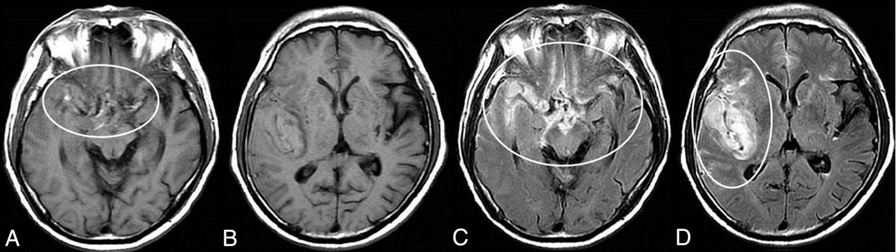
  • Fig 2.
    • Download figure
    • Open in new tab
    • Download powerpoint
    Fig 2.

    Typical neuroradiologic findings in a patient with minor leak before major SAH attack. Images are from an 80-year-old woman with a ruptured right middle cerebral artery aneurysm. T1WI on admission shows clearly iso- to mildly hyperintense blood obscuring the right Sylvian fissure, in addition to the more conspicuous bright T1 foci (A and B, circles). These findings indicate subacute subarachnoid blood due to a minor leak that occurred before the major attack. FLAIR images on admission show the SAH in the acute phase in the quadrigeminal cistern and left Sylvian fissure (C and D, circles), in addition to the BHSB on T1WI. We defined this as T1-FLAIR mismatch and used it as a neuroradiologic diagnosis of minor leak that occurred before major SAH.
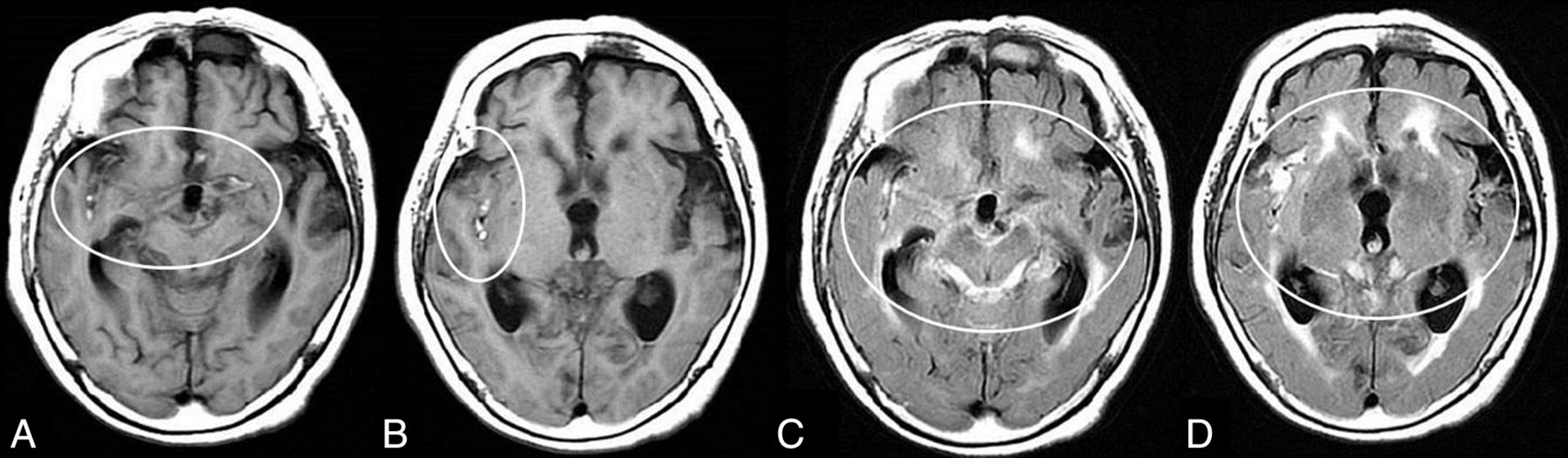
  • Fig 3.
    • Download figure
    • Open in new tab
    • Download powerpoint
    Fig 3.

    Typical neuroradiologic findings in a case with minor leak before a major SAH attack with intra-Sylvian hematoma. Images are from a 66-year-old woman with a ruptured right middle cerebral artery aneurysm. T1WI shows iso- to mildly hyperintense blood obscuring the right Sylvian fissure and suprasellar cistern, in addition to the more conspicuous bright T1 foci including the neighboring area of the ruptured aneurysm. An intra-Sylvian hematoma is depicted as an isointense signal (A circle, B). FLAIR images show the intra-Sylvian hematoma and SAH (right Sylvian fissure, suprasellar and right ambient cistern) as a high-intensity signal, which indicates acute blood (C and D, circles).

Tables

  • Figures
    • View popup
    Table 1:

    Clinical features, neurologic status, and CT features on admissiona

    TotalT1-FLAIR MismatchP Value
    PositiveNegative
    No. of patients12743 (33.9)84 (66.1)
    Warning sign
        Positive1413 (30.2)1 (1.2)<.001
        Negative542 (4.7)52 (61.9)
        Unknown5928 (65.1)31 (36.9)
    Mean age (yr)61.5 ± 14.266.5 ± 13.258.9 ± 14.0.004
    Age range (yr)21–8940–8521–89
    Elderly patients
        Older than 80 years1210 (23.3)2 (2.4)<.001
    Female sex8932 (74.4)57 (67.9).541
    Rebleeding after admission2819 (44.2)9 (1.7)<.001
    WFNS grade on admission
        Grades I–II9728 (65.1)69 (82.1).028
        Grades IV–V2813 (30.2)15 (17.9).120
    Fisher group
        Group 38530 (69.8)55 (65.5).550
    Intracerebral hemorrhage2519 (44.2)6 (7.1)<.001
    Acute hydrocephalus6126 (60.5)35 (41.7).060
    Aneurysm site
        Anterior communicating artery419 (20.9)32 (38.1)
        Anterior cerebral artery84 (9.3)4 (4.8)
        Internal carotid artery3812 (27.9)26 (31.0)
        Middle cerebral artery3117 (39.5)14 (16.7)
        Posterior circulation808 (9.5).051
    Aneurysm size
        >5 mm7531 (72.1)44 (52.4).037
        >10 mm83 (7.0)5 (6.0)1.000
    • Note:—WFNS indicates World Federation of Neurological Surgeons.

    • ↵a Values are No. (%) unless otherwise stated. The “No. of patients” row shows the percentage of the total number of patients, whereas all other percentages in the “Positive” and “Negative” columns are the percentages of patients with positive and negative findings, respectively.

    • View popup
    Table 2:

    Surgery and subsequent eventsa

    TotalT1-FLAIR MismatchP Value
    PositiveNegative
    No. of patients12743 (33.9)84 (66.1)
    Aneurysm operation
        Craniotomy10536 (83.7)69 (82.1)1.000
        Coiling227 (16.3)15 (17.9)
    Delayed angiographic vasospasm3519 (44.2)16 (19.0).003
    DIND2216 (37.2)6 (7.1)<.001
        Infarction due to delayed vasospasm on DWI3017 (39.5)13 (15.5).003
        Chronic hydrocephalusb5423 (62.2)31 (37.8).029
    mRS score at 3 months
        >3–64128 (65.1)13 (15.5)<.001
    • Note:—DIND indicates delayed ischemic neurologic deficits.

    • ↵a The “No. of patients” row shows the percentage of the total number of patients, whereas all other percentages in the “Positive” and “Negative” columns are the percentages of patients with positive and negative findings, respectively.

    • ↵b The incidence of chronic hydrocephalus was calculated for the surviving patients.

    • View popup
    Table 3:

    Results of multivariate logistic regression analysis for the presence of a minor leak before admission diagnosed by T1-FLAIR mismatch

    Odds Ratio95% CIP Value
    Preoperative clinical factors
        Age older than 80 years8.4751.639–43.478.011
        Rebleeding5.2912.028–13.889.001
    Associated neuroradiologic findings on admission
        Intracerebral hemorrhage on CT7.1972.457–20.833<.001
    Postoperative factors
        mRS score 3–66.6902.548–17.564<.001
PreviousNext
Back to top

In this issue

American Journal of Neuroradiology: 36 (9)
American Journal of Neuroradiology
Vol. 36, Issue 9
1 Sep 2015
  • Table of Contents
  • Index by author
  • Complete Issue (PDF)
Advertisement
Print
Download PDF
Email Article

Thank you for your interest in spreading the word on American Journal of Neuroradiology.

NOTE: We only request your email address so that the person you are recommending the page to knows that you wanted them to see it, and that it is not junk mail. We do not capture any email address.

Enter multiple addresses on separate lines or separate them with commas.
Neuroradiologic Diagnosis of Minor Leak prior to Major SAH: Diagnosis by T1-FLAIR Mismatch
(Your Name) has sent you a message from American Journal of Neuroradiology
(Your Name) thought you would like to see the American Journal of Neuroradiology web site.
CAPTCHA
This question is for testing whether or not you are a human visitor and to prevent automated spam submissions.
Cite this article
S. Oda, M. Shimoda, A. Hirayama, M. Imai, F. Komatsu, H. Shigematsu, J. Nishiyama, M. Matsumae
Neuroradiologic Diagnosis of Minor Leak prior to Major SAH: Diagnosis by T1-FLAIR Mismatch
American Journal of Neuroradiology Sep 2015, 36 (9) 1616-1622; DOI: 10.3174/ajnr.A4325

Citation Manager Formats

  • BibTeX
  • Bookends
  • EasyBib
  • EndNote (tagged)
  • EndNote 8 (xml)
  • Medlars
  • Mendeley
  • Papers
  • RefWorks Tagged
  • Ref Manager
  • RIS
  • Zotero
0 Responses
Respond to this article
Share
Bookmark this article
Neuroradiologic Diagnosis of Minor Leak prior to Major SAH: Diagnosis by T1-FLAIR Mismatch
S. Oda, M. Shimoda, A. Hirayama, M. Imai, F. Komatsu, H. Shigematsu, J. Nishiyama, M. Matsumae
American Journal of Neuroradiology Sep 2015, 36 (9) 1616-1622; DOI: 10.3174/ajnr.A4325
del.icio.us logo Twitter logo Facebook logo Mendeley logo
  • Tweet Widget
  • Facebook Like
  • Google Plus One
Purchase

Jump to section

  • Article
    • Abstract
    • ABBREVIATIONS:
    • Materials and Methods
    • Results
    • Discussion
    • Conclusions
    • References
  • Figures & Data
  • Info & Metrics
  • Responses
  • References
  • PDF

Related Articles

  • No related articles found.
  • PubMed
  • Google Scholar

Cited By...

  • Complementary Roles of Dynamic Contrast-Enhanced MR Imaging and Postcontrast Vessel Wall Imaging in Detecting High-Risk Intracranial Aneurysms
  • A note of despair
  • Sentinel Headache: A Warning Sign Preceding Every Fourth Aneurysmal Subarachnoid Hemorrhage
  • Reply:
  • Crossref (15)
  • Google Scholar

This article has been cited by the following articles in journals that are participating in Crossref Cited-by Linking.

  • Unruptured intracranial aneurysms: An updated review of current concepts for risk factors, detection and management
    G. Boulouis, C. Rodriguez-Régent, E.C. Rasolonjatovo, W. Ben Hassen, D. Trystram, M. Edjlali-Goujon, J.-F. Meder, C. Oppenheim, O. Naggara
    Revue Neurologique 2017 173 9
  • Complementary Roles of Dynamic Contrast-Enhanced MR Imaging and Postcontrast Vessel Wall Imaging in Detecting High-Risk Intracranial Aneurysms
    H. Qi, X. Liu, P. Liu, W. Yuan, A. Liu, Y. Jiang, Y. Li, J. Sun, H. Chen
    American Journal of Neuroradiology 2019
  • Quantitative Susceptibility Mapping as a Possible Tool to Radiographically Diagnose Sentinel Headache Associated with Intracranial Aneurysm: Case Report
    Daichi Nakagawa, Cameron Cushing, Yasunori Nagahama, Lauren Allan, David Hasan
    World Neurosurgery 2017 103
  • Cerebrospinal fluid T1 value phantom reproduction at scan room temperature
    Akihiro Yamashiro, Masato Kobayashi, Takaaki Saito
    Journal of Applied Clinical Medical Physics 2019 20 7
  • Retrospective review of previous minor leak before major subarachnoid hemorrhage diagnosed by MRI as a predictor of occurrence of symptomatic delayed cerebral ischemia
    Shinri Oda, Masami Shimoda, Akihiro Hirayama, Masaaki Imai, Fuminari Komatsu, Hideaki Shigematsu, Jun Nishiyama, Kazuko Hotta, Mitsunori Matsumae
    Journal of Neurosurgery 2018 128 2
  • Sentinel Headache: A Warning Sign Preceding Every Fourth Aneurysmal Subarachnoid Hemorrhage
    H. Joswig, J.Y. Fournier, G. Hildebrandt, M.N. Stienen
    American Journal of Neuroradiology 2015 36 9
  • Should individual timeline and serial CT/MRI panels of all patients be presented in acute brain insult cohorts? A pilot study of 45 patients with decompressive craniectomy after aneurysmal subarachnoid hemorrhage
    Anniina H. Autio, Juho Paavola, Joona Tervonen, Maarit Lång, Terhi J. Huuskonen, Jukka Huttunen, Virve Kärkkäinen, Mikael von Und Zu Fraunberg, Antti E. Lindgren, Timo Koivisto, Jouni Kurola, Juha E. Jääskeläinen, Olli-Pekka Kämäräinen
    Acta Neurochirurgica 2023 165 11
  • Effect of Headache on the Pathologic Findings of Unruptured Cerebral Saccular Aneurysms
    Masaaki Hokari, Naoki Nakayama, Yusuke Shimoda, Kiyohiro Houkin
    World Neurosurgery 2017 103
  • On Aneurysmal Rupture and Rerupture
    Eelco F. M. Wijdicks
    Neurocritical Care 2018 29 2
  • Unrecognized Ruptured Intracranial Aneurysm Presenting as Cerebral Vasospasm-Induced Ischemic Stroke: A Case Report
    Joong-Goo Kim, Chul-Hoo Kang, Jay Chol Choi, Jong-Kook Rhim
    Neurointervention 2021 16 2

More in this TOC Section

ADULT BRAIN

  • Diagnostic Neuroradiology of Monoclonal Antibodies
  • Segmentation of Brain Metastases with BLAST
  • Cerebral ADC Changes in Fabry Disease
Show more ADULT BRAIN

INTERVENTIONAL

  • SAVE vs. Solumbra Techniques for Thrombectomy
  • Contrast-Induced Encephalopathy after NeuroIR
  • CT Perfusion&Reperfusion in Acute Ischemic Stroke
Show more INTERVENTIONAL

Similar Articles

Advertisement

Indexed Content

  • Current Issue
  • Accepted Manuscripts
  • Article Preview
  • Past Issues
  • Editorials
  • Editor's Choice
  • Fellows' Journal Club
  • Letters to the Editor
  • Video Articles

Cases

  • Case Collection
  • Archive - Case of the Week
  • Archive - Case of the Month
  • Archive - Classic Case

Special Collections

  • AJNR Awards
  • ASNR Foundation Special Collection
  • Most Impactful AJNR Articles
  • Photon-Counting CT
  • Spinal CSF Leak Articles (Jan 2020-June 2024)

More from AJNR

  • Trainee Corner
  • Imaging Protocols
  • MRI Safety Corner

Multimedia

  • AJNR Podcasts
  • AJNR Scantastics

Resources

  • Turnaround Time
  • Submit a Manuscript
  • Submit a Video Article
  • Submit an eLetter to the Editor/Response
  • Manuscript Submission Guidelines
  • Statistical Tips
  • Fast Publishing of Accepted Manuscripts
  • Graphical Abstract Preparation
  • Imaging Protocol Submission
  • Evidence-Based Medicine Level Guide
  • Publishing Checklists
  • Author Policies
  • Become a Reviewer/Academy of Reviewers
  • News and Updates

About Us

  • About AJNR
  • Editorial Board
  • Editorial Board Alumni
  • Alerts
  • Permissions
  • Not an AJNR Subscriber? Join Now
  • Advertise with Us
  • Librarian Resources
  • Feedback
  • Terms and Conditions
  • AJNR Editorial Board Alumni

American Society of Neuroradiology

  • Not an ASNR Member? Join Now

© 2025 by the American Society of Neuroradiology All rights, including for text and data mining, AI training, and similar technologies, are reserved.
Print ISSN: 0195-6108 Online ISSN: 1936-959X

Powered by HighWire