
Article Figures & Data
Figures
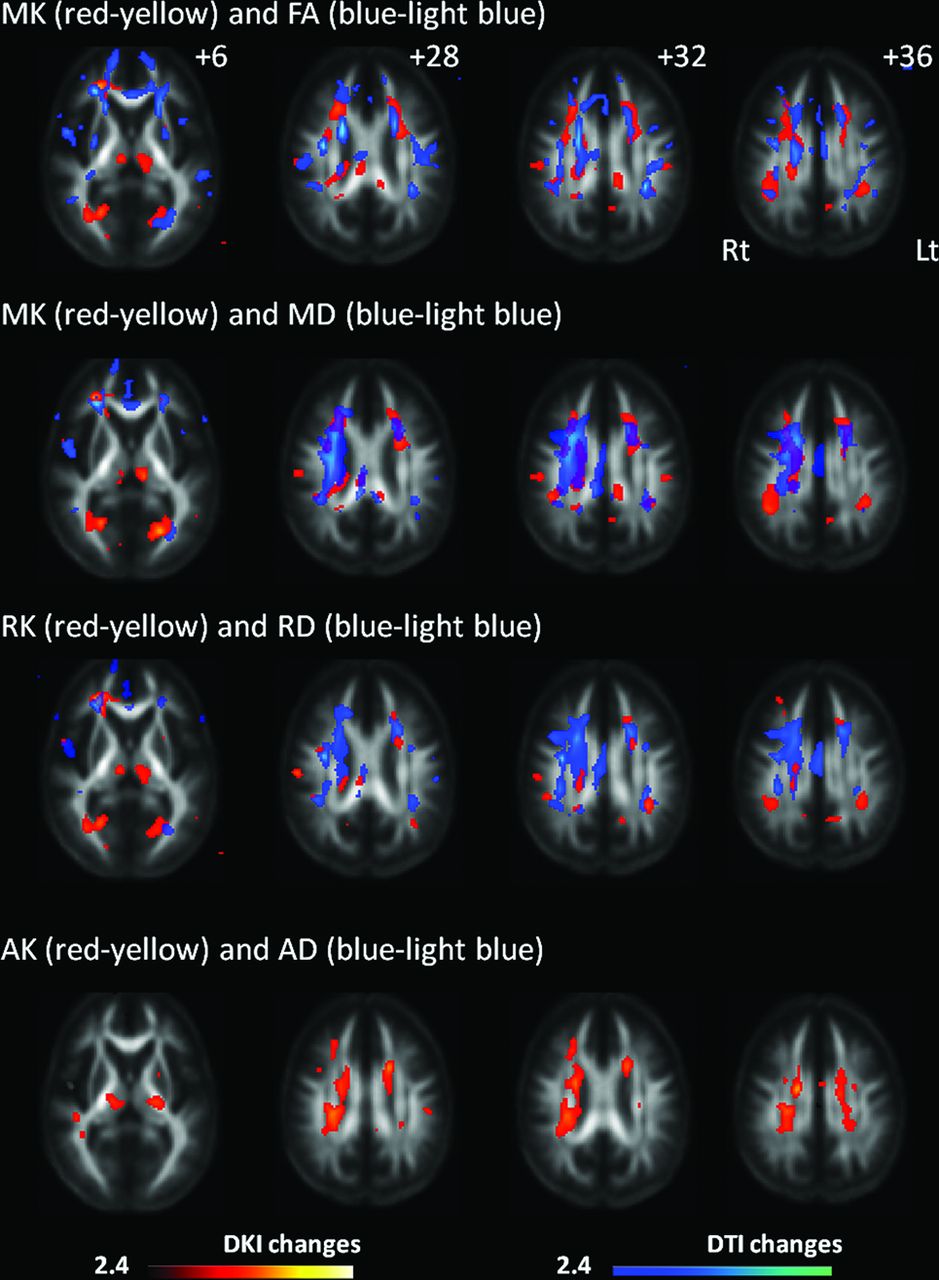
- Fig 1.
Changes in diffusional kurtosis imaging and diffusion tensor imaging parameters in Moyamoya disease shown in maps of 3 diffusional kurtosis parameters (mean kurtosis, radial kurtosis, and axial kurtosis) and 4 diffusion tensor parameters (fractional anisotropy, mean diffusivity, radial diffusivity, and axial diffusivity). Areas with significant changes in a combination of DKI/DTI parameters are as follows: decreased MK (red-yellow)/decreased FA (blue-light blue), decreased MK (red-yellow)/increased MD (blue-light blue), decreased RK (red-yellow)/increased RD (blue-light blue), and decreased AK (red-yellow)/increased and decreased AD (blue-light blue). Values from statistical parametric mapping analysis are projected onto axial sections of the average brain space of FA (z = 12, 28, 32, 36 mm). MK decrease is observed in the thalamus, a portion of the genu and body of the corpus callosum, corona radiata, frontoparietal subcortical white matter, and superior longitudinal fasciculus. RK decrease is observed in part of the frontoparietal subcortical white matter, thalamus, corona radiata, and occipital white matter. AK decrease is observed in the thalamus, temporo-occipital white matter, part of the SLF, and corona radiata. The radiologic convention is adopted, with the left side of the brain on the right side of axial panels. The color scale represents T values, with colored regions exceeding the significance threshold of P < .01 (T = 2.42) with a minimum cluster size of 50 voxels. Rt indicates right; Lt, left.
- Fig 2.
The bar graph indicates the number of the voxels with significant changes relative to the total number of white matter voxels in statistical parametric mapping comparing controls and patients with Moyamoya disease. Bar heights indicate decreases in MK/RK/AK/FA and increases in MD/RD/AD for 2 levels of threshold applied in group comparisons (blue, P < .01; red, P < .05, respectively).
- Fig 3.
Scatterplots indicating a significant correlation between neuropsychological test performance and diffusion parameters. Pearson moment-production correlation coefficient r and P values are demonstrated in each scatterplot. ROIs (green) for each parameter (AK, MK, FA, MD, and RD) are demonstrated with FA template images generated from 23 controls and 23 patients. A, Performance scores evaluated on the Wechsler Adult Intelligent Scale-III are significantly associated with axial kurtosis. Axial kurtosis is positively correlated with full-scale IQ (r = 0.42, P = .04) and subscores such as motor IQ (r = 0.49, P = .02), perceptual organization (r = 0.47, P = .03), and processing speed (r = 0.47, P = .03). B, Trail-Making Test, part B is inversely correlated with DKI/DTI parameters (MK; r = −0.44, P = .04; and FA; r = −0.49, P = .02) and positively correlated with DTI parameters (MD; r = 0.56, P = .007; and RD; r = 0.56, P = .007).


Tables
Control Moyamoya Disease P Value No. of subjects 23 23 – Age (mean) (range) (yr) 39.0 + 8.1 (25–56) 40.9 + 9.5 (21–58) .48 Sex (F/M) (No. of subjects) 13:10 17:6 .35 Risk factor (DM, HT, HL) (No. of subjects) 0 5 .049 Symptoms (No. of subjects) Asymptomatic – 13 TIA – 10 Note:—DM indicates diabetes mellitus; HT, hypertension; HL, hyperlipidemia.
Corpus Callosum Rt. SLF Lt. SLF CNT MMD CNT MMD CNT MMD r P r P r P r P r P r P MK vs FA 0.78 .000 0.80 .000 0.08 .715 0.60 .002 0.42 .047 0.47 .022 MK vs MD −0.90 .000 −0.86 .000 −0.60 .002 −0.68 .000 −0.41 .049 −0.49 .017 MK vs RD −0.90 .000 −0.87 .000 −0.54 .008 −0.73 .000 −0.49 .018 −0.55 .006 MK vs AD −0.87 .000 −0.82 .000 −0.50 .014 −0.46 .028 −0.19 .386 −0.35 .101 RK and RD −0.88 .000 −0.84 .000 −0.72 .000 −0.56 .005 −0.47 .025 −0.38 .072 AK and AD −0.75 .000 −0.64 .000 −0.44 .036 −0.61 .001 −0.47 .025 −0.68 .000 Note:—Rt. indicates right; Lt., left; CNT, controls; r; Pearson product-moment correlation coefficient.
↵a All P values, except .715, .386, 101, and ,072, indicate significant correlation between DKI and DTI parameters.
WCST Stroop CPT RST MK – .004b – .048b RK – .007b – – AK – .045c – .036c FA – .024b – – MD – .012b – – RD – .014b – – AD – .031c – – Note:—WCST indicates Wisconsin Card Sorting Test, Stroop; Stroop test.
↵a Data are P values. Correlations of diffusional kurtosis imaging and diffusion tensor imaging parameters with neuropsychological examinations were evaluated in patients with MMD. Three diffusional kurtosis parameters (MK, RK, and AK) and 4 diffusion tensor parameters (FA, MD, RD, and AD) were analyzed. Diffusion parameters and locations that demonstrated significant relationships with clinical variables follow.
↵b Two-tailed t test.
↵c One-tailed t test.


