
Article Figures & Data
Figures
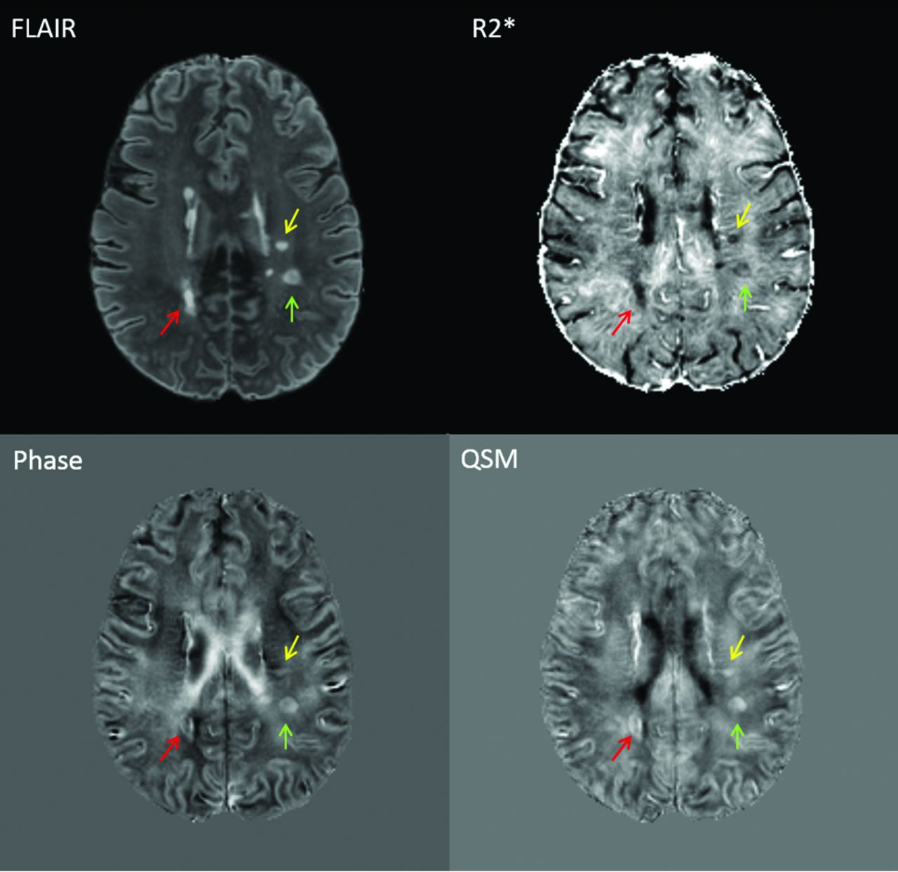
- Fig 1.
Examples of lesions seen on FLAIR, R2*, phase, and QSM. Each colored arrow indicates the same lesion seen on each of the 4 image contrasts. Lesions were initially identified on FLAIR images. Quantitative gray-scale values (ranging from black to white) for the above images are as follows: R2* = 0–283.78 Hz, phase = −48.69 to 40.00 Hz, QSM = −0.58 to 0.40 ppm.
- Fig 2.
Examples of common lesion patterns on R2*, phase, and QSM. Yellow arrows indicate lesions identified and shown here as samples of common lesion patterns found on each image contrast. Lesions were identified by the intensity of their core and outer rim when visually compared with surrounding white matter. Only a small portion of lesions were invisible on R2* (6%), whereas a larger proportion were invisible on phase (42%) and QSM (36%).
- Fig 3.
Quantitative comparison of lesion susceptibility values by disease subtype and level of disability. Box-and-whisker plots showing a quantitative comparison of mean lesion R2*, frequency (from phase), and relative susceptibility (from QSM) between subjects with RRMS and SPMS/PPMS (left column) and those with lower-versus-higher levels of disability based on the EDSS score (right column). Lesion R2* values were significantly lower in SPMS/PPMS and EDSS ≥ 5.0, and mean lesion relative susceptibility was lower in SPMS/PPMS. Lesion values were taken as the mean of all voxels within each lesion. Red lines indicate the median value for all lesions in each group. P values represent the results of Wilcoxon rank sum testing.
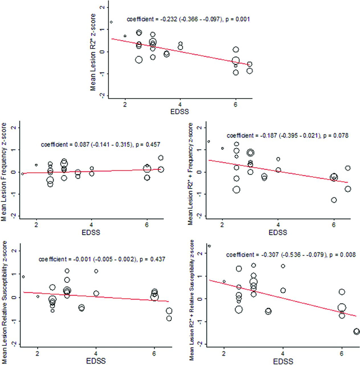
- Fig 4.
Relationship between EDSS and quantitative R2*, frequency, and relative susceptibility values. Shown are the results of linear mixed-model regression (adjusted for age and sex) for prediction of the quantitative MR imaging index value by the Expanded Disability Status Scale score as represented by a fitted-values plot. A significant inverse relationship was found between R2* and EDSS and for a combined index of R2* + relative susceptibility and EDSS. The open circles represent fitted values based on the fixed and random effects from the model, with each circle thus representing 1 subject and the size of the circle weighted for the number of lesions that particular subject contributed to the model. The regression coefficient for the fixed-effects portion of the model is shown in each panel, along with the P value for the significance of that coefficient. To place all quantitative values in an equivalent space, we converted all values to z score units (based on mean and SD from all lesions).


Tables
Demographics/Characteristics No. of subjects 24 No. of lesions 306 Lesions per subject analyzed (median) (range) 12 (2–29) Age (mean) (SD) (yr) 44.3 (10.0) Sex (No.) (%) Female 12 (50%) Male 12 (50%) Disease duration (mean) (SD) (yr) 11.2 (7.6) Clinical phenotype (No.) (%) Relapsing-remitting 21 (88%) Secondary-progressive 2 (8%) Primary-progressive 1 (4%) On MS treatment (No.) (%) 19 (79%) EDSS score (median) (range) 3.0 (1.5–6.5) MFIS score (mean) (SD) 37.7 (19.3) 9-HPT, dominant hand (mean) (SD) 23.0 (6.6) seconds 9-HPT, nondominant hand (mean) (SD) 27.7 (17.9) seconds Timed 25-ft walk (mean) (SD) 5.3 (2.4) seconds PASAT-3 score (mean) (SD) 46.3 (9.8) MSFC z score (mean) (SD) −0.27 (1.54) Note:—9-HPT indicates Nine Hole Peg Test; PASAT-3, Paced Auditory Serial Addition Test, 3-second delay.
- Table 2:
Hypothesized alterations in myelin and iron content in MS lesions associated with commonly observed R2*/QSM lesion contrast patterns
R2* Intensity QSM Intensity Alteration in Myelin Content Alteration in Iron Content Hypo Hyper ↓ ↔ Hypo Iso ↓ ↓ Iso Hyper ↓ ↑ Note:—Hypo indicates hypointensity; Hyper, hyperintensity; Iso, isointensity; ↑, increase; ↓, decrease; ↔, little-to-no change.


Jump to section
Related Articles
Cited By...
- Spherical Echo-Planar Time-resolved Imaging (sEPTI) for rapid 3D quantitative T2* and Susceptibility imaging
- Metabolic Insights into Iron Deposition in Relapsing-Remitting Multiple Sclerosis via 7T Magnetic Resonance Spectroscopic Imaging
- Quantitative MRI in Multiple Sclerosis: From Theory to Application
- Association of Slowly Expanding Lesions on MRI With Disability in People With Secondary Progressive Multiple Sclerosis
- QSMRim-Net: Imbalance-Aware Learning for Identification of Chronic Active Multiple Sclerosis Lesions on Quantitative Susceptibility Maps
- Disease correlates of quantitative susceptibility mapping rim lesions in multiple sclerosis
- Quantitative susceptibility mapping captures chronic multiple sclerosis rim lesions with greater myelin damage: Comparison with high-pass filtered phase MRI
- Absence of B Cells in Brainstem and White Matter Lesions Associates With Less Severe Disease and Absence of Oligoclonal Bands in MS
- Value of 3T Susceptibility-Weighted Imaging in the Diagnosis of Multiple Sclerosis
- Combining Quantitative Susceptibility Mapping with Automatic Zero Reference (QSM0) and Myelin Water Fraction Imaging to Quantify Iron-Related Myelin Damage in Chronic Active MS Lesions